DNA end resection

Last updated • 8 min readFrom Wikipedia, The Free Encyclopedia

DNA end resection, also called 5′–3′ degradation, is a biochemical process where the blunt end of a section of double-stranded DNA (dsDNA) is modified by cutting away some nucleotides from the 5' end to produce a 3' single-stranded sequence. [1] [2] The presence of a section of single-stranded DNA (ssDNA) allows the broken end of the DNA to line up accurately with a matching sequence, so that it can be accurately repaired. [1]

Contents

Mechanism of regulation of 5' resection of Mitotic and Telomeric DSBs. Mechanism of Telomeres and Mitotic DSBs.jpg
Mechanism of regulation of 5' resection of Mitotic and Telomeric DSBs.

Double-strand breaks (DSBs) can occur at any phase of the cell cycle causing DNA end resection and repair activities to take place, but they are also normal intermediates in mitosis recombination. [3] Furthermore, the natural ends of the linear chromosomes resemble DSBs, and although DNA breaks can cause damage to the integrity of genomic DNA, the natural ends are packed into complex specialized DNA protective packages called telomeres that prevent DNA repair activities. [3] [4] Telomeres and mitotic DSBs have different functionality, but both experience the same 5′–3′ degradation process.

Background

A double-strand break is a kind of DNA damage in which both strands in the double helix are severed. DSBs only occur during DNA replication of the cell cycle. Furthermore, DSBs can lead to genome rearrangements and instability. [3] Cases where two complementary strands are linked at the point of the DSB have potential to be catastrophic, such that the cell will not be able to complete mitosis when it next divides, and will either die or, in rare cases, undergo chromosomal loss, duplications, and even mutations. [5] [6] Three mechanisms exist to repair DSBs: non-homologous end joining (NHEJ), microhomology-mediated end joining (MMEJ), and homologous recombination HR. [7] [8] [9] Of these, only NHEJ does not rely on DNA end resection. [2]

Mechanism

Accurate repair of DSBs are essential in the upkeep of genome integrity. From the three mechanisms that exists to repair DSBs, NHEJ and HR repair mechanisms are the dominant pathways. [4] Several highly conservative proteins trigger the DNA Damage Checkpoint for detection of DSBs ensuing repair by either NHEJ or HR repair pathways. [3] NHEJ mechanism functions in ligating two different DSBs with high fidelity, while HR relies on a homologous template to repair DSB ends. [3] [4]

DNA end resection in the HR pathway only occurs at two specific phases: S and G2 phases. [4] [3] Since HR pathway requires sister chromatids for activation, this event only happens in the G2 and S phases of the cell cycle during replication. [3] [4] DSBs that have not begun DNA end resection can be ligated by NHEJ pathway, but resection of a few nucleotides inhibits the NHEJ pathway and commits' DNA repair by the HR pathway. [10] The NHEJ pathway is involved throughout the cell cycle, but it is critical to DNA repair during the G1 phase. [3] [10] In G1 phase there is no sister chromatids to repair DSBs via the HR pathway making the NHEJ pathway a critical repair mechanism. [3] [10]

Before resection can take place, the break needs to be detected. In animals, this detection is done by PARP1; [11] similar systems exist in other eukaryotes: in plants, PARP2 seems to play this role. [12] PARP binding then recruits the MRN complex to the breakage site. [13] This is a highly conserved complex consisting of Mre11, Rad50 and NBS1 (known as Nibrin [14] in mammals, or Xrs2 in yeast, where this complex is called the MRX complex).

Before resection can start, CtBP1-interacting protein (CtIP) needs to bind to the MRN complex so that the first phase of resection can begin, namely short-range end resection. [7] [15] [16] After phosphorylated CtIP binds, the Mre11 subunit is able to cut the 5'-terminated strand endonucleolytically, probably about 300 base pairs from the end, [15] [16] and then acts as a 3'→5' exonuclease to strip away the end of the 5' strand. [16]

Resection of telomere DSBs

Linear chromosomes are packed into complex specialized DNA protective packages called telomeres. [3] [4] The structure of telomeres is highly conserve and is organized in multiple short tandem DNA repeats. [3] Telomeres and DSBs have different functionality, such that telomeres prevent DNA repair activities. [3] During telomeric DNA replication in the S/G2 and G1 phases of the cell cycle, the 3' lagging strand leaves a short overhang called a G-tail. [4] [3] Telomeric DNA ends at the 3' G tail end because the 3' lagging strand extends without its complementary 5' C leading strand. The G tail provide a major function to telomeric DNA such that the G tails control telomere homeostasis. [4]

Telomeres in G1 phase

In the G1 phase of the cell cycle, the telomere-associated proteins RIF1, RIF2, and RAP2 bind to telomeric DNA and prevent access to the MRX complex. [4] [3] Such process in S. Cerevisiae for example is negatively regulated by this activity. The MRX complex and the Ku complex bind simultaneously and independently to DSBs ends. [3] [16] In the presence of the telomere-associated proteins, MRX fails to bind to the DSB ends while the Ku complex binds to DSB ends. [3] The bound Ku complex to the DSB ends protect the telomeres from nucleolytic degradation by exo1. [3] This results in an inhibition of telomerase elongation at the DSB ends and prevents further telomere action at the G1 phase of the cell cycle. [3] [4]

Telomeres in the late S/G2 phase

In the late S/G2 phase of the cell cycle, the telomere-associated proteins RIF1, RIF2, and RAP2 exhibit their inhibitory effect by binding to telomeric DNA. [3] In the Late S/G2 phase, the protein kinase CDK1 (cyclin-dependent) promotes telomeric resection. [4] [3] This control is exerted by cyclin-dependent kinases, which phosphorylate parts of the resection machinery. [15] This process alleviates the inhibitory effect of the telomere-associated proteins, and allows Cdc13 (a binding protein on both the lagging strand, and leading strand) to cover telomeric DNA. [15] The binding of cdc13 to DNA suppresses DNA damage checkpoint and allows resection to occur while allowing for telomerase elongation at the DSB ends. [3]

Resection of mitotic DSBs

One of the important regulatory controls in mitotic cells is deciding which specific DSB repair pathway to take. Once a DSB is detected, the highly conserved complexes are recruited by the DNA ends. [2] If the cell is in the G1 phase of the cell cycle, the complex Ku prevents resection to occur and triggers the NHEJ pathway factors. [3] DSBs in the NHEJ pathway are ligated, a step in the NHEJ pathway that requires DNA ligase activity of Dnl4-Lif1/XRCC4 heterodimer and the Nej1/XLF protein. [3] This process results in error-prone religation of DSB ends at the G1 phase of the cell cycle. [3]

If the cells are in S/G2 phase, mitotic DSBs are controlled through Cdk1 activity and involves phosphorylation of Sae2 Ser267. [4] [3] After phosphorylation occurs by Cdk1, MRX complex binds to dsDNA ends and generates short ssDNA that stretches in the 5' direction. [4] [3] The 5' ssDNA continues resection by the activity of the helicase enzyme, Sgs1 enzyme, and the nucleases Exo1 and Dna2. [17] Involvement of Sae2 Sar267 in DSB processing is highly conserved throughout eukaryotes, such that the Sae2 along with the MRX complex are involved in two major functions: single-strand annealing, and processing of hairpin DNA structures. [4] Like all ssDNA in the nucleus, the resected region is first coated by Replication protein A (RPA) complex, [18] [15] but RPA is then replaced with RAD51 to form a nucleoprotein filament which can take part in the search for a matching region, allowing HR to take place. [15] The 3' ssDNA coated by a RPA promotes the recruitment of Mec1. Mec1 further phosphorylates Sae2 along with cdk1. The resulting phosphorylation by Sae2 by Mec1 helps increase the effect of resection and this in turn leads to the DNA damage checkpoint activation. [4] [3]

Regulators

The pathway of choice in DNA repair is highly regulated to guarantee that cells in the S/G2 and G1 phase use the appropriate mechanism. Regulators in both the NHEJ and HR pathway mediate the appropriate DNA repair response pathway. [3] Furthermore, recent studies into DNA repair show that regulation of DNA end resection is governed by the activity of cdk1 in the cell replication cycle. [3] [19]

NHEJ pathway

DNA end resection is key in determining the correct pathway in NHEJ. For NHEJ pathway to occur, positive regulators such as the Ku and MRX complex mediate recruitment of other NHEJ-associated proteins such as Tel1, Lif1, Dnl4, and Nej1. [3] Since NHEJ does not rely on end resection, NHEJ could only happen in the G1 phase of the cell cycle. [3] [10] Both Ku and NHEJ-associated proteins prevent initiation of resection.

Resection ensures that DSBs are not repaired by NHEJ (which joins broken DNA ends together without ensuring that they match), but rather by methods based on homology (matching DNA sequences). Cyclin-dependent protein kinase such as cdk1 in yeast serves as a negative regulator of the NHEJ pathway. [3] Any activity associated with the presence of cyclin dependent protein kinases inhibit the NHEJ pathway [3]

Positive regulators

The presence of a ssDNA allows the broken end of the DNA to line up accurately with a matching sequence, so that it can be accurately repaired. [1] For HR pathway to occur in the S and G2 phases of the cell cycle, availability of a sister chromatid is required. [3] [4] 5′–3′ resection automatically links a DSB to the HR pathway. [2] Cyclin-dependent protein kinase such as cdk1 serve as a positive regulator of the HR pathway. [4] [3] This positive regulator promotes 5′–3′ nucleolytic degradation of DNA ends. Along with cdk1, the MRX complex, B1 cyclin, and Spo11-induced DSBs serve as a positive regulators to the HR pathway. [17] [19]

See also

Related Research Articles

<span class="mw-page-title-main">Cell cycle</span> Series of events and stages that result in cell division

The cell cycle, or cell-division cycle, is the series of events that take place in a cell that causes it to divide into two daughter cells. These events include the duplication of its DNA and some of its organelles, and subsequently the partitioning of its cytoplasm, chromosomes and other components into two daughter cells in a process called cell division.

<span class="mw-page-title-main">Cell division</span> Process by which living cells divide

Cell division is the process by which a parent cell divides into two daughter cells. Cell division usually occurs as part of a larger cell cycle in which the cell grows and replicates its chromosome(s) before dividing. In eukaryotes, there are two distinct types of cell division: a vegetative division (mitosis), producing daughter cells genetically identical to the parent cell, and a cell division that produces haploid gametes for sexual reproduction (meiosis), reducing the number of chromosomes from two of each type in the diploid parent cell to one of each type in the daughter cells. In Mitosis is a part of the cell cycle, in which, replicated chromosomes are separated into two new nuclei. Cell division gives rise to genetically identical cells in which the total number of chromosomes is maintained. In general, mitosis is preceded by the S stage of interphase and is followed by telophase and cytokinesis; which divides the cytoplasm, organelles, and cell membrane of one cell into two new cells containing roughly equal shares of these cellular components. The different stages of mitosis all together define the M phase of an animal cell cycle—the division of the mother cell into two genetically identical daughter cells. To ensure proper progression through the cell cycle, DNA damage is detected and repaired at various checkpoints throughout the cycle. These checkpoints can halt progression through the cell cycle by inhibiting certain cyclin-CDK complexes. Meiosis undergoes two divisions resulting in four haploid daughter cells. Homologous chromosomes are separated in the first division of meiosis, such that each daughter cell has one copy of each chromosome. These chromosomes have already been replicated and have two sister chromatids which are then separated during the second division of meiosis. Both of these cell division cycles are used in the process of sexual reproduction at some point in their life cycle. Both are believed to be present in the last eukaryotic common ancestor.

<span class="mw-page-title-main">Interphase</span> G1, S and G2 phases of the cell cycle

Interphase is the portion of the cell cycle that is not accompanied by visible changes under the microscope, and includes the G1, S and G2 phases. During interphase, the cell grows (G1), replicates its DNA (S) and prepares for mitosis (G2). A cell in interphase is not simply quiescent. The term quiescent would be misleading since a cell in interphase is very busy synthesizing proteins, copying DNA into RNA, engulfing extracellular material, processing signals, to name just a few activities. The cell is quiescent only in the sense of cell division. Interphase is the phase of the cell cycle in which a typical cell spends most of its life. Interphase is the 'daily living' or metabolic phase of the cell, in which the cell obtains nutrients and metabolizes them, grows, replicates its DNA in preparation for mitosis, and conducts other "normal" cell functions.

<span class="mw-page-title-main">DNA repair</span> Cellular mechanism

DNA repair is a collection of processes by which a cell identifies and corrects damage to the DNA molecules that encodes its genome. In human cells, both normal metabolic activities and environmental factors such as radiation can cause DNA damage, resulting in tens of thousands of individual molecular lesions per cell per day. Many of these lesions cause structural damage to the DNA molecule and can alter or eliminate the cell's ability to transcribe the gene that the affected DNA encodes. Other lesions induce potentially harmful mutations in the cell's genome, which affect the survival of its daughter cells after it undergoes mitosis. As a consequence, the DNA repair process is constantly active as it responds to damage in the DNA structure. When normal repair processes fail, and when cellular apoptosis does not occur, irreparable DNA damage may occur, including double-strand breaks and DNA crosslinkages. This can eventually lead to malignant tumors, or cancer as per the two hit hypothesis.

<span class="mw-page-title-main">S phase</span> DNA replication phase of the cell cycle, between G1 and G2 phase

S phase (Synthesis phase) is the phase of the cell cycle in which DNA is replicated, occurring between G1 phase and G2 phase. Since accurate duplication of the genome is critical to successful cell division, the processes that occur during S-phase are tightly regulated and widely conserved.

<span class="mw-page-title-main">Non-homologous end joining</span> Pathway that repairs double-strand breaks in DNA

Non-homologous end joining (NHEJ) is a pathway that repairs double-strand breaks in DNA. It is called "non-homologous" because the break ends are directly ligated without the need for a homologous template, in contrast to homology directed repair (HDR), which requires a homologous sequence to guide repair. NHEJ is active in both non-dividing and proliferating cells, while HDR is not readily accessible in non-dividing cells. The term "non-homologous end joining" was coined in 1996 by Moore and Haber.

G<sub>2</sub> phase Second growth phase in the eukaryotic cell cycle, prior to mitosis

G2 phase, Gap 2 phase, or Growth 2 phase, is the third subphase of interphase in the cell cycle directly preceding mitosis. It follows the successful completion of S phase, during which the cell’s DNA is replicated. G2 phase ends with the onset of prophase, the first phase of mitosis in which the cell’s chromatin condenses into chromosomes.

<span class="mw-page-title-main">Homologous recombination</span> Genetic recombination between identical or highly similar strands of genetic material

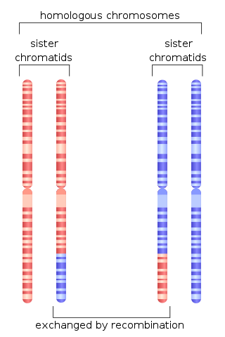
Homologous recombination is a type of genetic recombination in which genetic information is exchanged between two similar or identical molecules of double-stranded or single-stranded nucleic acids.

<span class="mw-page-title-main">Cell cycle checkpoint</span> Control mechanism in the eukaryotic cell cycle

Cell cycle checkpoints are control mechanisms in the eukaryotic cell cycle which ensure its proper progression. Each checkpoint serves as a potential termination point along the cell cycle, during which the conditions of the cell are assessed, with progression through the various phases of the cell cycle occurring only when favorable conditions are met. There are many checkpoints in the cell cycle, but the three major ones are: the G1 checkpoint, also known as the Start or restriction checkpoint or Major Checkpoint; the G2/M checkpoint; and the metaphase-to-anaphase transition, also known as the spindle checkpoint. Progression through these checkpoints is largely determined by the activation of cyclin-dependent kinases by regulatory protein subunits called cyclins, different forms of which are produced at each stage of the cell cycle to control the specific events that occur therein.

<span class="mw-page-title-main">Ku (protein)</span>

Ku is a dimeric protein complex that binds to DNA double-strand break ends and is required for the non-homologous end joining (NHEJ) pathway of DNA repair. Ku is evolutionarily conserved from bacteria to humans. The ancestral bacterial Ku is a homodimer. Eukaryotic Ku is a heterodimer of two polypeptides, Ku70 (XRCC6) and Ku80 (XRCC5), so named because the molecular weight of the human Ku proteins is around 70 kDa and 80 kDa. The two Ku subunits form a basket-shaped structure that threads onto the DNA end. Once bound, Ku can slide down the DNA strand, allowing more Ku molecules to thread onto the end. In higher eukaryotes, Ku forms a complex with the DNA-dependent protein kinase catalytic subunit (DNA-PKcs) to form the full DNA-dependent protein kinase, DNA-PK. Ku is thought to function as a molecular scaffold to which other proteins involved in NHEJ can bind, orienting the double-strand break for ligation.

Cyclin A is a member of the cyclin family, a group of proteins that function in regulating progression through the cell cycle. The stages that a cell passes through that culminate in its division and replication are collectively known as the cell cycle Since the successful division and replication of a cell is essential for its survival, the cell cycle is tightly regulated by several components to ensure the efficient and error-free progression through the cell cycle. One such regulatory component is cyclin A which plays a role in the regulation of two different cell cycle stages.

<span class="mw-page-title-main">TP53BP1</span> Protein-coding gene in the species Homo sapiens

Tumor suppressor p53-binding protein 1 also known as p53-binding protein 1 or 53BP1 is a protein that in humans is encoded by the TP53BP1 gene.

<span class="mw-page-title-main">Homology directed repair</span>

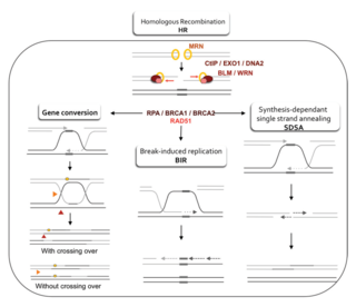
Homology-directed repair (HDR) is a mechanism in cells to repair double-strand DNA lesions. The most common form of HDR is homologous recombination. The HDR mechanism can only be used by the cell when there is a homologous piece of DNA present in the nucleus, mostly in G2 and S phase of the cell cycle. Other examples of homology-directed repair include single-strand annealing and breakage-induced replication. When the homologous DNA is absent, another process called non-homologous end joining (NHEJ) takes place instead.

The MRX complex is a heterotrimeric protein complex consisting of Mre11, Rad50, and Xrs2. It is a budding yeast homolog of the mammalian Mre11-Rad50-Nbs1 (MRN) DNA damage repair complex.

Microhomology-mediated end joining (MMEJ), also known as alternative nonhomologous end-joining (Alt-NHEJ) is one of the pathways for repairing double-strand breaks in DNA. As reviewed by McVey and Lee, the foremost distinguishing property of MMEJ is the use of microhomologous sequences during the alignment of broken ends before joining, thereby resulting in deletions flanking the original break. MMEJ is frequently associated with chromosome abnormalities such as deletions, translocations, inversions and other complex rearrangements.

<span class="mw-page-title-main">G2-M DNA damage checkpoint</span>

The G2-M DNA damage checkpoint is an important cell cycle checkpoint in eukaryotic organisms that ensures that cells don't initiate mitosis until damaged or incompletely replicated DNA is sufficiently repaired. Cells with a defective G2-M checkpoint will undergo apoptosis or death after cell division if they enter the M phase before repairing their DNA. The defining biochemical feature of this checkpoint is the activation of M-phase cyclin-CDK complexes, which phosphorylate proteins that promote spindle assembly and bring the cell to metaphase.

Shelterin is a protein complex known to protect telomeres in many eukaryotes from DNA repair mechanisms, as well as to regulate telomerase activity. In mammals and other vertebrates, telomeric DNA consists of repeating double-stranded 5'-TTAGGG-3' (G-strand) sequences along with the 3'-AATCCC-5' (C-strand) complement, ending with a 50-400 nucleotide 3' (G-strand) overhang. Much of the final double-stranded portion of the telomere forms a T-loop (Telomere-loop) that is invaded by the 3' (G-strand) overhang to form a small D-loop (Displacement-loop).

Cell cycle withdrawal refers to the natural stoppage of cell cycle during cell division. When cells divide, there are many internal or external factors that would lead to a stoppage of division. This stoppage could be permanent or temporary, and could occur in any one of the four cycle phases, depending on the status of cells or the activities they are undergoing. During the process, all cell duplication process, including mitosis, meiosis as well as DNA replication, will be paused. The mechanisms involve the proteins and DNA sequences inside cells.

Telomeres, the caps on the ends of eukaryotic chromosomes, play critical roles in cellular aging and cancer. An important facet to how telomeres function in these roles is their involvement in cell cycle regulation.

<span class="mw-page-title-main">Double-strand break repair model</span>

A double-strand break repair model refers to the various models of pathways that cells undertake to repair double strand-breaks (DSB). DSB repair is an important cellular process, as the accumulation of unrepaired DSB could lead to chromosomal rearrangements, tumorigenesis or even cell death. In human cells, there are two main DSB repair mechanisms: Homologous recombination (HR) and non-homologous end joining (NHEJ). HR relies on undamaged template DNA as reference to repair the DSB, resulting in the restoration of the original sequence. NHEJ modifies and ligates the damaged ends regardless of homology. In terms of DSB repair pathway choice, most mammalian cells appear to favor NHEJ rather than HR. This is because the employment of HR may lead to gene deletion or amplification in cells which contains repetitive sequences. In terms of repair models in the cell cycle, HR is only possible during the S and G2 phases, while NHEJ can occur throughout whole process. These repair pathways are all regulated by the overarching DNA damage response mechanism. Besides HR and NHEJ, there are also other repair models which exists in cells. Some are categorized under HR, such as synthesis-dependent strain annealing, break-induced replication, and single-strand annealing; while others are an entirely alternate repair model, namely, the pathway microhomology-mediated end joining (MMEJ).

References

  1. 1 2 3 Jimeno S, Mejías-Navarro F, Prados-Carvajal R, Huertas P (2019). "Controlling the balance between chromosome break repair pathways". Advances in Protein Chemistry and Structural Biology. Elsevier. 115: 95–134. doi:10.1016/bs.apcsb.2018.10.004. ISBN   9780128155592. PMID   30798939. S2CID   73459973.
  2. 1 2 3 4 Liu T, Huang J (June 2016). "DNA End Resection: Facts and Mechanisms". Genomics, Proteomics & Bioinformatics. 14 (3): 126–130. doi:10.1016/j.gpb.2016.05.002. PMC   4936662 . PMID   27240470.
  3. 1 2 3 4 5 6 7 8 9 10 11 12 13 14 15 16 17 18 19 20 21 22 23 24 25 26 27 28 29 30 31 32 33 34 35 36 Longhese MP, Bonetti D, Manfrini N, Clerici M (September 2010). "Mechanisms and regulation of DNA end resection". The EMBO Journal. 29 (17): 2864–2874. doi:10.1038/emboj.2010.165. PMC   2944052 . PMID   20647996.
  4. 1 2 3 4 5 6 7 8 9 10 11 12 13 14 15 16 17 Mimitou EP, Symington LS (September 2009). "DNA end resection: many nucleases make light work". DNA Repair. 8 (9): 983–995. doi:10.1016/j.dnarep.2009.04.017. PMC   2760233 . PMID   19473888.
  5. Bjorksten J, Acharya PV, Ashman S, Wetlaufer DB (July 1971). "Gerogenic fractions in the tritiated rat". Journal of the American Geriatrics Society. 19 (7): 561–574. doi:10.1111/j.1532-5415.1971.tb02577.x. PMID   5106728. S2CID   33154242.
  6. Acharya PV (1972). "The isolation and partial characterization of age-correlated oligo-deoxyribo-ribonucleotides with covalently linked aspartyl-glutamyl polypeptides". Johns Hopkins Medical Journal. Supplement (1): 254–260. PMID   5055816.
  7. 1 2 Huertas P (January 2010). "DNA resection in eukaryotes: deciding how to fix the break". Nature Structural & Molecular Biology. 17 (1): 11–16. doi:10.1038/nsmb.1710. PMC   2850169 . PMID   20051983.
  8. Liang L, Deng L, Chen Y, Li GC, Shao C, Tischfield JA (September 2005). "Modulation of DNA end joining by nuclear proteins". The Journal of Biological Chemistry. 280 (36): 31442–31449. doi: 10.1074/jbc.M503776200 . PMID   16012167.
  9. Watson JD, Baker TA, Bell SP, Gann A, Levine M, Losick R, eds. (2004). Molecular Biology of the Gene (5th ed.). Pearson Benjamin Cummings; CSHL Press. Ch. 9, 10. OCLC   936762772.
  10. 1 2 3 4 Daley JM, Laan RL, Suresh A, Wilson TE (August 2005). "DNA joint dependence of pol X family polymerase action in nonhomologous end joining". The Journal of Biological Chemistry. 280 (32): 29030–29037. doi: 10.1074/jbc.M505277200 . PMID   15964833.
  11. Ray Chaudhuri A, Nussenzweig A (October 2017). "The multifaceted roles of PARP1 in DNA repair and chromatin remodelling". Nature Reviews. Molecular Cell Biology. 18 (10): 610–621. doi:10.1038/nrm.2017.53. PMC   6591728 . PMID   28676700.
  12. Song J, Keppler BD, Wise RR, Bent AF (May 2015). "PARP2 Is the Predominant Poly(ADP-Ribose) Polymerase in Arabidopsis DNA Damage and Immune Responses". PLOS Genetics. 11 (5): e1005200. doi: 10.1371/journal.pgen.1005200 . PMC   4423837 . PMID   25950582.
  13. Haince JF, McDonald D, Rodrigue A, Déry U, Masson JY, Hendzel MJ, Poirier GG (January 2008). "PARP1-dependent kinetics of recruitment of MRE11 and NBS1 proteins to multiple DNA damage sites". The Journal of Biological Chemistry. 283 (2): 1197–1208. doi: 10.1074/jbc.M706734200 . PMID   18025084.
  14. Uhrhammer N, Bay JO, Gatti RA (October 2002). "NBN (Nijmegen breakage syndrome 1)". Atlas of Genetics and Cytogenetics in Oncology and Haematology - NBS1. Archived from the original on 2006-09-29. Retrieved 2008-02-12.
  15. 1 2 3 4 5 6 Casari E, Rinaldi C, Marsella A, Gnugnoli M, Colombo CV, Bonetti D, Longhese MP (2019). "Processing of DNA Double-Strand Breaks by the MRX Complex in a Chromatin Context". Frontiers in Molecular Biosciences. 6: 43. doi: 10.3389/fmolb.2019.00043 . PMC   6567933 . PMID   31231660.
  16. 1 2 3 4 Pinto C, Anand R, Cejka P (2018). "Methods to Study DNA End Resection II: Biochemical Reconstitution Assays". Mechanisms of DNA Recombination and Genome Rearrangements: Methods to Study Homologous Recombination. Methods in Enzymology. Vol. 600. pp. 67–106. doi:10.1016/bs.mie.2017.11.009. ISBN   9780128144299. PMID   29458776.
  17. 1 2 Xue C, Wang W, Crickard JB, Moevus CJ, Kwon Y, Sung P, Greene EC (March 2019). "Regulatory control of Sgs1 and Dna2 during eukaryotic DNA end resection". Proceedings of the National Academy of Sciences of the United States of America. 116 (13): 6091–6100. Bibcode:2019PNAS..116.6091X. doi: 10.1073/pnas.1819276116 . PMC   6442620 . PMID   30850524.
  18. Chen C, ed. (2013). New research directions in DNA repair. Croatia: InTech. ISBN   978-953-51-1114-6. OCLC   957280914.
  19. 1 2 Poon RY (2016-01-01). "Mitotic Catastrophe". In Bradshaw RA, Stahl PD (eds.). Encyclopedia of Cell Biology. Waltham: Academic Press. pp. 399–403. ISBN   978-0-12-394796-3.