Mastocytosis, a type of mast cell disease, is a rare disorder affecting both children and adults caused by the accumulation of functionally defective mast cells and CD34+ mast cell precursors.

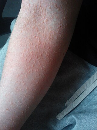
Hives, also known as urticaria, is a kind of skin rash with red, raised, itchy bumps. Hives may burn or sting. The patches of rash may appear on different body parts, with variable duration from minutes to days, and does not leave any long-lasting skin change. Fewer than 5% of cases last for more than six weeks. The condition frequently recurs.
Darier's disease (DD) is a rare, genetic skin disorder. It is an autosomal dominant disorder, that is, if one parent has DD, there is a 50% chance than a child will inherit DD. It was first reported by French dermatologist Ferdinand-Jean Darier in 1889.

A seborrheic keratosis is a non-cancerous (benign) skin tumour that originates from cells, namely keratinocytes, in the outer layer of the skin called the epidermis. Like liver spots, seborrheic keratoses are seen more often as people age.
Aquagenic pruritus is a skin condition characterized by the development of severe, intense, prickling-like epidermal itching without observable skin lesions and evoked by contact with water.

Urticaria pigmentosa (also known as generalized eruption of cutaneous mastocytosis (childhood type) ) is the most common form of cutaneous mastocytosis. It is a rare disease caused by excessive numbers of mast cells in the skin that produce hives or lesions on the skin when irritated.

Physical urticaria is a distinct subgroup of urticaria (hives) that are induced by an exogenous physical stimulus rather than occurring spontaneously. There are seven subcategories that are recognized as independent diseases. Physical urticaria is known to be painful, itchy and physically unappealing; it can recur for months to years.

The Koebner phenomenon or Köbner phenomenon, also called the Koebner response or the isomorphic response, attributed to Heinrich Köbner, is the appearance of skin lesions on lines of trauma. The Koebner phenomenon may result from either a linear exposure or irritation. Conditions demonstrating linear lesions after a linear exposure to a causative agent include: molluscum contagiosum, warts and toxicodendron dermatitis. Warts and molluscum contagiosum lesions can be spread in linear patterns by self-scratching ("auto-inoculation"). Toxicodendron dermatitis lesions are often linear from brushing up against the plant. Causes of the Koebner phenomenon that are secondary to scratching rather than an infective or chemical cause include vitiligo, psoriasis, lichen planus, lichen nitidus, pityriasis rubra pilaris, and keratosis follicularis.

Cholinergic urticaria or also known as (CholU) and CU, is a rare form of hives (urticaria) that is triggered by an elevation in body temperature, breaking a sweat, or exposure to heat. It is also sometimes called exercise-induced urticaria or heat hives. The condition is considered to be one of the many rarest forms of allergies known to medical science.

Gestational pemphigoid (GP) is a rare autoimmune variant of the skin disease bullous pemphigoid, and first appears in pregnancy. It presents with tense blisters, small bumps, hives and intense itching, usually starting around the navel before spreading to limbs in mid-pregnancy or shortly after delivery. The head, face and mouth are not usually affected.

Solar urticaria (SU) is a rare condition in which exposure to ultraviolet or UV radiation, or sometimes even visible light, induces a case of urticaria or hives that can appear in both covered and uncovered areas of the skin. It is classified as a type of physical urticaria. The classification of disease types is somewhat controversial. One classification system distinguished various types of SU based on the wavelength of the radiation that causes the breakout; another classification system is based on the type of allergen that initiates a breakout.
Aquagenic urticaria, also known as water allergy and water urticaria, is a form of physical urticaria in which hives develop on the skin after contact with water, regardless of its temperature. The condition typically results from contact with water of any type, temperature or additive.

Solitary mastocytoma, also known as cutaneous mastocytoma, may be present at birth or may develop during the first weeks of life, originating as a brown macule that urticates on stroking. Solitary mastocytoma is a round, erythematous, indurated lesion measuring 1-5 cm in diameter. It can be mildly itchy or asymptomatic and develops over time. Predilection is the head and neck, followed by the trunk, extremities, and flexural areas.
Telangiectasia macularis eruptiva perstans (TMEP) is persistent, pigmented, asymptomatic eruption of macules usually less than 0.5 cm in diameter with a slightly reddish-brown tinge.
One of the most prevalent forms of adverse drug reactions is cutaneous reactions, with drug-induced urticaria ranking as the second most common type, preceded by drug-induced exanthems. Urticaria, commonly known as hives, manifests as weals, itching, burning, redness, swelling, and angioedema—a rapid swelling of lower skin layers, often more painful than pruritic. These symptoms may occur concurrently, successively, or independently. Typically, when a drug triggers urticaria, symptoms manifest within 24 hours of ingestion, aiding in the identification of the causative agent. Urticaria symptoms usually subside within 1–24 hours, while angioedema may take up to 72 hours to resolve completely.
Adrenergic urticaria is a skin condition characterized by an eruption consisting of small (1-5mm) red macules and papules with a pale halo, appearing within 10 to 15 min after emotional upset. There have been 10 cases described in medical literature, and involve a trigger followed by a rise in catecholamine and IgE. Treatment involves propranolol and trigger avoidance.
Senile pruritus is one of the most common conditions in the elderly or people over 65 years of age with an emerging itch that may be accompanied with changes in temperature and textural characteristics. In the elderly, xerosis, is the most common cause for an itch due to the degradation of the skin barrier over time. However, the cause of senile pruritus is not clearly known. Diagnosis is based on an elimination criteria during a full body examination that can be done by either a dermatologist or non-dermatologist physician.

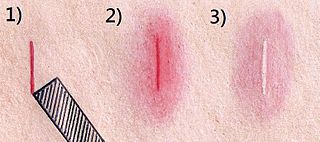
The triple response of Lewis is a cutaneous response that occurs from firm stroking of the skin. This produces an initial red line, followed by a flare around that line, and then finally a wheal.

Chronic spontaneous urticaria(CSU) also known as Chronic idiopathic urticaria(CIU) is defined by the presence of wheals, angioedema, or both for more than six weeks. The most common symptoms of chronic spontaneous urticaria are angioedema and hives that are accompanied by itchiness.
Cutaneous manifestations of COVID-19 are characteristic signs or symptoms of the Coronavirus disease 2019 that occur in the skin. The American Academy of Dermatology reports that skin lesions such as morbilliform, pernio, urticaria, macular erythema, vesicular purpura, papulosquamous purpura and retiform purpura are seen in people with COVID-19. Pernio-like lesions were more common in mild disease while retiform purpura was seen only in critically ill patients. The major dermatologic patterns identified in individuals with COVID-19 are urticarial rash, confluent erythematous/morbilliform rash, papulovesicular exanthem, chilbain-like acral pattern, livedo reticularis and purpuric "vasculitic" pattern. Chilblains and Multisystem inflammatory syndrome in children are also cutaneous manifestations of COVID-19.


