Related Research Articles
This is a glossary of medical terms related to communication disorders which are psychological or medical conditions that could have the potential to affect the ways in which individuals can hear, listen, understand, speak and respond to others.

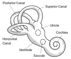
The inner ear is the innermost part of the vertebrate ear. In vertebrates, the inner ear is mainly responsible for sound detection and balance. In mammals, it consists of the bony labyrinth, a hollow cavity in the temporal bone of the skull with a system of passages comprising two main functional parts:

Ménière's disease (MD) is a disease of the inner ear that is characterized by potentially severe and incapacitating episodes of vertigo, tinnitus, hearing loss, and a feeling of fullness in the ear. Typically, only one ear is affected initially, but over time, both ears may become involved. Episodes generally last from 20 minutes to a few hours. The time between episodes varies. The hearing loss and ringing in the ears can become constant over time.

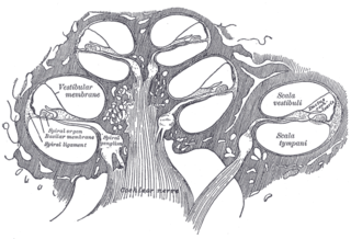
The cochlea is the part of the inner ear involved in hearing. It is a spiral-shaped cavity in the bony labyrinth, in humans making 2.75 turns around its axis, the modiolus. A core component of the cochlea is the organ of Corti, the sensory organ of hearing, which is distributed along the partition separating the fluid chambers in the coiled tapered tube of the cochlea.
A balance disorder is a disturbance that causes an individual to feel unsteady, for example when standing or walking. It may be accompanied by feelings of giddiness, or wooziness, or having a sensation of movement, spinning, or floating. Balance is the result of several body systems working together: the visual system (eyes), vestibular system (ears) and proprioception. Degeneration or loss of function in any of these systems can lead to balance deficits.

The vestibular system, in vertebrates, is a sensory system that creates the sense of balance and spatial orientation for the purpose of coordinating movement with balance. Together with the cochlea, a part of the auditory system, it constitutes the labyrinth of the inner ear in most mammals.

Endolymph is the fluid contained in the membranous labyrinth of the inner ear. The major cation in endolymph is potassium, with the values of sodium and potassium concentration in the endolymph being 0.91 mM and 154 mM, respectively. It is also called Scarpa's fluid, after Antonio Scarpa.

Otology is a branch of medicine which studies normal, pathological anatomy and physiology of the ear, as well as their diseases, diagnosis and treatment. Otologic surgery generally refers to surgery of the middle ear and mastoid related to chronic otitis media, such as tympanoplasty, or ear drum surgery, ossiculoplasty, or surgery of the hearing bones, and mastoidectomy. Otology also includes surgical treatment of conductive hearing loss, such as stapedectomy surgery for otosclerosis.

Betahistine, sold under the brand name Serc among others, is an anti-vertigo medication. It is commonly prescribed for balance disorders or to alleviate vertigo symptoms. It was first registered in Europe in 1970 for the treatment of Ménière's disease, but current evidence does not support its efficacy in treating it.

In the inner ear, stereocilia are the mechanosensing organelles of hair cells, which respond to fluid motion in numerous types of animals for various functions, including hearing and balance. They are about 10–50 micrometers in length and share some similar features of microvilli. The hair cells turn the fluid pressure and other mechanical stimuli into electric stimuli via the many microvilli that make up stereocilia rods. Stereocilia exist in the auditory and vestibular systems.

Vertigo is a condition in which a person has the sensation that they are moving, or that objects around them are moving, when they are not. Often it feels like a spinning or swaying movement. It may be associated with nausea, vomiting, perspiration, or difficulties walking. It is typically worse when the head is moved. Vertigo is the most common type of dizziness.

Perilymph is an extracellular fluid located within the inner ear. It is found within the scala tympani and scala vestibuli of the cochlea. The ionic composition of perilymph is comparable to that of plasma and cerebrospinal fluid. The major cation in perilymph is sodium, with the values of sodium and potassium concentration in the perilymph being 138 mM and 6.9 mM, respectively. It is also named Cotunnius' liquid and liquor cotunnii for Domenico Cotugno.

The vestibular membrane, vestibular wall or Reissner's membrane is a membrane inside the cochlea of the inner ear. It separates the cochlear duct from the vestibular duct. It helps to transmit vibrations from fluid in the vestibular duct to the cochlear duct. Together with the basilar membrane, it creates a compartment in the cochlea filled with endolymph, which is important for the function of the spiral organ of Corti. It allows nutrients to travel from the perilymph to the endolymph of the membranous labyrinth. It may be damaged in Ménière's disease. It is named after the German anatomist Ernst Reissner.

Autoimmune inner ear disease (AIED) was first defined by Dr. Brian McCabe in a landmark paper describing an autoimmune loss of hearing. The disease results in progressive sensorineural hearing loss (SNHL) that acts bilaterally and asymmetrically, and sometimes affects an individual's vestibular system. AIED is used to describe any disorder in which the inner ear is damaged as a result of an autoimmune response. Some examples of autoimmune disorders that have presented with AIED are Cogan's syndrome, relapsing polychondritis, systemic lupus erythematosus, granulomatosis with polyangiitis, polyarteritis nodosa, Sjogren's syndrome, and Lyme disease.
EAST syndrome is a syndrome consisting of epilepsy, ataxia, sensorineural deafness and salt-wasting renal tubulopathy. The tubulopathy in this condition predispose to hypokalemic metabolic alkalosis with normal blood pressure. Hypomagnesemia may also be present.

Large vestibular aqueduct is a structural deformity of the inner ear. Enlargement of this duct is one of the most common inner ear deformities and is commonly associated with hearing loss during childhood. The term was first discovered in 1791 by Mondini when he was completing a temporal bone dissection. It was then defined by Valvassori and Clemis as a vestibular aqueduct that is greater than or equal to 2.0 mm at the operculum and/or greater than or equal to 1.0 mm at the midpoint. Some use the term enlarged vestibular aqueduct syndrome, but this is felt by others to be erroneous as it is a clinical finding which can occur in several syndromes.
Neurotology or neuro-otology is a subspecialty of otolaryngology—head and neck surgery, also known as ENT medicine. Neuro-otology is closely related to otology, clinical neurology and neurosurgery.

An endolymphatic sac tumor (ELST) is a very uncommon papillary epithelial neoplasm arising within the endolymphatic sac or endolymphatic duct. This tumor shows a very high association with Von Hippel–Lindau syndrome (VHL).
Dark cells are specialized nonsensory epithelial cells found on either side of the vestibular organs and lining the endolymphatic space. These dark-cell areas in the vestibular organ are structures involved in the production of endolymph, an inner ear fluid, secreting potassium towards the endolymphatic fluid. Dark cells take part in fluid homeostasis to preserve the unique high-potassium and low-sodium content of the endolymph and also maintain the calcium homeostasis of the inner ear.
Cochlear hydrops is a condition of the inner ear involving a pathological increase of fluid affecting the cochlea. This results in swelling that can lead to hearing loss or changes in hearing perception. It is a form of endolymphatic hydrops and related to Ménière's disease. Cochlear hydrops refers to a case of inner-ear hydrops that only involves auditory symptoms and does not cause vestibular issues.
References
- 1 2 Gürkov R, Pyykö I, Zou J, Kentala E (2016). "What is Menière's disease? A contemporary re-evaluation of endolymphatic hydrops". Journal of Neurology. 263 Suppl 1: 71–81. doi:10.1007/s00415-015-7930-1. PMC 4833790 . PMID 27083887.
- ↑ Butman, JA; Kim, HJ; Baggenstos, M; Ammerman, JM; Dambrosia, J; Patsalides, A; Patronas, NJ; Oldfield, EH; Lonser, RR (4 July 2007). "Mechanisms of morbid hearing loss associated with tumors of the endolymphatic sac in von Hippel-Lindau disease". JAMA. 298 (1): 41–8. doi:10.1001/jama.298.1.41. PMID 17609489.
- ↑ Salt AN, Plontke SK (2010). "Endolymphatic hydrops: pathophysiology and experimental models". Otolaryngologic Clinics of North America. 43 (5): 971–83. doi:10.1016/j.otc.2010.05.007. PMC 2923478 . PMID 20713237.
- ↑ Boegle, R; Gerb, J; Kierig, E; Becker-Bense, S; Ertl-Wagner, B; Dieterich, M; Kirsch, V (2021-04-21). "Intravenous Delayed Gadolinium-Enhanced MR Imaging of the Endolymphatic Space: A Methodological Comparative Study". Frontiers in Neurology. 2021 (12). doi: 10.3389/fneur.2021.647296 . PMC 8100585 . PMID 33967941.
- 1 2 "Endolymphatic hydrops". Ménière’s Society. Retrieved 3 November 2020.
- ↑ "Eating Well with Meniere's Disease". Medical University of South Carolina . Retrieved 3 November 2020.