Fasciclin 2 (Fas2 or FasII) [1] is a 95 kilodalton cell membrane glycoprotein in the immunoglobulin (Ig) – related superfamily of cell adhesion molecules (CAMs). [2] It was first identified in the developing grasshopper embryo, seen dynamically expressed on a subset of fasciculating axons in the central nervous system (CNS), functioning as a neuronal recognition molecule in the regulation of selective axon fasciculation. [2] Subsequently, fasII was cloned and has mainly been studied in the fruit fly ( Drosophila melanogaster ). [2] Its extracellular structure consists of two Fibronectin type III domains and five Ig-like C2 domains, having structural homology to the neural cell adhesion molecule (NCAM) found in vertebrates. [2] Alternative splicing of fasII gives rise to its expression in three major isoforms, including a membrane-associated form that is attached to the outer leaflet of the plasma membrane via a glycophosphatidylinositol (GPI anchor) linkage and two integral transmembrane forms. [2] The larger transmembrane form has an amino acid motif contained in its cytoplasmic domain that is rich in proline, glutamic acid, serine and threonine residues (PEST sequence). [2] The fasciclin 1 (Fas1) and fasciclin 3 (Fas3) genes in Drosophila also code for cell adhesion proteins in the nervous system but do not show any structural or functional similarities with NCAM. [2]
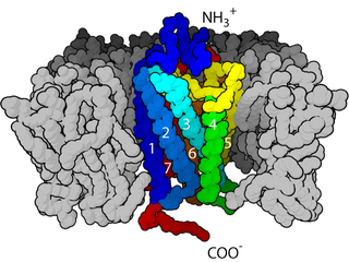
The cell membrane is a biological membrane that separates the interior of all cells from the outside environment which protects the cell from its environment consisting of a lipid bilayer with embedded proteins. The cell membrane controls the movement of substances in and out of cells and organelles. In this way, it is selectively permeable to ions and organic molecules. In addition, cell membranes are involved in a variety of cellular processes such as cell adhesion, ion conductivity and cell signalling and serve as the attachment surface for several extracellular structures, including the cell wall, the carbohydrate layer called the glycocalyx, and the intracellular network of protein fibers called the cytoskeleton. In the field of synthetic biology, cell membranes can be artificially reassembled.
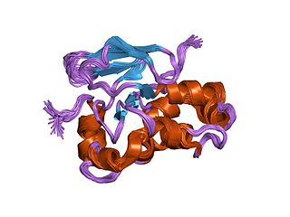
Glycoproteins are proteins which contain oligosaccharide chains (glycans) covalently attached to amino acid side-chains. The carbohydrate is attached to the protein in a cotranslational or posttranslational modification. This process is known as glycosylation. Secreted extracellular proteins are often glycosylated.
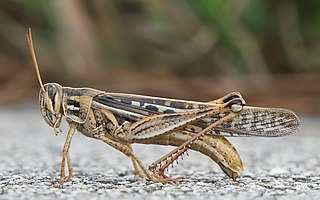
Grasshoppers are a group of insects belonging to the suborder Caelifera. They are among what is probably the most ancient living group of chewing herbivorous insects, dating back to the early Triassic around 250 million years ago.
FasII is initially expressed selectively localized to basolateral junctions during the process of oogenesis, where it functions to establish polarity in inner polar cells of epithelium-derived border cells. [2] During embryogenesis, fasII is dynamically expressed on a subset of axon fascicles in longitudinal nervous system pathways, [3] including the MP1 tract. [2] Here, fasII (and other attractive/repulsive environmental cues such as semaphorins and other morphogens) functions as a framework for pathfinding choices of newly extending axons. [2] This is achieved through trans-homophilic fasII-mediated adhesion and subsequent activation of downstream intracellular signaling pathways involving mitogen-activated protein kinase (MAPK) and regulation of intracellular calcium levels. [2] Later, fasII is expressed on growth cones of axons in other tracts including embryonic peripheral nervous system (PNS) motor neurons. [2] Only the transmembrane isoforms are expressed by neurons, while the GPI-linked form is expressed by non-neuronal cells (mainly glial cells), where it functions as a substrate for growth cones of extending axons, directing adhesion and axon guidance. [2] FasII is also expressed by clusters of differentiating neuroblasts at early stages of neurogenesis where its function is not fully understood but might be involved in proneural gene induction. [2]
Oogenesis, ovogenesis, or oögenesis is the differentiation of the ovum into a cell competent to further develop when fertilized. It is developed from the primary oocyte by maturation. Oogenesis is initiated in the embryonic stage.
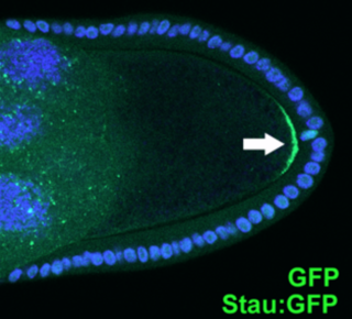
Cell polarity refers to spatial differences in shape, structure, and function within a cell. Almost all cell types exhibit some form of polarity, which enables them to carry out specialized functions. Classical examples of polarized cells are described below, including epithelial cells with apical-basal polarity, neurons in which signals propagate in one direction from dendrites to axons, and migrating cells. Furthermore, cell polarity is important during many types of asymmetric cell division to set up functional asymmetries between daughter cells.
Epithelium is one of the four basic types of animal tissue, along with connective tissue, muscle tissue and nervous tissue. Epithelial tissues line the outer surfaces of organs and blood vessels throughout the body, as well as the inner surfaces of cavities in many internal organs. An example is the epidermis, the outermost layer of the skin.
Other roles for fasII include delineating two axonal pathways in the adult ocellar sensory system (OSS) via its expression on ocellar pioneer (OP) neurons where it acts to promote neurite outgrowth from primary neurons (along with neuroglian) by activating fibroblast growth factor receptor (FGFR) signaling. [2] In addition, fasII has been shown to be involved in synaptic target selection, stabilization and remodeling along with several proteins such as netrins, semaphorins and other Ig-CAMs. [2]
A neurite or neuronal process refers to any projection from the cell body of a neuron. This projection can be either an axon or a dendrite. The term is frequently used when speaking of immature or developing neurons, especially of cells in culture, because it can be difficult to tell axons from dendrites before differentiation is complete.
The fibroblast growth factor receptors are, as their name implies, receptors that bind to members of the fibroblast growth factor family of proteins. Some of these receptors are involved in pathological conditions. For example, a point mutation in FGFR3 can lead to achondroplasia.
The human homolog is STAB2.
Integrins are transmembrane receptors that facilitate cell-extracellular matrix (ECM) adhesion. Upon ligand binding, integrins activate signal transduction pathways that mediate cellular signals such as regulation of the cell cycle, organization of the intracellular cytoskeleton, and movement of new receptors to the cell membrane. The presence of integrins allows rapid and flexible responses to events at the cell surface.
A neurotransmitter receptor is a membrane receptor protein that is activated by a neurotransmitter. Chemicals on the outside of the cell, such as a neurotransmitter, can bump into the cell's membrane and along the membrane we can find receptors. If a neurotransmitter bumps into its corresponding receptor, they will bind and can trigger other events to occur inside the cell. Therefore, a membrane receptor is part of the molecular machinery that allows cells to communicate with one another. A neurotransmitter receptor is a class of receptors that specifically binds with neurotransmitters as opposed to other molecules.
Synaptogenesis is the formation of synapses between neurons in the nervous system. Although it occurs throughout a healthy person's lifespan, an explosion of synapse formation occurs during early brain development, known as exuberant synaptogenesis. Synaptogenesis is particularly important during an individual's critical period, during which there is a certain degree of synaptic pruning due to competition for neural growth factors by neurons and synapses. Processes that are not used, or inhibited during their critical period will fail to develop normally later on in life.
Cell adhesion molecules (CAMs) are a subset of cell adhesion proteins located on the cell surface involved in binding with other cells or with the extracellular matrix (ECM) in the process called cell adhesion. In essence, cell adhesion molecules help cells stick to each other and to their surroundings. Cell adhesion is a crucial component in maintaining tissue structure and function. In fully developed animals, these molecules play an integral role in creating force and movement and consequently ensure that organs are able to execute their functions. In addition to serving as "molecular glue", cell adhesion is important in affecting cellular mechanisms of growth, contact inhibition, and apoptosis. Oftentimes aberrant expression of CAMs will result in pathologies ranging from frostbite to cancer.
L1, also known as L1CAM, is a transmembrane protein member of the L1 protein family, encoded by the L1CAM gene. This protein, of 200-220 kDa, is a neuronal cell adhesion molecule with a strong implication in cell migration, adhesion, neurite outgrowth, myelination and neuronal differentiation. It also plays a key role in treatment-resistant cancers due to its function. It was first identified in 1984 by M. Schachner who found the protein in post-mitotic mice neurons.
Neural cell adhesion molecule (NCAM), also called CD56, is a homophilic binding glycoprotein expressed on the surface of neurons, glia and skeletal muscle. Although CD56 is often considered a marker of neural lineage commitment due to its discovery site, CD56 expression is also found in, among others, the hematopoietic system. Here, the expression of CD56 is most stringently associated with, but certainly not limited to, natural killer cells. CD56 has been detected on other lymphoid cells, including gamma delta (γδ) Τ cells and activated CD8+ T cells, as well as on dendritic cells. NCAM has been implicated as having a role in cell–cell adhesion, neurite outgrowth, synaptic plasticity, and learning and memory.
DSCAM and Dscam are both abbreviations for Down syndrome cell adhesion molecule. In humans, DSCAM refers to a gene that encodes one of several protein isoforms.
Axon guidance is a subfield of neural development concerning the process by which neurons send out axons to reach the correct targets. Axons often follow very precise paths in the nervous system, and how they manage to find their way so accurately is being researched.
Semaphorins are a class of secreted and membrane proteins that were originally identified as axonal growth cone guidance molecules. They primarily act as short-range inhibitory signals and signal through multimeric receptor complexes. Semaphorins are usually cues to deflect axons from inappropriate regions, especially important in neural system development. The major class of proteins that act as their receptors are called plexins, with neuropilins as their co-receptors in many cases. The main receptors for semaphorins are plexins, which have established roles in regulating Rho-family GTPases. Recent work shows that plexins can also influence R-Ras, which, in turn, can regulate integrins. Such regulation is probably a common feature of semaphorin signalling and contributes substantially to our understanding of semaphorin biology.
Myelin-associated glycoprotein is a type 1 transmembrane protein glycoprotein localized in periaxonal Schwann cell and oligodendrocyte membranes, where it plays a role in glial-axonal interactions. MAG is a member of the SIGLEC family of proteins and is a functional ligand of the NOGO-66 receptor, NgR. MAG is believed to be involved in myelination during nerve regeneration in the PNS and is vital for the long-term survival of the myelinated axons following myelinogenesis. In the CNS MAG is one of three main myelin-associated inhibitors of axonal regeneration after injury, making it an important protein for future research on neurogenesis in the CNS.
Neuropilin is a protein receptor active in neurons.
A Plexin is a protein which acts as a receptor for Semaphorin family signaling proteins. It is classically known for its expression on the surface of axon growth cones and involvement in signal transduction to steer axon growth away from the source of Semaphorin. Plexin also has implications in development of other body systems by activating GTPase enzymes to induce a number of intracellular biochemical changes leading to a variety of downstream effects.
Semaphorin-4D (SEMA4D) also known as Cluster of Differentiation 100 (CD100), is a protein of the semaphorin family that in humans is encoded by the SEMA4D gene.
In molecular biology, the fasciclin domain is an extracellular domain of about 140 amino acid residues. It has been suggested that the FAS1 domain represents an ancient cell adhesion domain common to plants and animals; related FAS1 domains are also found in bacteria.
Slit-Robo is the name of a cell signaling pathway with many diverse functions including axon guidance and angiogenesis.
The growth cone is a highly dynamic structure of the developing neuron, changing directionality in response to different secreted and contact-dependent guidance cues; it navigates through the developing nervous system in search of its target. The migration of the growth cone is mediated through the interaction of numerous trophic and tropic factors; netrins, slits, ephrins and semaphorins are four well-studied tropic cues (Fig.1). The growth cone is capable of modifying its sensitivity to these guidance molecules as it migrates to its target; this sensitivity regulation is an important theme seen throughout development.
Cellular adhesions can be defined as proteins or protein aggregates that form mechanical and chemical linkages between the intracellular and extracellular space. Adhesions serve several critical processes including cell migration, signal transduction, tissue development and repair. Due to this functionality, adhesions and adhesion molecules have been a topic of study within the scientific community. Specifically, it has been found that adhesions are involved in tissue development, plasticity, and memory formation within the central nervous system (CNS), and may prove vital in the generation of CNS-specific therapeutics.
This page describes the process of synapse stabilization mediated by cell adhesion molecules. To see related articles please see the pages on Synaptogenesis, Synaptic plasticity, Cell adhesion molecule, Development of the nervous system.
Tight junction proteins (TJs) are molecules situated at the tight junction of epithelia, endothelia and myelinated cell. This mutliprotein junctional complex has a regulatory function in passage of ionts, water and solutes through the paracellular pathway. It can also coordinate the motion of lipids and proteins between the apical and basolateral surfaces of the plasma membrane. Thereby tight junction conducts signaling molecules, that influence the differentiation, proliferation and polarity of cells. So tight junction plays a key role in maintenance of osmotic balance and trans-cellular transport of tissue specific molecules. Nowadays is known more than 40 different proteins, that are involved in these selective TJ channels.