
A motor neuron is a neuron whose cell body is located in the motor cortex, brainstem or the spinal cord, and whose axon (fiber) projects to the spinal cord or outside of the spinal cord to directly or indirectly control effector organs, mainly muscles and glands. There are two types of motor neuron – upper motor neurons and lower motor neurons. Axons from upper motor neurons synapse onto interneurons in the spinal cord and occasionally directly onto lower motor neurons. The axons from the lower motor neurons are efferent nerve fibers that carry signals from the spinal cord to the effectors. Types of lower motor neurons are alpha motor neurons, beta motor neurons, and gamma motor neurons.

Muscle spindles are stretch receptors within the body of a skeletal muscle that primarily detect changes in the length of the muscle. They convey length information to the central nervous system via afferent nerve fibers. This information can be processed by the brain as proprioception. The responses of muscle spindles to changes in length also play an important role in regulating the contraction of muscles, for example, by activating motor neurons via the stretch reflex to resist muscle stretch.
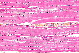
Striated muscle tissue is a muscle tissue that features repeating functional units called sarcomeres. The presence of sarcomeres manifests as a series of bands visible along the muscle fibers, which is responsible for the striated appearance observed in microscopic images of this tissue. There are two types of striated muscle:

The fusiform gyrus, also known as the lateral occipitotemporal gyrus,is part of the temporal lobe and occipital lobe in Brodmann area 37. The fusiform gyrus is located between the lingual gyrus and parahippocampal gyrus above, and the inferior temporal gyrus below. Though the functionality of the fusiform gyrus is not fully understood, it has been linked with various neural pathways related to recognition. Additionally, it has been linked to various neurological phenomena such as synesthesia, dyslexia, and prosopagnosia.
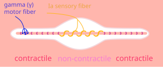
A type Ia sensory fiber, or a primary afferent fiber is a type of afferent nerve fiber. It is the sensory fiber of a stretch receptor called the muscle spindle found in muscles, which constantly monitors the rate at which a muscle stretch changes. The information carried by type Ia fibers contributes to the sense of proprioception.
Lower motor neurons (LMNs) are motor neurons located in either the anterior grey column, anterior nerve roots or the cranial nerve nuclei of the brainstem and cranial nerves with motor function. Many voluntary movements rely on spinal lower motor neurons, which innervate skeletal muscle fibers and act as a link between upper motor neurons and muscles. Cranial nerve lower motor neurons also control some voluntary movements of the eyes, face and tongue, and contribute to chewing, swallowing and vocalization. Damage to the lower motor neurons can lead to flaccid paralysis, absent deep tendon reflexes and muscle atrophy.
A gamma motor neuron, also called gamma motoneuron, or fusimotor neuron, is a type of lower motor neuron that takes part in the process of muscle contraction, and represents about 30% of (Aγ) fibers going to the muscle. Like alpha motor neurons, their cell bodies are located in the anterior grey column of the spinal cord. They receive input from the reticular formation of the pons in the brainstem. Their axons are smaller than those of the alpha motor neurons, with a diameter of only 5 μm. Unlike the alpha motor neurons, gamma motor neurons do not directly adjust the lengthening or shortening of muscles. However, their role is important in keeping muscle spindles taut, thereby allowing the continued firing of alpha neurons, leading to muscle contraction. These neurons also play a role in adjusting the sensitivity of muscle spindles.

The spinocerebellar tract is a nerve tract originating in the spinal cord and terminating in the same side (ipsilateral) of the cerebellum.

The dentate nucleus is a cluster of neurons, or nerve cells, in the central nervous system that has a dentate – tooth-like or serrated – edge. It is located within the deep white matter of each cerebellar hemisphere, and it is the largest single structure linking the cerebellum to the rest of the brain. It is the largest and most lateral, or farthest from the midline, of the four pairs of deep cerebellar nuclei, the others being the globose and emboliform nuclei, which together are referred to as the interposed nucleus, and the fastigial nucleus. The dentate nucleus is responsible for the planning, initiation and control of voluntary movements. The dorsal region of the dentate nucleus contains output channels involved in motor function, which is the movement of skeletal muscle, while the ventral region contains output channels involved in nonmotor function, such as conscious thought and visuospatial function.
Intrafusal muscle fibers are skeletal muscle fibers that serve as specialized sensory organs (proprioceptors). They detect the amount and rate of change in length of a muscle. They constitute the muscle spindle, and are innervated by both sensory (afferent) and motor (efferent) fibers.
The rectus femoris muscle is one of the four quadriceps muscles of the human body. The others are the vastus medialis, the vastus intermedius, and the vastus lateralis. All four parts of the quadriceps muscle attach to the patella by the quadriceps tendon.

The stretch reflex, or more accurately "muscle stretch reflex", is a muscle contraction in response to stretching a muscle. The function of the reflex is generally thought to be maintaining the muscle at a constant length but the response is often coordinated across multiple muscles and even joints. The older term deep tendon reflex is now criticized as misleading. Tendons have little to do with the response, and some muscles with stretch reflexes have no tendons. Rather, muscle spindles detect a stretch and convey the information to the central nervous system.

Alpha (α) motor neurons (also called alpha motoneurons), are large, multipolar lower motor neurons of the brainstem and spinal cord. They innervate extrafusal muscle fibers of skeletal muscle and are directly responsible for initiating their contraction. Alpha motor neurons are distinct from gamma motor neurons, which innervate intrafusal muscle fibers of muscle spindles.

A bipolar neuron, or bipolar cell, is a type of neuron characterized by having both an axon and a dendrite extending from the soma in opposite directions. These neurons are predominantly found in the retina and olfactory system. The embryological period encompassing weeks seven through eight marks the commencement of bipolar neuron development. Many bipolar cells are specialized sensory neurons for the transmission of sense. As such, they are part of the sensory pathways for smell, sight, taste, hearing, touch, balance and proprioception. The other shape classifications of neurons include unipolar, pseudounipolar and multipolar. During embryonic development, pseudounipolar neurons begin as bipolar in shape but become pseudounipolar as they mature.

Proprioception is the sense of self-movement, force, and body position.

Phenacovolva rosea, also known as the rosy spindle cowry, is a species of sea snail, a marine gastropod mollusc in the family Ovulidae, the ovulids, cowry allies or false cowries. It lives and feeds on fan, whip and bush-type gorgonians.
Beta motor neurons, also called beta motoneurons, are a kind of lower motor neuron, along with alpha motor neurons and gamma motor neurons. Beta motor neurons innervate intrafusal fibers of muscle spindles with collaterals to extrafusal fibers - a type of slow twitch fiber. Also, axons of alpha, beta, and gamma motor neurons become myelinated. Moreover, these efferent neurons originate from the anterior grey column of the spinal cord and travel to skeletal muscles. However, the larger diameter alpha motor fibers require higher conduction velocity than beta and gamma.
Lugaro cells are primary sensory interneurons of the cerebellum, that have an inhibitory function. They are fusiform, having a spindle shape that tapers at each end. They were first described by Ernesto Lugaro in the early 20th century. Lugaro cells are found just beneath the layer of Purkinje cells between the molecular layer and the granular layer. They have thick principal dendrites coming from opposite poles of their bodies. These dendrites are very long and travel along the boundary between the Purkinje layer and the granular layer. They seem to contact from 5 to 15 Purkinje cells in a horizontal direction. They can also sense stimuli close to the Purkinje cells and their dendrites form a large receptive area that monitors the environment near to the Purkinje cells. Whilst their dendrites make contact with the Purkinje cells they also receive inputs from branches of the Purkinje axons by which they seem to have a sampling and integration role.

Anatomical terminology is used to uniquely describe aspects of skeletal muscle, cardiac muscle, and smooth muscle such as their actions, structure, size, and location.

