Glutaminolysis

Last updated

Glutaminolysis ( glutamine + -lysis ) is a series of biochemical reactions by which the amino acid glutamine is lysed to glutamate, aspartate, CO2, pyruvate, lactate, alanine and citrate. [1] [2]

Contents

The glutaminolytic pathway

Glutaminolysis partially recruits reaction steps from the citric acid cycle and the malate-aspartate shuttle.

Reaction steps from glutamine to α-ketoglutarate

The conversion of the amino acid glutamine to α-ketoglutarate takes place in two reaction steps:

Conversion of glutamine to a-ketoglutarate Glutaminolysisengl1.png
Conversion of glutamine to α-ketoglutarate

1. Hydrolysis of the amino group of glutamine yielding glutamate and ammonium. Catalyzing enzyme: glutaminase (EC 3.5.1.2)

2. Glutamate can be excreted or can be further metabolized to α-ketoglutarate.

For the conversion of glutamate to α-ketoglutarate three different reactions are possible:

Catalyzing enzymes:

Recruited reaction steps of the citric acid cycle and malate aspartate shuttle

The glutaminolytic pathway. Figure legend: blue color = reaction steps of the citric acid cycle; brown color = reaction steps of the malate aspartate shuttle; green color = enzymes overexpressed in tumors. 1 = glutaminase, 2 = GOT, 3 = a-ketoglutarate dehydrogenase, 4 = succinate dehydrogenase, 5 = fumarase, 6 = malate dehydrogenase, 7a = cytosolic malic enzyme, 7b = mitochondrial malic enzyme, 8 = citrate synthase, 9 = aconitase, 10 = lactate dehydrogenase Glutaminolysisengl2.png
The glutaminolytic pathway. Figure legend: blue color = reaction steps of the citric acid cycle; brown color = reaction steps of the malate aspartate shuttle; green color = enzymes overexpressed in tumors. 1 = glutaminase, 2 = GOT, 3 = α-ketoglutarate dehydrogenase, 4 = succinate dehydrogenase, 5 = fumarase, 6 = malate dehydrogenase, 7a = cytosolic malic enzyme, 7b = mitochondrial malic enzyme, 8 = citrate synthase, 9 = aconitase, 10 = lactate dehydrogenase

catalyzing enzyme: α-ketoglutarate dehydrogenase complex

catalyzing enzyme: succinyl-CoA-synthetase, EC 6.2.1.4

catalyzing enzyme: succinate dehydrogenase, EC 1.3.5.1

catalyzing enzyme: fumarase, EC 4.2.1.2

catalyzing enzyme: malate dehydrogenase, EC 1.1.1.37 (component of the malate aspartate shuttle)

catalyzing enzyme: citrate synthase, EC 2.3.3.1

Reaction steps from malate to pyruvate and lactate

The conversion of malate to pyruvate and lactate is catalyzed by

according to the following equations:

Intracellular compartmentalization of the glutaminolytic pathway

The reactions of the glutaminolytic pathway take place partly in the mitochondria and to some extent in the cytosol (compare the metabolic scheme of the glutaminolytic pathway).

An important energy source in tumor cells

Glutaminolysis takes place in all proliferating cells, [3] such as lymphocytes, thymocytes, colonocytes, adipocytes and especially in tumor cells. [1] Glutaminolysis has been targeted for therapeutic purposes. [4] In tumor cells the citric acid cycle is truncated due to an inhibition of the enzyme aconitase (EC 4.2.1.3) by high concentrations of reactive oxygen species (ROS) [5] [6] Aconitase catalyzes the conversion of citrate to isocitrate. On the other hand, tumor cells over express phosphate dependent glutaminase and NAD(P)-dependent malate decarboxylase, [7] [8] [9] [10] which in combination with the remaining reaction steps of the citric acid cycle from α-ketoglutarate to citrate impart the possibility of a new energy producing pathway, the degradation of the amino acid glutamine to glutamate, aspartate, pyruvate CO2, lactate and citrate.

Besides glycolysis in tumor cells glutaminolysis is another main pillar for energy production. High extracellular glutamine concentrations stimulate tumor growth and are essential for cell transformation. [9] [11] On the other hand, a reduction of glutamine correlates with phenotypical and functional differentiation of the cells. [12]

Energy efficacy of glutaminolysis in tumor cells


Due to low glutamate dehydrogenase and glutamate pyruvate transaminase activities, in tumor cells the conversion of glutamate to alpha-ketoglutarate mainly takes place via glutamate oxaloacetate transaminase. [13]

Advantages of glutaminolysis in tumor cells

See also

Related Research Articles

<span class="mw-page-title-main">Citric acid cycle</span> Interconnected biochemical reactions releasing energy

The citric acid cycle—also known as the Krebs cycle, Szent-Györgyi-Krebs cycle or the TCA cycle (tricarboxylic acid cycle)—is a series of biochemical reactions to release the energy stored in nutrients through the oxidation of acetyl-CoA derived from carbohydrates, fats, and proteins. The chemical energy released is available under the form of ATP. The Krebs cycle is used by organisms that respire (as opposed to organisms that ferment) to generate energy, either by anaerobic respiration or aerobic respiration. In addition, the cycle provides precursors of certain amino acids, as well as the reducing agent NADH, that are used in numerous other reactions. Its central importance to many biochemical pathways suggests that it was one of the earliest components of metabolism. Even though it is branded as a 'cycle', it is not necessary for metabolites to follow only one specific route; at least three alternative segments of the citric acid cycle have been recognized.

<span class="mw-page-title-main">Glycolysis</span> Series of interconnected biochemical reactions

Glycolysis is the metabolic pathway that converts glucose into pyruvate and, in most organisms, occurs in the liquid part of cells. The free energy released in this process is used to form the high-energy molecules adenosine triphosphate (ATP) and reduced nicotinamide adenine dinucleotide (NADH). Glycolysis is a sequence of ten reactions catalyzed by enzymes.

A dehydrogenase is an enzyme belonging to the group of oxidoreductases that oxidizes a substrate by reducing an electron acceptor, usually NAD+/NADP+ or a flavin coenzyme such as FAD or FMN. Like all catalysts, they catalyze reverse as well as forward reactions, and in some cases this has physiological significance: for example, alcohol dehydrogenase catalyzes the oxidation of ethanol to acetaldehyde in animals, but in yeast it catalyzes the production of ethanol from acetaldehyde.

<span class="mw-page-title-main">Acetyl-CoA</span> Chemical compound

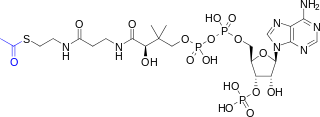
Acetyl-CoA is a molecule that participates in many biochemical reactions in protein, carbohydrate and lipid metabolism. Its main function is to deliver the acetyl group to the citric acid cycle to be oxidized for energy production.

<span class="mw-page-title-main">Glutamate dehydrogenase</span> Hexameric enzyme

Glutamate dehydrogenase is an enzyme observed in both prokaryotes and eukaryotic mitochondria. The aforementioned reaction also yields ammonia, which in eukaryotes is canonically processed as a substrate in the urea cycle. Typically, the α-ketoglutarate to glutamate reaction does not occur in mammals, as glutamate dehydrogenase equilibrium favours the production of ammonia and α-ketoglutarate. Glutamate dehydrogenase also has a very low affinity for ammonia, and therefore toxic levels of ammonia would have to be present in the body for the reverse reaction to proceed. However, in brain, the NAD+/NADH ratio in brain mitochondria encourages oxidative deamination. In bacteria, the ammonia is assimilated to amino acids via glutamate and aminotransferases. In plants, the enzyme can work in either direction depending on environment and stress. Transgenic plants expressing microbial GLDHs are improved in tolerance to herbicide, water deficit, and pathogen infections. They are more nutritionally valuable.

<span class="mw-page-title-main">Oxaloacetic acid</span> Organic compound

Oxaloacetic acid (also known as oxalacetic acid or OAA) is a crystalline organic compound with the chemical formula HO2CC(O)CH2CO2H. Oxaloacetic acid, in the form of its conjugate base oxaloacetate, is a metabolic intermediate in many processes that occur in animals. It takes part in gluconeogenesis, the urea cycle, the glyoxylate cycle, amino acid synthesis, fatty acid synthesis and the citric acid cycle.

<span class="mw-page-title-main">Malate dehydrogenase</span> Class of enzymes

Malate dehydrogenase (EC 1.1.1.37) (MDH) is an enzyme that reversibly catalyzes the oxidation of malate to oxaloacetate using the reduction of NAD+ to NADH. This reaction is part of many metabolic pathways, including the citric acid cycle. Other malate dehydrogenases, which have other EC numbers and catalyze other reactions oxidizing malate, have qualified names like malate dehydrogenase (NADP+).

In molecular biology, biosynthesis is a multi-step, enzyme-catalyzed process where substrates are converted into more complex products in living organisms. In biosynthesis, simple compounds are modified, converted into other compounds, or joined to form macromolecules. This process often consists of metabolic pathways. Some of these biosynthetic pathways are located within a single cellular organelle, while others involve enzymes that are located within multiple cellular organelles. Examples of these biosynthetic pathways include the production of lipid membrane components and nucleotides. Biosynthesis is usually synonymous with anabolism.

<span class="mw-page-title-main">Isocitrate dehydrogenase</span> Class of enzymes

Isocitrate dehydrogenase (IDH) (EC 1.1.1.42) and (EC 1.1.1.41) is an enzyme that catalyzes the oxidative decarboxylation of isocitrate, producing alpha-ketoglutarate (α-ketoglutarate) and CO2. This is a two-step process, which involves oxidation of isocitrate (a secondary alcohol) to oxalosuccinate (a ketone), followed by the decarboxylation of the carboxyl group beta to the ketone, forming alpha-ketoglutarate. In humans, IDH exists in three isoforms: IDH3 catalyzes the third step of the citric acid cycle while converting NAD+ to NADH in the mitochondria. The isoforms IDH1 and IDH2 catalyze the same reaction outside the context of the citric acid cycle and use NADP+ as a cofactor instead of NAD+. They localize to the cytosol as well as the mitochondrion and peroxisome.

<span class="mw-page-title-main">Mitochondrial matrix</span> Space within the inner membrane of the mitochondrion

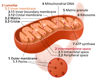
In the mitochondrion, the matrix is the space within the inner membrane. The word "matrix" stems from the fact that this space is viscous, compared to the relatively aqueous cytoplasm. The mitochondrial matrix contains the mitochondrial DNA, ribosomes, soluble enzymes, small organic molecules, nucleotide cofactors, and inorganic ions.[1] The enzymes in the matrix facilitate reactions responsible for the production of ATP, such as the citric acid cycle, oxidative phosphorylation, oxidation of pyruvate, and the beta oxidation of fatty acids.

The oxoglutarate dehydrogenase complex (OGDC) or α-ketoglutarate dehydrogenase complex is an enzyme complex, most commonly known for its role in the citric acid cycle.

<span class="mw-page-title-main">Malate–aspartate shuttle</span> Biochemical system for transporting electrons produced during glycolysis

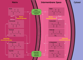
The malate–aspartate shuttle is a biochemical system for translocating electrons produced during glycolysis across the semipermeable inner membrane of the mitochondrion for oxidative phosphorylation in eukaryotes. These electrons enter the electron transport chain of the mitochondria via reduction equivalents to generate ATP. The shuttle system is required because the mitochondrial inner membrane is impermeable to NADH, the primary reducing equivalent of the electron transport chain. To circumvent this, malate carries the reducing equivalents across the membrane.

<span class="mw-page-title-main">Amino acid synthesis</span> The set of biochemical processes by which amino acids are produced

Amino acid biosynthesis is the set of biochemical processes by which the amino acids are produced. The substrates for these processes are various compounds in the organism's diet or growth media. Not all organisms are able to synthesize all amino acids. For example, humans can synthesize 11 of the 20 standard amino acids. These 11 are called the non-essential amino acids).

<span class="mw-page-title-main">Reverse Krebs cycle</span>

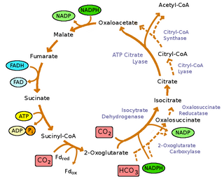
The reverse Krebs cycle is a sequence of chemical reactions that are used by some bacteria to produce carbon compounds from carbon dioxide and water by the use of energy-rich reducing agents as electron donors.

The mitochondrial shuttles are biochemical transport systems used to transport reducing agents across the inner mitochondrial membrane. NADH as well as NAD+ cannot cross the membrane, but it can reduce another molecule like FAD and [QH2] that can cross the membrane, so that its electrons can reach the electron transport chain.

In enzymology, a glutamine-pyruvate transaminase is an enzyme that catalyzes the chemical reaction

<span class="mw-page-title-main">GOT2</span> Mitochondrial enzyme involved in amino acid metabolism

Aspartate aminotransferase, mitochondrial is an enzyme that in humans is encoded by the GOT2 gene. Glutamic-oxaloacetic transaminase is a pyridoxal phosphate-dependent enzyme which exists in cytoplasmic and inner-membrane mitochondrial forms, GOT1 and GOT2, respectively. GOT plays a role in amino acid metabolism and the urea and Kreb's cycle. Also, GOT2 is a major participant in the malate-aspartate shuttle, which is a passage from the cytosol to the mitochondria. The two enzymes are homodimeric and show close homology. GOT2 has been seen to have a role in cell proliferation, especially in terms of tumor growth.

<span class="mw-page-title-main">Malate dehydrogenase 2</span> Enzyme that oxidizes malate to oxaloacetate in Krebs cycle

Malate dehydrogenase, mitochondrial also known as malate dehydrogenase 2 is an enzyme that in humans is encoded by the MDH2 gene.

In biochemistry, the glutamate–glutamine cycle is a cyclic metabolic pathway which maintains an adequate supply of the neurotransmitter glutamate in the central nervous system. Neurons are unable to synthesize either the excitatory neurotransmitter glutamate, or the inhibitory GABA from glucose. Discoveries of glutamate and glutamine pools within intercellular compartments led to suggestions of the glutamate–glutamine cycle working between neurons and astrocytes. The glutamate/GABA–glutamine cycle is a metabolic pathway that describes the release of either glutamate or GABA from neurons which is then taken up into astrocytes. In return, astrocytes release glutamine to be taken up into neurons for use as a precursor to the synthesis of either glutamate or GABA.

<span class="mw-page-title-main">Citrate–malate shuttle</span> Series of chemical reactions

The citrate-malate shuttle is a series of chemical reactions, commonly referred to as a biochemical cycle or system, that transports acetyl-CoA in the mitochondrial matrix across the inner and outer mitochondrial membranes for fatty acid synthesis. Mitochondria are enclosed in a double membrane. As the inner mitochondrial membrane is impermeable to acetyl-CoA, the shuttle system is essential to fatty acid synthesis in the cytosol. It plays an important role in the generation of lipids in the liver.

References

  1. 1 2 Krebs, HA; Bellamy D (1960). "The interconversion of glutamic acid and aspartic acid in respiring tissues". The Biochemical Journal. 75 (3): 523–529. doi:10.1042/bj0750523. PMC   1204504 . PMID   14411856.
  2. Jin, L; Alesi, GN; Kang, S (14 July 2016). "Glutaminolysis as a target for cancer therapy". Oncogene. 35 (28): 3619–25. doi:10.1038/onc.2015.447. PMC   5225500 . PMID   26592449.
  3. 1 2 Fernandez-de-Cossio-Diaz, Jorge; Vazquez, Alexei (2017-10-18). "Limits of aerobic metabolism in cancer cells". Scientific Reports. 7 (1): 13488. Bibcode:2017NatSR...713488F. doi:10.1038/s41598-017-14071-y. ISSN   2045-2322. PMC   5647437 . PMID   29044214.
  4. "Enhancing the efficacy of glutamine metabolism inhibitors in cancer therapy". Trends in Cancer.
  5. Gardner, PR; Raineri I; Epstein LB; White CW (1995). "Superoxide radical and iron modulate aconitase activity in mammalian cells". Journal of Biological Chemistry. 270 (22): 13399–13405. doi: 10.1074/jbc.270.22.13399 . PMID   7768942.
  6. Kim, KH; Rodriguez AM; Carrico PM; Melendez JA (2001). "Potential mechanisms for the inhibition of tumor cell growth by manganese superoxide dismutase". Antioxidants & Redox Signaling. 3 (3): 361–373. doi:10.1089/15230860152409013. PMID   11491650.
  7. Matsuno, T; Goto I (1992). "Glutaminase and glutamine synthetase activities in human cirrhotic liver and hepatocellular carcinoma". Cancer Research. 52 (5): 1192–1194. PMID   1346587.
  8. 1 2 Aledo JC, Segura JA, Medina MA, Alonso FJ, Núñez de Castro I, Márquez J (1994). "Phosphate-activated glutaminase expression during tumor development". FEBS Letters. 341 (1): 39–42. doi: 10.1016/0014-5793(94)80236-X . PMID   8137919. S2CID   12702894.
  9. 1 2 Lobo C, Ruiz-Bellido MA, Aledo JC, Márquez J, Núñez De Castro I, Alonso FJ (2000). "Inhibition of glutaminase expression by antisense mRNA decreases growth and tumourigenicity of tumour cells". Biochemical Journal. 348 (2): 257–261. doi:10.1042/0264-6021:3480257. PMC   1221061 . PMID   10816417.
  10. Mazurek, S; Grimm H; Oehmke M; Weisse G; Teigelkamp S; Eigenbrodt E (2000). "Tumor M2-PK and glutaminolytic enzymes in the metabolic shift of tumor cells". Anticancer Research. 20 (6D): 5151–5154. PMID   11326687.
  11. Turowski, GA; Rashid Z; Hong F; Madri JA; Basson MD (1994). "Glutamine modulates phenotype and stimulates proliferation in human colon cancer cell lines". Cancer Research. 54 (22): 5974–5980. PMID   7954430.
  12. Spittler, A; Oehler R; Goetzinger P; Holzer S; Reissner CM; Leutmezer J; Rath V; Wrba F; Fuegger R; Boltz-Nitulescu G; Roth E (1997). "Low glutamine concentrations induce phenotypical and functional differentiation of U937 myelomonocytic cells". The Journal of Nutrition. 127 (11): 2151–2157. doi: 10.1093/jn/127.11.2151 . PMID   9349841.
  13. Matsuno, T (1991). "Pathway of glutamate oxidation and its regulation in HuH13 line of human hepatoma cells". Journal of Cellular Physiology. 148 (2): 290–294. doi:10.1002/jcp.1041480215. PMID   1679060. S2CID   30893440.
  14. Stephen J. Ralph; Rafael Moreno-Sánchez; Jiri Neuzil; Sara Rodríguez-Enríquez (24 August 2011). "Inhibitors of Succinate: Quinone Reductase/Complex II Regulate Production of Mitochondrial Reactive Oxygen Species and Protect Normal Cells from Ischemic Damage but Induce Specific Cancer Cell Death" . Pharmaceutical Research . 28 (2695): 2695–2730. doi:10.1007/s11095-011-0566-7. PMID   21863476. S2CID   21836546 . Retrieved 1 November 2021. Unlike aconitase, glutaminolysis is relatively insensitive to ROS levels.
  15. Parlo, RA; Coleman PS (1984). "Enhanced rate of citrate export from cholesterol-rich hepatoma mitochondria. The truncated Krebs cycle and other metabolic ramifications of mitochondrial membrane cholesterol". The Journal of Biological Chemistry. 259 (16): 9997–10003. doi: 10.1016/S0021-9258(18)90917-8 . PMID   6469976.
  16. Mazurek, S; Grimm H; Boschek CB; Vaupel P; Eigenbrodt E (2002). "Pyruvate kinase type M2: a crossroad in the tumor metabolome". The British Journal of Nutrition. 87: S23–S29. doi: 10.1079/BJN2001455 . PMID   11895152.
  17. Eck, HP; Drings P; Dröge W (1989). "Plasma glutamate levels, lymphocyte reactivity and death in patients with bronchial carcinoma". Journal of Cancer Research and Clinical Oncology. 115 (6): 571–574. doi:10.1007/BF00391360. PMID   2558118. S2CID   23057794.
  18. Grimm, H; Tibell A; Norrlind B; Blecher C; Wilker S; Schwemmle K (1994). "Immunoregulation by parental lipids: impact of the n-3 to n-6 fatty acid ratio". Journal of Parenteral and Enteral Nutrition. 18 (5): 417–421. doi:10.1177/0148607194018005417. PMID   7815672.
  19. Jiang, WG; Bryce RP; Hoorobin DF (1998). "Essential fatty acids: molecular and cellular basis of their anti-cancer action and clinical implications". Critical Reviews in Oncology/Hematology. 27 (3): 179–209. doi:10.1016/S1040-8428(98)00003-1. PMID   9649932.