Tumor M2-PK

Last updated

Tumor M2-PK is a synonym for the dimeric form of the pyruvate kinase isoenzyme type M2 (PKM2), a key enzyme within tumor metabolism. Tumor M2-PK can be elevated in many tumor types, rather than being an organ-specific tumor marker such as PSA. Increased stool (fecal) levels are being investigated as a method of screening for colorectal tumors, and EDTA plasma levels are undergoing testing for possible application in the follow-up of various cancers.

Contents

Sandwich ELISAs based on two monoclonal antibodies which specifically recognize Tumor M2-PK (the dimeric form of M2-PK) are available for the quantification of Tumor M2-PK in stool and EDTA-plasma samples respectively. As a biomarker, the amount of Tumor M2-PK in stool and EDTA-plasma reflects the specific metabolic status of the tumors.

Early detection of colorectal tumors and polyps

M2-PK, as measured in feces, is a potential tumor marker for colorectal cancer. When measured in feces with a cutoff value of 4 U/ml, its sensitivity has been estimated to be 85% (with a 95% confidence interval of 65 to 96%) for colon cancer and 56% (confidence interval 41–74%) for rectal cancer. [1] Its specificity is 95%. [2]

The M2-PK test is not dependent on occult blood (ELISA method), so it can detect bleeding or non-bleeding bowel cancer and also polyps with high sensitivity and high specificity with no false negative, but false positives may occur. [3]

Most people are more willing to accept non-invasive preventive medical check-ups. Therefore, the measurement of tumor M2-PK in stool samples, with follow-up by colonoscopy to clarify the tumor M2-PK positive results, may prove to be an advance in the early detection of colorectal carcinomas. The CE marked M2-PK Test is available in form of an ELISA test for quantitative results or as point of care test to receive results within minutes.

Tumor M2-PK is also useful to diagnose lung cancer and better than SCC and NSE tumor markers. [4] With renal cell carcinoma (RCC), the M2PK test has sensitivity of 66.7 percent for metastatic RCC and 27.5 percent for nonmetastatic RCC, but M2PK test cannot detect transitional cell carcinoma of the bladder, prostate cancer and benign prostatic hyperplasia. [5]

Cancer follow-up

Studies from various international working groups have revealed a significantly increased amount of Tumor M2-PK in EDTA-plasma samples of patients with renal, lung, breast, cervical and gastrointestinal tumors (oesophagus, stomach, pancreas, colon, rectum), as well as melanoma, which correlated with the tumor stage.

The combination of Tumor M2-PK with the appropriate classical tumor marker, such as CEA for bowel cancer, CA 19-9 for pancreatic cancer and CA 72-4 for gastric cancer, significantly increases the sensitivity to detect various cancers.

An important application of the Tumor M2-PK test in EDTA-plasma is for follow-up during tumor therapy, to monitor the success or failure of the chosen treatment, as well as predicting the chances of a “cure” and survival.

If Tumor M2-PK levels decrease during therapy and then remain low after therapy it points towards successful treatment. An increase in the Tumor M2-PK values during or after therapy points towards relapse and/or metastasis.

Increased Tumor M2-PK values can sometimes also occur in severe inflammatory diseases, which must be excluded by differential diagnosis.

Tetrameric and dimeric PKM2

Pyruvate kinase catalyzes the last step within the glycolytic sequence, the dephosphorylation of phosphoenolpyruvate to pyruvate and is responsible for net energy production within the glycolytic pathway. Depending upon the different metabolic functions of the tissues, different isoenzymes of pyruvate kinase are expressed.

M2-PK (PKM2) is the predominant pyruvate kinase isoform in proliferating cells, such as fibroblasts, embryonic cells and adult stem cells and most human tissue, including lung, bladder, kidney and thymus; M2-PK is upgregulated in many human tumors. [6]

M2-PK can occur in two different forms in proliferating cells:

The tetrameric form of M2-PK has a high affinity to its substrate, phosphoenolpyruvate (PEP), and is highly active at physiological PEP concentrations. Furthermore, the tetrameric form of M2-PK is associated with several other glycolytic enzymes within the so-called glycolytic enzyme complex. Due to the close proximity of the enzymes, the association within the glycolytic enzyme complex leads to a highly effective conversion of glucose to lactate. When M2-PK is mainly in the highly active tetrameric form, which is the case in most normal cells, glucose is mostly converted to lactate, with the attendant production of energy.

In contrast, the dimeric form of M2-PK has a low affinity for phosphoenolpyruvate, being nearly inactive at physiological PEP concentrations. When M2-PK is mainly in the dimeric form, which is the case in tumor cells, all phosphometabolites above pyruvate kinase accumulate and are channelled into synthetic processes which branch off from glycolytic intermediates, such as nucleic acids, phospholipids and amino acids, important cell building blocks for highly proliferating cells such as tumor cells.

Metabolic consequences of the tetrameric and dimeric forms of M2-PK M2-PKglycolysiseng.png
Metabolic consequences of the tetrameric and dimeric forms of M2-PK

As a consequence of the key position of pyruvate kinase within glycolysis, the tetramer : dimer ratio of M2-PK determines whether glucose carbons are converted to pyruvate and lactate, along with the production of energy (tetrameric form), or channelled into synthetic processes (dimeric form). In tumor cells M2-PK is mainly in the dimeric form. Therefore, the dimeric form of M2-PK has been termed Tumor M2-PK.

The dimerization of M2-PK in tumor cells is induced by the direct interaction of M2-PK with different oncoproteins.

However, the tetramer : dimer ratio of M2-PK is not constant.

Oxygen starvation or highly accumulated glycolytic intermediates, such as fructose 1,6-bisphosphate (fructose 1,6-P2) or the amino acid serine, induce the reassociation of the dimeric form of M2-PK to the tetrameric form. Consequently, due to the activation of M2-PK, glucose is converted to pyruvate and lactate under the production of energy until the fructose 1,6-P2 levels drop below a certain threshold value, which allows the dissociation of the tetrameric form of M2-PK to the dimeric form. Thereafter, the cycle of oscillation starts again when the fructose 1,6-P2 levels reach a certain upper threshold value which induces the tetramerization of M2-PK.

When M2-PK is mainly in the less active dimeric form, energy is produced by the degradation of the amino acid glutamine to aspartate, pyruvate and lactate, which is termed glutaminolysis.

In tumor cells the increased rate of lactate production in the presence of oxygen is termed the Warburg effect.

Mutations

For the first time pyruvate kinase M2 enzyme was reported with two missense mutations, H391Y and K422R, found in cells from Bloom syndrome patients, prone to develop cancer. Results show that despite the presence of mutations in the inter-subunit contact domain, the K422R and H391Y mutant proteins maintained their homotetrameric structure, similar to the wild-type protein, but showed a loss of activity of 75 and 20%, respectively. H391Y showed a 6-fold increase in affinity for its substrate phosphoenolpyruvate and behaved like a non-allosteric protein with compromised cooperative binding. However, the affinity for phosphoenolpyruvate was lost significantly in K422R. Unlike K422R, H391Y showed enhanced thermal stability, stability over a range of pH values, a lesser effect of the allosteric inhibitor Phe, and resistance toward structural alteration upon binding of the activator (fructose 1,6-bisphosphate) and inhibitor (Phe). Both mutants showed a slight shift in the pH optimum from 7.4 to 7.0. [7] The co-expression of homotetrameric wild type and mutant PKM2 in the cellular milieu resulting in the interaction between the two at the monomer level was substantiated further by in vitro experiments. The cross-monomer interaction significantly altered the oligomeric state of PKM2 by favoring dimerisation and heterotetramerization. In silico study provided an added support in showing that hetero-oligomerization was energetically favorable. The hetero-oligomeric populations of PKM2 showed altered activity and affinity, and their expression resulted in an increased growth rate of Escherichia coli as well as mammalian cells, along with an increased rate of polyploidy. These features are known to be essential to tumor progression. [8]

Potential multi-functional protein

[9]

See also

Related Research Articles

<span class="mw-page-title-main">Glycolysis</span> Catabolic pathway

Glycolysis is the metabolic pathway that converts glucose into pyruvate, and in most organisms, occurs in the liquid part of cells, the cytosol. The free energy released in this process is used to form the high-energy molecules adenosine triphosphate (ATP) and reduced nicotinamide adenine dinucleotide (NADH). Glycolysis is a sequence of ten reactions catalyzed by enzymes.

Gluconeogenesis (GNG) is a metabolic pathway that results in the generation of glucose from certain non-carbohydrate carbon substrates. It is a ubiquitous process, present in plants, animals, fungi, bacteria, and other microorganisms. In vertebrates, gluconeogenesis occurs mainly in the liver and, to a lesser extent, in the cortex of the kidneys. It is one of two primary mechanisms – the other being degradation of glycogen (glycogenolysis) – used by humans and many other animals to maintain blood sugar levels, avoiding low levels (hypoglycemia). In ruminants, because dietary carbohydrates tend to be metabolized by rumen organisms, gluconeogenesis occurs regardless of fasting, low-carbohydrate diets, exercise, etc. In many other animals, the process occurs during periods of fasting, starvation, low-carbohydrate diets, or intense exercise.

<span class="mw-page-title-main">Tumor hypoxia</span> Situation where tumor cells have been deprived of oxygen

Tumor hypoxia is the situation where tumor cells have been deprived of oxygen. As a tumor grows, it rapidly outgrows its blood supply, leaving portions of the tumor with regions where the oxygen concentration is significantly lower than in healthy tissues. Hypoxic microenvironements in solid tumors are a result of available oxygen being consumed within 70 to 150 μm of tumour vasculature by rapidly proliferating tumor cells thus limiting the amount of oxygen available to diffuse further into the tumor tissue. In order to support continuous growth and proliferation in challenging hypoxic environments, cancer cells are found to alter their metabolism. Furthermore, hypoxia is known to change cell behavior and is associated with extracellular matrix remodeling and increased migratory and metastatic behavior.

<span class="mw-page-title-main">Phosphofructokinase 1</span> Class of enzymes

Phosphofructokinase-1 (PFK-1) is one of the most important regulatory enzymes of glycolysis. It is an allosteric enzyme made of 4 subunits and controlled by many activators and inhibitors. PFK-1 catalyzes the important "committed" step of glycolysis, the conversion of fructose 6-phosphate and ATP to fructose 1,6-bisphosphate and ADP. Glycolysis is the foundation for respiration, both anaerobic and aerobic. Because phosphofructokinase (PFK) catalyzes the ATP-dependent phosphorylation to convert fructose-6-phosphate into fructose 1,6-bisphosphate and ADP, it is one of the key regulatory steps of glycolysis. PFK is able to regulate glycolysis through allosteric inhibition, and in this way, the cell can increase or decrease the rate of glycolysis in response to the cell's energy requirements. For example, a high ratio of ATP to ADP will inhibit PFK and glycolysis. The key difference between the regulation of PFK in eukaryotes and prokaryotes is that in eukaryotes PFK is activated by fructose 2,6-bisphosphate. The purpose of fructose 2,6-bisphosphate is to supersede ATP inhibition, thus allowing eukaryotes to have greater sensitivity to regulation by hormones like glucagon and insulin.

<span class="mw-page-title-main">Pyruvate kinase</span> Class of enzymes

Pyruvate kinase is the enzyme involved in the last step of glycolysis. It catalyzes the transfer of a phosphate group from phosphoenolpyruvate (PEP) to adenosine diphosphate (ADP), yielding one molecule of pyruvate and one molecule of ATP. Pyruvate kinase was inappropriately named before it was recognized that it did not directly catalyze phosphorylation of pyruvate, which does not occur under physiological conditions. Pyruvate kinase is present in four distinct, tissue-specific isozymes in animals, each consisting of particular kinetic properties necessary to accommodate the variations in metabolic requirements of diverse tissues.

<span class="mw-page-title-main">Pyruvate kinase deficiency</span> Medical condition

Pyruvate kinase deficiency is an inherited metabolic disorder of the enzyme pyruvate kinase which affects the survival of red blood cells. Both autosomal dominant and recessive inheritance have been observed with the disorder; classically, and more commonly, the inheritance is autosomal recessive. Pyruvate kinase deficiency is the second most common cause of enzyme-deficient hemolytic anemia, following G6PD deficiency.

A tumor marker is a biomarker found in blood, urine, or body tissues that can be elevated by the presence of one or more types of cancer. There are many different tumor markers, each indicative of a particular disease process, and they are used in oncology to help detect the presence of cancer. An elevated level of a tumor marker can indicate cancer; however, there can also be other causes of the elevation.

<span class="mw-page-title-main">Aldolase A</span> Mammalian protein found in Homo sapiens

Aldolase A, also known as fructose-bisphosphate aldolase, is an enzyme that in humans is encoded by the ALDOA gene on chromosome 16.

In oncology, the Warburg effect is the observation that most cancer cells produce energy predominantly not through the 'usual' citric acid cycle and oxidative phosphorylation in the mitochondria as observed in normal cells, but through a less efficient process of 'aerobic glycolysis' consisting of a high level of glucose uptake and glycolysis followed by lactic acid fermentation taking place in the cytosol, not the mitochondria, even in the presence of abundant oxygen. This observation was first published by Otto Heinrich Warburg, who was awarded the 1931 Nobel Prize in Physiology for his "discovery of the nature and mode of action of the respiratory enzyme". The precise mechanism and therapeutic implications of the Warburg effect, however, remain unclear.

<span class="mw-page-title-main">Pyruvate dehydrogenase lipoamide kinase isozyme 1</span> Protein-coding gene in the species Homo sapiens

Pyruvate dehydrogenase lipoamide kinase isozyme 1, mitochondrial is an enzyme that in humans is encoded by the PDK1 gene. It codes for an isozyme of pyruvate dehydrogenase kinase (PDK).

<span class="mw-page-title-main">Tumor metabolome</span>

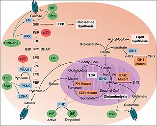
The study of the tumor metabolism, also known as tumor metabolome describes the different characteristic metabolic changes in tumor cells. The characteristic attributes of the tumor metabolome are high glycolytic enzyme activities, the expression of the pyruvate kinase isoenzyme type M2, increased channeling of glucose carbons into synthetic processes, such as nucleic acid, amino acid and phospholipid synthesis, a high rate of pyrimidine and purine de novo synthesis, a low ratio of Adenosine triphosphate and Guanosine triphosphate to Cytidine triphosphate and Uridine triphosphate, low Adenosine monophosphate levels, high glutaminolytic capacities, release of immunosuppressive substances and dependency on methionine.

<span class="mw-page-title-main">Fructose 1,6-bisphosphate</span> Chemical compound

Fructose 1,6-bisphosphate, also known as Harden-Young ester, is fructose sugar phosphorylated on carbons 1 and 6. The β-D-form of this compound is common in cells. Upon entering the cell, most glucose and fructose is converted to fructose 1,6-bisphosphate.

Glucose-1,6-bisphosphate synthase is a type of enzyme called a phosphotransferase and is involved in mammalian starch and sucrose metabolism. It catalyzes the transfer of a phosphate group from 1,3-bisphosphoglycerate to glucose-1-phosphate, yielding 3-phosphoglycerate and glucose-1,6-bisphosphate.

<span class="mw-page-title-main">ARAF</span> Protein-coding gene in humans

Serine/threonine-protein kinase A-Raf or simply A-Raf is an enzyme that in humans is encoded by the ARAF gene. A-Raf is a member of the Raf kinase family of serine/threonine-specific protein kinases.

<span class="mw-page-title-main">Lactate dehydrogenase</span> Class of enzymes

Lactate dehydrogenase (LDH or LD) is an enzyme found in nearly all living cells. LDH catalyzes the conversion of pyruvate to lactate and back, as it converts NAD+ to NADH and back. A dehydrogenase is an enzyme that transfers a hydride from one molecule to another.

Glutaminolysis (glutamine + -lysis) is a series of biochemical reactions by which the amino acid glutamine is lysed to glutamate, aspartate, CO2, pyruvate, lactate, alanine and citrate.

<span class="mw-page-title-main">PKM2</span> Protein-coding gene in the species Homo sapiens

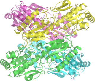
Pyruvate kinase isozymes M1/M2 (PKM1/M2), also known as pyruvate kinase muscle isozyme (PKM), pyruvate kinase type K, cytosolic thyroid hormone-binding protein (CTHBP), thyroid hormone-binding protein 1 (THBP1), or opa-interacting protein 3 (OIP3), is an enzyme that in humans is encoded by the PKM2 gene.

<span class="mw-page-title-main">M2-PK Test</span> Screening for colorectal cancers and polyps

The M2-PK Test is a non-invasive screening method for the early detection of colorectal cancers and polyps which are known to be the precursors of colorectal cancer. The M2-PK Test which is used for stool analysis is available either as fully quantitative ELISA Test or as a rapid test that can be performed by any general practitioner without the need of a laboratory or any additional equipment.

The lactate shuttle hypothesis describes the movement of lactate intracellularly and intercellularly. The hypothesis is based on the observation that lactate is formed and utilized continuously in diverse cells under both anaerobic and aerobic conditions. Further, lactate produced at sites with high rates of glycolysis and glycogenolysis can be shuttled to adjacent or remote sites including heart or skeletal muscles where the lactate can be used as a gluconeogenic precursor or substrate for oxidation. The hypothesis was proposed by professor George Brooks of the University of California at Berkeley.

Zhimin (James) Lu is a Chinese-American biologist and oncologist. He is a professor, Kuancheng Wang Distinguished Chair, and Dean of Institute of Translational Medicine at Zhejiang University. Prior to joining Zhejiang University in 2019, he was the Ruby E. Rutherford Distinguished Professor and the director of Cancer Metabolism Program at the University of Texas MD Anderson Cancer Center.

References

  1. Haug, U.; Rothenbacher, D.; Wente, M. N.; Seiler, C. M.; Stegmaier, C.; Brenner, H. (2007). "Tumour M2-PK as a stool marker for colorectal cancer: Comparative analysis in a large sample of unselected older adults vs colorectal cancer patients". British Journal of Cancer. 96 (9): 1329–34. doi:10.1038/sj.bjc.6603712. PMC   2360192 . PMID   17406361.
  2. Tonus, Carolin; Sellinger, M.; Koss, K.; Neupert, G. (2012). "Faecal pyruvate kinase isoenzyme type M2 for colorectal cancer screening: A meta-analysis". World Journal of Gastroenterology. 18 (30): 4004–4011. doi: 10.3748/wjg.v18.i30.4004 . PMC   3419997 . PMID   22912551.
  3. "Tumor M2-PK Stool Test" . Retrieved June 8, 2013.
  4. Oremek, G; Kukshaĭte, R; Sapoutzis, N; Ziolkovski, P (2007). "The significance of TU M2-PK tumor marker for lung cancer diagnostics". Klinicheskaia Meditsina. 85 (7): 56–58. PMID   17882813.
  5. Roigas J, Schulze G, Raytarowski S, Jung K, Schnorr D, Loening SA. (September 2001). "Tumor M2 pyruvate kinase in plasma of patients with urological tumors". Tumour Biol. 22 (5): 282–5. doi:10.1159/000050628. PMID   11553857. S2CID   46855687.{{cite journal}}: CS1 maint: multiple names: authors list (link)
  6. Bluemlein K, Grüning NM, Feichtinger RG, Lehrach H, Kofler B, Ralser M (2011). "No evidence for a shift in pyruvate kinase PKM1 to PKM2 expression during tumorigenesis". Oncotarget. 2 (5): 393–400. doi:10.18632/oncotarget.278. PMC   3248187 . PMID   21789790.
  7. Akhtar K, Gupta V, Koul A, Alam N, Bhat R, Bamezai RN (May 2009). "Differential behavior of missense mutations in the intersubunit contact domain of the human pyruvate kinase M2 isozyme". J. Biol. Chem. 284 (18): 11971–81. doi: 10.1074/jbc.M808761200 . PMC   2673266 . PMID   19265196.
  8. Gupta V, Kalaiarasan P, Faheem M, Singh N, Iqbal MA, Bamezai RN (May 2010). "Dominant negative mutations affect oligomerization of human pyruvate kinase M2 isozyme and promote cellular growth and polyploidy". J. Biol. Chem. 285 (22): 16864–73. doi: 10.1074/jbc.M109.065029 . PMC   2878009 . PMID   20304929.
  9. Gupta V, Bamezai RN (Sep 2010). "Human pyruvate kinase-M2: "A multi-functional protein"". Protein Sci. 19 (11): 2031–44. doi:10.1002/pro.505. PMC   3005776 . PMID   20857498.

Stool

Plasma

Scientific background