Ribose-phosphate diphosphokinase

Last updated
Ribose-phosphate diphosphokinase
2h06.jpg
Phosphoribosyl pyrophosphate synthase 1, hexamer, Human
Identifiers
EC no. 2.7.6.1
CAS no. 9031-46-3
Databases
IntEnz IntEnz view
BRENDA BRENDA entry
ExPASy NiceZyme view
KEGG KEGG entry
MetaCyc metabolic pathway
PRIAM profile
PDB structures RCSB PDB PDBe PDBsum
Gene Ontology AmiGO / QuickGO
Search
PMC articles
PubMed articles
NCBI proteins
phosphoribosyl pyrophosphate synthetase 1
Identifiers
SymbolPRPS1
NCBI gene 5631
HGNC 9462
OMIM 311850
RefSeq NM_002764
UniProt P60891
Other data
EC number 2.7.6.1
Locus Chr. X q21-q27
Search for
Structures Swiss-model
Domains InterPro
phosphoribosyl pyrophosphate synthetase 2
Identifiers
SymbolPRPS2
NCBI gene 5634
HGNC 9465
OMIM 311860
RefSeq NM_002765
UniProt P11908
Other data
EC number 2.7.6.1
Locus Chr. X pter-q21
Search for
Structures Swiss-model
Domains InterPro

Ribose-phosphate diphosphokinase (or phosphoribosyl pyrophosphate synthetase or ribose-phosphate pyrophosphokinase) is an enzyme that converts ribose 5-phosphate into phosphoribosyl pyrophosphate (PRPP). [1] [2] It is classified under EC 2.7.6.1.

Contents

The enzyme is involved in the synthesis of nucleotides (purines and pyrimidines), cofactors NAD and NADP, and amino acids histidine and tryptophan, [1] [2] [3] linking these biosynthetic processes to the pentose phosphate pathway, from which the substrate ribose 5-phosphate is derived. Ribose 5-phosphate is produced by the HMP Shunt Pathway from Glucose-6-Phosphate. The product phosphoribosyl pyrophosphate acts as an essential component of the purine salvage pathway and the de novo synthesis of purines. Dysfunction of the enzyme would thereby undermine purine metabolism. Ribose-phosphate pyrophosphokinase exists in bacteria, plants, and animals, and there are three isoforms of human ribose-phosphate pyrophosphokinase. [2] In humans, the genes encoding the enzyme are located on the X chromosome. [2]

Reaction mechanism

Overall reaction for phosphoribosyl pyrophosphate synthetase Phosphoribosyl pyrophosphate synthetase equation.png
Overall reaction for phosphoribosyl pyrophosphate synthetase

Ribose-phosphate diphosphokinase transfers the diphosphoryl group from Mg-ATP (Mg2+ coordinated to ATP) to ribose 5-phosphate. [2] The enzymatic reaction begins with the binding of ribose 5-phosphate, followed by binding of Mg-ATP to the enzyme. In the transition state upon binding of both substrates, the diphosphate is transferred. The enzyme first releases AMP before releasing the product phosphoribosyl pyrophosphate. [4] Experiments using oxygen 18 labelled water demonstrate that the reaction mechanism proceeds with the nucleophilic attack of the anomeric hydroxyl group of ribose 5-phosphate on the beta-phosphorus of ATP in an SN2 reaction. [5]

SN2 mechanism of phosphoribosyl pyrophosphate synthetase Phosphoribosyl pyrophosphate synthetase mechanism.png
SN2 mechanism of phosphoribosyl pyrophosphate synthetase

Structure

PyMol rendering of one subunit of the enzyme phosphoribosyl pyrophosphate synthetase I (human). Flexible loop colored in green; ribose 5-phosphate binding region colored in blue. One subunit of the enzyme phosphoribosyl pyrophosphate synthase I (human).png
PyMol rendering of one subunit of the enzyme phosphoribosyl pyrophosphate synthetase I (human). Flexible loop colored in green; ribose 5-phosphate binding region colored in blue.
PyMol rendering of phosphoribosyl pyrophosphate synthetase I (human) as a homodimer, formed by two subunits (red and blue). Three homodimers form the active enzyme complex. Homodimer of phosphoribosyl pyrophosphate synthase I (human).png
PyMol rendering of phosphoribosyl pyrophosphate synthetase I (human) as a homodimer, formed by two subunits (red and blue). Three homodimers form the active enzyme complex.

Crystallization and X-ray diffraction studies elucidated the structure of the enzyme, which was isolated by cloning, protein expression, and purification techniques. One subunit of ribose-phosphate diphosphokinase consists of 318 amino acids; the active enzyme complex consists of three homodimers (or six subunits, a hexamer). The structure of one subunit is a five-stranded parallel beta sheet (the central core) surrounded by four alpha helices at the N-terminal domain and five alpha helices at the C-terminal domain, with two short anti-parallel beta-sheets extending from the core. [2] The catalytic site of the enzyme binds ATP and ribose 5-phosphate. The flexible loop (Phe92–Ser108), pyrophosphate binding loop (Asp171–Gly174), and flag region (Val30–Ile44 from an adjacent subunit) comprise the ATP binding site, located at the interface between two domains of one subunit. The flexible loop is so named because of its large variability in conformation. [6] The ribose 5-phosphate binding site consists of residues Asp220–Thr228, located in the C-terminal domain of one subunit. [2] [6] The allosteric site, which binds ADP, consists of amino acid residues from three subunits. [2]

Function

The product of this reaction, phosphoribosyl pyrophosphate (PRPP), is used in numerous biosynthesis (de novo and salvage) pathways. PRPP provides the ribose sugar in de novo synthesis of purines and pyrimidines, used in the nucleotide bases that form RNA and DNA. PRPP reacts with orotate to form orotidylate, which can be converted to uridylate (UMP). UMP can then be converted to the nucleotide cytidine triphosphate (CTP). The reaction of PRPP, glutamine, and ammonia forms 5-Phosphoribosyl-1-amine, a precursor to inosinate (IMP), which can ultimately be converted to adenosine triphosphate (ATP) or guanosine triphosphate (GTP). PRPP plays a role in purine salvage pathways by reacting with free purine bases to form adenylate, guanylate, and inosinate. [7] [8] PRPP is also used in the synthesis of NAD: the reaction of PRPP with nicotinic acid yields the intermediate nicotinic acid mononucleotide. [9]

Regulation

Ribose-phosphate diphosphokinase requires Mg2+ for activity; the enzyme acts only on ATP coordinated with Mg2+. Ribose-phosphate diphosphokinase is regulated by phosphorylation and allostery. It is activated by phosphate and inhibited by ADP; it is suggested that phosphate and ADP compete for the same regulatory site. At normal concentrations, phosphate activates the enzyme by binding to its allosteric regulatory site. However, at high concentrations, phosphate is shown to have an inhibitory effect by competing with the substrate ribose 5-phosphate for binding at the active site. ADP is the key allosteric inhibitor of ribose-phosphate diphosphokinase. It has been shown that at lower concentrations of the substrate ribose 5-phosphate, ADP may inhibit the enzyme competitively. Ribose-phosphate pyrophosphokinase is also inhibited by some of its downstream biosynthetic products. [2] [6]

Role in disease

Because its product is a key compound in many biosynthetic pathways, ribose-phosphate diphosphokinase is involved in some rare disorders and X-linked recessive diseases. Mutations that lead to super-activity (increased enzyme activity or de-regulation of the enzyme) result in purine and uric acid overproduction. Super-activity symptoms include gout, sensorineural hearing loss, [10] weak muscle tone (hypotonia), impaired muscle coordination (ataxia), hereditary peripheral neuropathy, [11] and neurodevelopmental disorder. [12] [13] [14] Mutations that lead to loss-of-function in ribose-phosphate diphosphokinase result in Charcot-Marie-Tooth disease and Arts syndrome. [15]

Related Research Articles

<span class="mw-page-title-main">Nucleotide</span> Biological molecules that form the building blocks of nucleic acids

Nucleotides are organic molecules composed of a nitrogenous base, a pentose sugar and a phosphate. They serve as monomeric units of the nucleic acid polymers – deoxyribonucleic acid (DNA) and ribonucleic acid (RNA), both of which are essential biomolecules within all life-forms on Earth. Nucleotides are obtained in the diet and are also synthesized from common nutrients by the liver.

<span class="mw-page-title-main">Adenosine monophosphate</span> Chemical compound

Adenosine monophosphate (AMP), also known as 5'-adenylic acid, is a nucleotide. AMP consists of a phosphate group, the sugar ribose, and the nucleobase adenine. It is an ester of phosphoric acid and the nucleoside adenosine. As a substituent it takes the form of the prefix adenylyl-.

<span class="mw-page-title-main">Ribonucleotide</span> Nucleotide containing ribose as its pentose component

In biochemistry, a ribonucleotide is a nucleotide containing ribose as its pentose component. It is considered a molecular precursor of nucleic acids. Nucleotides are the basic building blocks of DNA and RNA. Ribonucleotides themselves are basic monomeric building blocks for RNA. Deoxyribonucleotides, formed by reducing ribonucleotides with the enzyme ribonucleotide reductase (RNR), are essential building blocks for DNA. There are several differences between DNA deoxyribonucleotides and RNA ribonucleotides. Successive nucleotides are linked together via phosphodiester bonds.

A nucleoside triphosphate is a nucleoside containing a nitrogenous base bound to a 5-carbon sugar, with three phosphate groups bound to the sugar. They are the molecular precursors of both DNA and RNA, which are chains of nucleotides made through the processes of DNA replication and transcription. Nucleoside triphosphates also serve as a source of energy for cellular reactions and are involved in signalling pathways.

In molecular biology, biosynthesis is a multi-step, enzyme-catalyzed process where substrates are converted into more complex products in living organisms. In biosynthesis, simple compounds are modified, converted into other compounds, or joined to form macromolecules. This process often consists of metabolic pathways. Some of these biosynthetic pathways are located within a single cellular organelle, while others involve enzymes that are located within multiple cellular organelles. Examples of these biosynthetic pathways include the production of lipid membrane components and nucleotides. Biosynthesis is usually synonymous with anabolism.

<span class="mw-page-title-main">Inosinic acid</span> Chemical compound

Inosinic acid or inosine monophosphate (IMP) is a nucleotide. Widely used as a flavor enhancer, it is typically obtained from chicken byproducts or other meat industry waste. Inosinic acid is important in metabolism. It is the ribonucleotide of hypoxanthine and the first nucleotide formed during the synthesis of purine nucleotides. It can also be formed by the deamination of adenosine monophosphate by AMP deaminase. It can be hydrolysed to inosine.

<span class="mw-page-title-main">Adenine phosphoribosyltransferase</span> Mammalian protein found in Homo sapiens

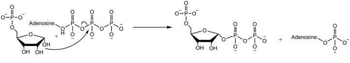
Adenine phosphoribosyltransferase (APRTase) is an enzyme encoded by the APRT gene, found in humans on chromosome 16. It is part of the Type I PRTase family and is involved in the nucleotide salvage pathway, which provides an alternative to nucleotide biosynthesis de novo in humans and most other animals. In parasitic protozoa such as giardia, APRTase provides the sole mechanism by which AMP can be produced. APRTase deficiency contributes to the formation of kidney stones (urolithiasis) and to potential kidney failure.

<span class="mw-page-title-main">Phosphoribosyl pyrophosphate</span> Chemical compound

Phosphoribosyl pyrophosphate (PRPP) is a pentose phosphate. It is a biochemical intermediate in the formation of purine nucleotides via inosine-5-monophosphate, as well as in pyrimidine nucleotide formation. Hence it is a building block for DNA and RNA. The vitamins thiamine and cobalamin, and the amino acid tryptophan also contain fragments derived from PRPP. It is formed from ribose 5-phosphate (R5P) by the enzyme ribose-phosphate diphosphokinase:

<span class="mw-page-title-main">Nucleic acid metabolism</span> Process

Nucleic acid metabolism is a collective term that refers to the variety of chemical reactions by which nucleic acids are either synthesized or degraded. Nucleic acids are polymers made up of a variety of monomers called nucleotides. Nucleotide synthesis is an anabolic mechanism generally involving the chemical reaction of phosphate, pentose sugar, and a nitrogenous base. Degradation of nucleic acids is a catabolic reaction and the resulting parts of the nucleotides or nucleobases can be salvaged to recreate new nucleotides. Both synthesis and degradation reactions require multiple enzymes to facilitate the event. Defects or deficiencies in these enzymes can lead to a variety of diseases.

<span class="mw-page-title-main">Ribose 5-phosphate</span> Chemical compound

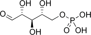
Ribose 5-phosphate (R5P) is both a product and an intermediate of the pentose phosphate pathway. The last step of the oxidative reactions in the pentose phosphate pathway is the production of ribulose 5-phosphate. Depending on the body's state, ribulose 5-phosphate can reversibly isomerize to ribose 5-phosphate. Ribulose 5-phosphate can alternatively undergo a series of isomerizations as well as transaldolations and transketolations that result in the production of other pentose phosphates as well as fructose 6-phosphate and glyceraldehyde 3-phosphate.

Phosphoribosylformylglycinamidine cyclo-ligase is the fifth enzyme in the de novo synthesis of purine nucleotides. It catalyzes the reaction to form 5-aminoimidazole ribotide (AIR) from formylglycinamidine-ribonucleotide FGAM. This reaction closes the ring and produces a 5-membered imidazole ring of the purine nucleus (AIR):

<span class="mw-page-title-main">Orotate phosphoribosyltransferase</span>

Orotate phosphoribosyltransferase (OPRTase) or orotic acid phosphoribosyltransferase is an enzyme involved in pyrimidine biosynthesis. It catalyzes the formation of orotidine 5'-monophosphate (OMP) from orotate and phosphoribosyl pyrophosphate. In yeast and bacteria, orotate phosphoribosyltransferase is an independent enzyme with a unique gene coding for the protein, whereas in mammals and other multicellular organisms, the catalytic function is carried out by a domain of the bifunctional enzyme UMP synthase (UMPS).

Purine metabolism refers to the metabolic pathways to synthesize and break down purines that are present in many organisms.

<span class="mw-page-title-main">Phosphoribosylamine</span> Chemical compound

Phosphoribosylamine (PRA) is a biochemical intermediate in the formation of purine nucleotides via inosine-5-monophosphate, and hence is a building block for DNA and RNA. The vitamins thiamine and cobalamin also contain fragments derived from PRA.

<span class="mw-page-title-main">Amidophosphoribosyltransferase</span> Mammalian protein found in Homo sapiens

Amidophosphoribosyltransferase (ATase), also known as glutamine phosphoribosylpyrophosphate amidotransferase (GPAT), is an enzyme responsible for catalyzing the conversion of 5-phosphoribosyl-1-pyrophosphate (PRPP) into 5-phosphoribosyl-1-amine (PRA), using the amine group from a glutamine side-chain. This is the committing step in de novo purine synthesis. In humans it is encoded by the PPAT gene. ATase is a member of the purine/pyrimidine phosphoribosyltransferase family.

<span class="mw-page-title-main">Glycine—tRNA ligase</span> Protein-coding gene in the species Homo sapiens

Glycine—tRNA ligase also known as glycyl–tRNA synthetase is an enzyme that in humans is encoded by the GARS1 gene.

<span class="mw-page-title-main">Phosphoribosylaminoimidazolesuccinocarboxamide synthase</span> Class of enzymes

In molecular biology, the protein domain SAICAR synthase is an enzyme which catalyses a reaction to create SAICAR. In enzymology, this enzyme is also known as phosphoribosylaminoimidazolesuccinocarboxamide synthase. It is an enzyme that catalyzes the chemical reaction

<span class="mw-page-title-main">ATP phosphoribosyltransferase</span> Class of enzymes

In enzymology, an ATP phosphoribosyltransferase is an enzyme that catalyzes the chemical reaction

<span class="mw-page-title-main">5-Aminoimidazole ribotide</span> Chemical compound

5′-Phosphoribosyl-5-aminoimidazole is a biochemical intermediate in the formation of purine nucleotides via inosine-5-monophosphate, and hence is a building block for DNA and RNA. The vitamins thiamine and cobalamin also contain fragments derived from AIR. It is an intermediate in the adenine pathway and is synthesized from 5′-phosphoribosylformylglycinamidine by AIR synthetase.

<span class="mw-page-title-main">Arts syndrome</span> Medical condition

Arts syndrome is a rare metabolic disorder that causes serious neurological problems in males due to a malfunction of the PRPP synthetase 1 enzyme. Arts Syndrome is part of a spectrum of PRPS-1 related disorders with reduced activity of the enzyme that includes Charcot–Marie–Tooth disease and X-linked non-syndromic sensorineural deafness.

References

  1. 1 2 Visentin LP, Hasnain S, Gallin W (July 1977). "Ribosomal protein S1/S1A in bacteria". FEBS Lett. 79 (2): 258–63. doi: 10.1016/0014-5793(77)80799-0 . PMID   330231. S2CID   38926849.
  2. 1 2 3 4 5 6 7 8 9 Li S, Lu Y, Peng B, Ding J (January 2007). "Crystal structure of human phosphoribosylpyrophosphate synthetase 1 reveals a novel allosteric site". Biochem. J. 401 (1): 39–47. doi:10.1042/BJ20061066. PMC   1698673 . PMID   16939420.
  3. Tang W, Li X, Zhu Z, Tong S, Li X, Zhang X, Teng M, Niu L (May 2006). "Expression, purification, crystallization and preliminary X-ray diffraction analysis of human phosphoribosyl pyrophosphate synthetase 1 (PRS1)". Acta Crystallographica Section F. 62 (Pt 5): 432–4. doi:10.1107/S1744309106009067. PMC   2219982 . PMID   16682768.
  4. Fox IH, Kelley WN (April 1972). "Human phosphoribosylpyrophosphate synthetase. Kinetic mechanism and end product inhibition". J. Biol. Chem. 247 (7): 2126–31. doi: 10.1016/S0021-9258(19)45500-2 . PMID   4335863.
  5. Miller GA, Rosenzweig S, Switzer RL (December 1975). "Oxygen-18 studies of the mechanism of pyrophosphoryl group transfer catalyzed by phosphoribosylpyrophosphate synthetase". Arch. Biochem. Biophys. 171 (2): 732–6. doi:10.1016/0003-9861(75)90086-7. PMID   173242.
  6. 1 2 3 Eriksen TA, Kadziola A, Bentsen AK, Harlow KW, Larsen S (April 2000). "Structural basis for the function of Bacillus subtilis phosphoribosyl-pyrophosphate synthetase". Nat. Struct. Biol. 7 (4): 303–8. doi:10.1038/74069. PMID   10742175. S2CID   23115108.
  7. Fox IH, Kelley WN (March 1971). "Phosphoribosylpyrophosphate in man: biochemical and clinical significance". Ann. Intern. Med. 74 (3): 424–33. doi:10.7326/0003-4819-74-3-424. PMID   4324023.
  8. Jeremy M. Berg; John L. Tymoczko; Lubert Stryer; Gregory J. Gatto Jr. (2012). Biochemistry (7th ed.). New York: W.H. Freeman. ISBN   978-1429229364.
  9. Rongvaux A, Andris F, Van Gool F, Leo O (July 2003). "Reconstructing eukaryotic NAD metabolism". BioEssays. 25 (7): 683–90. doi:10.1002/bies.10297. PMID   12815723.
  10. Liu X, Han D, Li J, Han B, Ouyang X, Cheng J, Li X, Jin Z, Wang Y, Bitner-Glindzicz M, Kong X, Xu H, Kantardzhieva A, Eavey RD, Seidman CE, Seidman JG, Du LL, Chen ZY, Dai P, Teng M, Yan D, Yuan H (January 2010). "Loss-of-function mutations in the PRPS1 gene cause a type of nonsyndromic X-linked sensorineural deafness, DFN2". Am. J. Hum. Genet. 86 (1): 65–71. doi:10.1016/j.ajhg.2009.11.015. PMC   2801751 . PMID   20021999.
  11. Kim HJ, Sohn KM, Shy ME, Krajewski KM, Hwang M, Park JH, Jang SY, Won HH, Choi BO, Hong SH, Kim BJ, Suh YL, Ki CS, Lee SY, Kim SH, Kim JW (September 2007). "Mutations in PRPS1, which encodes the phosphoribosyl pyrophosphate synthetase enzyme critical for nucleotide biosynthesis, cause hereditary peripheral neuropathy with hearing loss and optic neuropathy (cmtx5)". Am. J. Hum. Genet. 81 (3): 552–8. doi:10.1086/519529. PMC   1950833 . PMID   17701900.
  12. Becker MA, Smith PR, Taylor W, Mustafi R, Switzer RL (November 1995). "The genetic and functional basis of purine nucleotide feedback-resistant phosphoribosylpyrophosphate synthetase superactivity". J. Clin. Invest. 96 (5): 2133–41. doi:10.1172/JCI118267. PMC   185862 . PMID   7593598.
  13. Zoref E, De Vries A, Sperling O (November 1975). "Mutant feedback-resistant phosphoribosylpyrophosphate synthetase associated with purine overproduction and gout. Phosphoribosylpyrophosphate and purine metabolism in cultured fibroblasts". J. Clin. Invest. 56 (5): 1093–9. doi:10.1172/JCI108183. PMC   301970 . PMID   171280.
  14. "Phosphoribosylpyrophosphate synthetase superactivity". Lister Hill National Center for Biomedical Communications. Retrieved 25 February 2014.
  15. Synofzik M, Müller Vom Hagen J, Haack TB, Wilhelm C, Lindig T, Beck-Wödl S, Nabuurs SB, van Kuilenburg AB, de Brouwer AP, Schöls L (2014). "X-linked Charcot-Marie-Tooth disease, Arts syndrome, and prelingual non-syndromic deafness form a disease continuum: evidence from a family with a novel PRPS1 mutation". Orphanet J Rare Dis. 9 (1): 24. doi: 10.1186/1750-1172-9-24 . PMC   3931488 . PMID   24528855.