Hemoglobinometer

Last updated
Hemoglobinometer
Sahli's graduated hemoglobin tube and comparator.jpg
Sahli's hemoglobinometer
Specialty Haematology, pathology
ICD-10-PCS D58.2 [1] R71.0 [2]
ICD-9-CM 282.7 [3]
MedlinePlus 003645

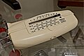
A hemoglobinometer or haemoglobinometer (British English) is a medical device used to measure hemoglobin concentration in blood. [4] It can operate by spectrophotometric measurement of hemoglobin concentration. Portable hemoglobinometers provide easy and convenient measurement of hematological variables, especially in areas where clinic laboratories are unavailable. [5]

Contents

As per guidelines of National AIDS Control Organisation (NACO) for accurate results & mass screening,[ citation needed ] analysis using hemoglobinometer is a recommended method used for absorbance measurement of whole blood at Hb/HbO2/Isobestic point,[ citation needed ] based on microcuvette technology such as HemoCue 301 [6] and Mokshit-Chanda-AM005A. [7]

Devices

See also

Related Research Articles

<span class="mw-page-title-main">Hemoglobin</span> Metalloprotein that binds with oxygen

Hemoglobin is a protein containing iron that facilitates the transport of oxygen in red blood cells. Almost all vertebrates contain hemoglobin, with the exception of the fish family Channichthyidae and the tissues of some invertebrate animals. Hemoglobin in the blood carries oxygen from the respiratory organs to the other tissues of the body, where it releases the oxygen to enable aerobic respiration which powers the animal's metabolism. A healthy human has 12 to 20 grams of hemoglobin in every 100 mL of blood. Hemoglobin is a metalloprotein, a chromoprotein, and globulin.

<span class="mw-page-title-main">Hemoglobinopathy</span> Medical condition

Hemoglobinopathy is the medical term for a group of inherited blood disorders and diseases that primarily affect red blood cells. They are single-gene disorders and, in most cases, they are inherited as autosomal co-dominant traits.

<span class="mw-page-title-main">Thalassemia</span> Medical condition

Thalassemias are inherited blood disorders that result in abnormal hemoglobin. Symptoms depend on the type of thalassemia and can vary from none to severe. Often there is mild to severe anemia as thalassemia can affect the production of red blood cells and also affect how long the red blood cells live. Symptoms of anemia include feeling tired and having pale skin. Other symptoms of thalassemia include bone problems, an enlarged spleen, yellowish skin, pulmonary hypertension, and dark urine. Slow growth may occur in children. Symptoms and presentations of thalassemia can change over time.

<span class="mw-page-title-main">Cyanosis</span> Decreased oxygen in the blood

Cyanosis is the change of body tissue color to a bluish-purple hue, as a result of decrease in the amount of oxygen bound to the hemoglobin in the red blood cells of the capillary bed. Cyanosis is apparent usually in the body tissues covered with thin skin, including the mucous membranes, lips, nail beds, and ear lobes. Some medications may cause discoloration such as medications containing amiodarone or silver. Furthermore, mongolian spots, large birthmarks, and the consumption of food products with blue or purple dyes can also result in the bluish skin tissue discoloration and may be mistaken for cyanosis. Appropriate physical examination and history taking is a crucial part to diagnose cyanosis. Management of cyanosis involves treating the main cause, as cyanosis isn’t a disease, it is a symptom.

<span class="mw-page-title-main">Fetal hemoglobin</span> Oxygen carrier protein in the human fetus

Fetal hemoglobin, or foetal haemoglobin is the main oxygen carrier protein in the human fetus. Hemoglobin F is found in fetal red blood cells, and is involved in transporting oxygen from the mother's bloodstream to organs and tissues in the fetus. It is produced at around 6 weeks of pregnancy and the levels remain high after birth until the baby is roughly 2–4 months old. Hemoglobin F has a different composition than adult forms of hemoglobin, allowing it to bind oxygen more strongly; this in turn enables the developing fetus to retrieve oxygen from the mother's bloodstream, which occurs through the placenta found in the mother's uterus.

<span class="mw-page-title-main">Pulse oximetry</span> Measurement of blood oxygen saturation

Pulse oximetry is a noninvasive method for monitoring a person's blood oxygen saturation. Peripheral oxygen saturation (SpO2) readings are typically within 2% accuracy of the more accurate reading of arterial oxygen saturation (SaO2) from arterial blood gas analysis. But the two are correlated well enough that the safe, convenient, noninvasive, inexpensive pulse oximetry method is valuable for measuring oxygen saturation in clinical use.

The Fick principle states that blood flow to an organ can be calculated using a marker substance if the following information is known:

Glycated hemoglobin is a form of hemoglobin (Hb) that is chemically linked to a sugar. Most monosaccharides, including glucose, galactose and fructose, spontaneously bond with hemoglobin when present in the bloodstream. However, glucose is less likely to do so than galactose and fructose, which may explain why glucose is used as the primary metabolic fuel in humans.

<span class="mw-page-title-main">Hemoglobin A</span> 4f CC w I/ pop m onf

Hemoglobin A (HbA), also known as adult hemoglobin, hemoglobin A1 or α2β2, is the most common human hemoglobin tetramer, accounting for over 97% of the total red blood cell hemoglobin. Hemoglobin is an oxygen-binding protein, found in erythrocytes, which transports oxygen from the lungs to the tissues. Hemoglobin A is the most common adult form of hemoglobin and exists as a tetramer containing two alpha subunits and two beta subunits (α2β2). Hemoglobin A2 (HbA2) is a less common adult form of hemoglobin and is composed of two alpha and two delta-globin subunits. This hemoglobin makes up 1-3% of hemoglobin in adults.


Computed tomography laser mammography (CTLM) is the trademark of Imaging Diagnostic Systems, Inc. for its optical tomographic technique for female breast imaging.

<span class="mw-page-title-main">Functional near-infrared spectroscopy</span> Optical technique for monitoring brain activity

Functional near-infrared spectroscopy (fNIRS) is an optical brain monitoring technique which uses near-infrared spectroscopy for the purpose of functional neuroimaging. Using fNIRS, brain activity is measured by using near-infrared light to estimate cortical hemodynamic activity which occur in response to neural activity. Alongside EEG, fNIRS is one of the most common non-invasive neuroimaging techniques which can be used in portable contexts. The signal is often compared with the BOLD signal measured by fMRI and is capable of measuring changes both in oxy- and deoxyhemoglobin concentration, but can only measure from regions near the cortical surface. fNIRS may also be referred to as Optical Topography (OT) and is sometimes referred to simply as NIRS.

<span class="mw-page-title-main">Alpha-thalassemia</span> Thalassemia involving the genes HBA1and HBA2 hemoglobin genes

Alpha-thalassemia is a form of thalassemia involving the genes HBA1 and HBA2. Thalassemias are a group of inherited blood conditions which result in the impaired production of hemoglobin, the molecule that carries oxygen in the blood. Normal hemoglobin consists of two alpha chains and two beta chains; in alpha-thalassemia, there is a quantitative decrease in the amount of alpha chains, resulting in fewer normal hemoglobin molecules. Furthermore, alpha-thalassemia leads to the production of unstable beta globin molecules which cause increased red blood cell destruction. The degree of impairment is based on which clinical phenotype is present.

Fructosamines are compounds that result from glycation reactions between a sugar and a primary amine, followed by isomerization via the Amadori rearrangement. Biologically, fructosamines are recognized by fructosamine-3-kinase, which may trigger the degradation of advanced glycation end-products. Fructosamine can also refer to the specific compound 1-amino-1-deoxy-D-fructose (isoglucosamine), first synthesized by Nobel laureate Hermann Emil Fischer in 1886.

Hemoglobin Barts, abbreviated Hb Barts, is an abnormal type of hemoglobin that consists of four gamma globins. It is moderately insoluble, and therefore accumulates in the red blood cells. Hb Barts has an extremely high affinity for oxygen, so it cannot release oxygen to the tissue. Therefore, this makes it an inefficient oxygen carrier. As an embryo develops, it begins to produce alpha-globins at weeks 5–6 of development. When both of the HBA1 and HBA2 genes which code for alpha globins becomes dysfunctional, the affected fetuses will have difficulty in synthesizing a functional hemoglobin. As a result, gamma chains will accumulate and form four gamma globins. These gamma globins bind to form hemoglobin Barts. It is produced in the disease alpha-thalassemia and in the most severe of cases, it is the only form of hemoglobin in circulation. In this situation, a fetus will develop hydrops fetalis and normally die before or shortly after birth, unless intrauterine blood transfusion is performed.

<span class="mw-page-title-main">Hemoglobin subunit alpha</span> Human hemoglobin protein

Hemoglobin subunit alpha, Hemoglobin, alpha 1, is a hemoglobin protein that in humans is encoded by the HBA1 gene.

<span class="mw-page-title-main">Hemoglobin Lepore syndrome</span> Medical condition

Hemoglobin Lepore syndrome is typically an asymptomatic hemoglobinopathy, which is caused by an autosomal recessive genetic mutation. The Hb Lepore variant, consisting of two normal alpha globin chains (HBA) and two delta-beta globin fusion chains which occurs due to a "crossover" between the delta (HBD) and beta globin (HBB) gene loci during meiosis and was first identified in the Lepore family, an Italian-American family, in 1958. There are three varieties of Hb Lepore, Washington, Baltimore and Hollandia. All three varieties show similar electrophoretic and chromatographic properties and hematological findings bear close resemblance to those of the beta-thalassemia trait; a blood disorder that reduces the production of the iron-containing protein hemoglobin which carries oxygen to cells and which may cause anemia.

Radiometer is a Danish multinational company which develops, manufactures and markets solutions for blood sampling, blood gas analysis, transcutaneous monitoring, immunoassay testing and the related IT management systems. The company was founded in 1935 in Copenhagen, Denmark by Børge Aagaard Nielsen and Carl Schrøder. It has over 3,200 employees and direct representation in more than 32 countries. Corporate headquarters remain in Copenhagen.

Hemoglobin H (Hb H)Disease, also called alpha-thalassemia intermedia, is a disease affecting hemoglobin, the oxygen carrying molecule within red blood cells. It is a form of Alpha-thalassemia which most commonly occurs due to deletion of 3 out of 4 of the α-globin genes.


Hemoglobin O-Arab or Haemoglobin O-Arab is a rare alternation of Hemoglobin or Haemoglobin, characterised with the presence of β^121Glu → Lys. Mutations of heterozygotes for Hb O-Arab have been reported in Saudi Arabia, North Africa, Sudan, the Mediterranean and the United States. Diagnosis of Hb O-Arab requires liquid chromatography on both cellulose acetate and citrate agar, due to co-migrating with Hb C at acidic pH. When combined with Hemoglobin S it causes a severe form of Sickle cell disease known as Hemoglobin S/O-Arab. Detection of Hb O-Arab can be carried out with a blood test, identifying the carries of hemoglobinopathies, so as to inform patients their chances of producing an affected child and ensure appropriate guidance is given.

<span class="mw-page-title-main">Hemoglobin M disease</span> Medical condition

Hemoglobin M disease is a rare form of hemoglobinopathy, characterized by the presence of hemoglobin M (HbM) and elevated methemoglobin (metHb) level in blood. HbM is an altered form of hemoglobin (Hb) due to point mutation occurring in globin-encoding genes, mostly involving tyrosine substitution for proximal (F8) or distal (E7) histidine residues. HbM variants are inherited as autosomal dominant disorders and have altered oxygen affinity. The pathophysiology of hemoglobin M disease involves heme iron autoxidation promoted by heme pocket structural alteration.

References

  1. "ICD-10-CM 2022 Diagnosis Code D58.2: Other hemoglobinopathies". Archived from the original on 13 June 2021. Retrieved 4 March 2022.
  2. "ICD-10-CM 2022 Diagnosis Code R71.0: Precipitous drop in hematocrit". Archived from the original on 16 April 2021. Retrieved 5 March 2022.
  3. "ICD-9-CM 2015 Diagnosis Code 282.7 : Other hemoglobinopathies". Archived from the original on 2 February 2022. Retrieved 5 March 2022.
  4. Schmalzel, JL; Steinke, JM; Randal, VT; Shepherd, AP (2 October 1989). "An optical hemoglobinometer for whole blood". The American Journal of Physiology. San Antonio: University of Texas Health Science Center. 257 (4 Pt 2): H1306–11. doi:10.1152/ajpheart.1989.257.4.H1306. PMID   2801989. Archived from the original on 4 March 2022. Retrieved 10 June 2011.
  5. Noiri, E.; Kobayashi, N.; Takamura, Y.; Iijima, T.; Takagi, T.; Doi, K.; Nakao, A.; Yamamoto, T.; Takeda, S.; Fujita, T. (4 December 2005). "Pulse total-hemoglobinometer provides accurate noninvasive monitoring". Critical Care Medicine. University of Tokyo. 33 (12): 2831–5. doi:10.1097/01.ccm.0000190430.96750.51. PMID   16352948. S2CID   46153309. Archived from the original on 4 March 2022. Retrieved 10 June 2011.
  6. "Point-of-care anemia screening - HemoCue® Hb 301 System - HemoCue". www.hemocue.in. Archived from the original on 15 May 2021. Retrieved 4 March 2022.
  7. "Pathological Equipment | Blood Storage Refrigerator | Mokshit Corporation in Chhattisgarh, India". Archived from the original on 20 February 2020. Retrieved 4 March 2022.