Intraoperative radiation therapy

Last updated
Intraoperative radiation therapy
Other namesIORT
ICD-9-CM 92.4

Intraoperative radiation therapy (IORT) is radiation therapy that is administered during surgery directly in the operating room (hence intraoperative).

Contents

Usually therapeutic levels of radiation are delivered to the tumor bed while the area is exposed during surgery. IORT is typically a component in the multidisciplinary treatment of locally advanced and recurrent cancer, in combination with external beam radiation, surgery, and chemotherapy. As a growing trend in recent years, IORT can also be used in earlier stage cancers such as prostate and breast cancer.

Medical uses

IORT was found to be useful and feasible in the multidisciplinary management of many solid tumors but further studies are needed to determine the benefit more precisely. [1] Single-institution experiences have suggested a role of IORT e.g. in brain tumors and cerebral metastases, locally advanced and recurrent rectal cancer, skin cancer, retroperitoneal sarcoma, pancreatic cancer, and selected gynaecologic and genitourinary malignancies. For local recurrences, irradiation with IORT is, besides brachytherapy, the only radiotherapeutic option if repeated EBRT is no longer possible. Generally, the normal tissue tolerance does not allow a second full-dose course of EBRT, even after years. [2] [3]

Breast cancer

On 25 July 2014, the UK National Institute for Health and Care Excellence (NICE) gave provisional recommendation for the use of TARGIT IORT with Intrabeam in the UK National Health Service. [4] The 2015 update of guidelines of the Association of Gynecological Oncology (AGO), an autonomous community of the German Society of Gynecology and Obstetrics (DGGG) and the German Cancer Society includes TARGIT IORT during lumpectomy as a recommended option for women with a T1, Grade 1 or 2, ER positive breast cancer. [5]

Rationale

The rationale for IORT is to deliver a high dose of radiation precisely to the targeted area with minimal exposure of surrounding tissues which are displaced or shielded during the IORT. Conventional radiation techniques such as external beam radiotherapy (EBRT) following surgical removal of the tumor have several drawbacks: The tumor bed where the highest dose should be applied is frequently missed due to the complex localization of the wound cavity even when modern radiotherapy planning is used. Additionally, the usual delay between the surgical removal of the tumor and EBRT may allow a repopulation of the tumor cells. These potentially harmful effects can be avoided by delivering the radiation more precisely to the targeted tissues leading to immediate sterilization of residual tumor cells. Another aspect is that wound fluid has a stimulating effect on tumor cells. IORT was found to inhibit the stimulating effects of wound fluid. [6]

Methods

Several methods are used to deliver IORT. IORT can be delivered using electron beams (electron IORT), orthovoltage (250–300 kV) X-rays (X-ray IORT), high-dose-rate brachytherapy (HDR-IORT), or low-energy (50 kV) x-rays (low-energy IORT).

Electron IORT

While IORT was first used in clinical practice in 1905, [7] [8] the modern era of IORT began with the introduction of electron IORT in the mid-1960s by transporting patients from the OR after the tumor was removed to the radiation department to receive their electron IORT. [9] [10] Electron IORT has the advantages of being able to carefully control the depth of radiation penetration while providing a very uniform dose to the tumor bed. Applied with energies in the range of 3 MeV to 12 MeV, electron IORT can treat to depths of up to 4 cm over areas as large as 300 cm² (i.e. a 10 cm diameter circle) and takes only 1–3 minutes to deliver the prescribed radiation dose. A few hospitals built shielded operation rooms in which a conventional linear accelerator was installed to deliver the IORT radiation. This eliminated the complex logistics involved with patient transportation, but was so costly that only a few hospitals were able to use this approach. The breakthrough came in 1997, with the introduction of a miniaturized, self-shielded, mobile linear accelerator (Mobetron, IntraOp Corporation, US) [11] and a mobile but unshielded linear accelerator (Novac, Liac–SIT, Italy). More than 75,000 patients have been treated with electron IORT, almost half of them since the introduction of mobile electron IORT technology.

X-ray IORT

Early practitioners of IORT treated primarily abdominal malignancies using superficial X-rays (75–125 kV) and later orthovoltage x-rays (up to 300 kV in energy) prior to the advent of technology that enabled high-energy electrons. For the first 75 years, X-ray IORT was used mostly for palliation, but there were a few anecdotal reports of long-term survivors. In the early 1980s, when the use of electron IORT was increasing and showed promising results for certain indications, a handful of hospitals installed othovoltage units in lightly shielded ORs to see if this lower cost approach could achieve comparable results to that of electron IORT. This approach was less costly than building a shielded OR for an electron IORT unit and eliminated the logistics involved with patient transportation. However, it had a number of problems that limited its appeal. X-ray IORT has a poor uniformity of dose as a function of depth of penetration, the radiation does not stop at a pre-defined depth but continues to deposit radiation to underlying structures, and can do damage to boney structures if too high a dose is delivered. Despite its long use (since the 1930s), fewer than 1000 patients have been treated with this approach, and it is no longer offered at most centers. [12]

HDR-IORT

This technique was developed in the late 1980s in an attempt to combine the dosimetric advantages of high-dose rate brachytherapy with the challenges of treating some complex anatomic surfaces with IORT. It has the advantage of being lower cost than dedicated electron IORT systems, since many radiation centers already have an HDR system that can be transported to the OR when HDR-IORT is needed. HDR-IORT can also treat very large and convoluted surfaces. However, it does require a shielded OR or a shielded room in the OR complex to deliver the HDR-IORT. [13] The depth of penetration is very limited, typically either ½ cm to 1 cm depth, sometimes requiring extensive surgery due to the limited penetration of the radiation. Treatments tend to be 40 minutes or longer, resulting in greater OR time, more anesthesia and greater blood loss when compared to electron IORT. There are about 10 to 20 active centers using HDR-IORT for locally advanced and recurrent disease, and approximately 2000 patients have received this treatment, mostly for colorectal cancer, head and neck cancer, and gynecologic cancer.

Low-energy IORT (50 kV)

Intrabeam, [14] (Carl Zeiss AG, Germany) received FDA and CE approval in 1999 and is a miniature mobile X-ray source which emits low-energy X-ray radiation (max. 50 kV) in isotropic distribution. Due to the higher ionization density caused by soft X-ray radiation in the tissue, the relative biological effectiveness (RBE) of low-energy X-rays on tumor cells is higher when compared to high-energy X-rays or gamma rays which are delivered by linear accelerators. [15] The radiation which is produced by low-energy mobile radiation systems has a limited range. For this reason, conventional walls are regarded sufficient to stop the radiation scatter produced in the operating room and no extra measures for radiation protection are necessary. This makes IORT accessible for more hospitals. Targeted intra-operative radiotherapy is a low-energy IORT technique. Evaluation of the long-term outcomes in patients who were treated with TARGIT-IORT for breast cancer confirmed that it is as effective as whole breast external beam radiotherapy in controlling cancer, and also reduces deaths from other causes [16] as shown in a large international randomised clinical trial published in the British Medical Journal. [17]

See also

Related Research Articles

<span class="mw-page-title-main">Radiation therapy</span> Therapy using ionizing radiation, usually to treat cancer

Radiation therapy or radiotherapy, often abbreviated RT, RTx, or XRT, is a treatment using ionizing radiation, generally provided as part of cancer therapy to either kill or control the growth of malignant cells. It is normally delivered by a linear particle accelerator. Radiation therapy may be curative in a number of types of cancer if they are localized to one area of the body, and have not spread to other parts. It may also be used as part of adjuvant therapy, to prevent tumor recurrence after surgery to remove a primary malignant tumor. Radiation therapy is synergistic with chemotherapy, and has been used before, during, and after chemotherapy in susceptible cancers. The subspecialty of oncology concerned with radiotherapy is called radiation oncology. A physician who practices in this subspecialty is a radiation oncologist.

<span class="mw-page-title-main">External beam radiotherapy</span> Treatment of cancer with ionized radiation

External beam radiation therapy (EBRT) is a compound word that refers to the use of a collimated beam of ionizing radiation from outside the body to treat a disease.

<span class="mw-page-title-main">Brachytherapy</span> Type of radiation therapy

Brachytherapy is a form of radiation therapy where a sealed radiation source is placed inside or next to the area requiring treatment. Brachy is Greek for short. Brachytherapy is commonly used as an effective treatment for cervical, prostate, breast, esophageal and skin cancer and can also be used to treat tumours in many other body sites. Treatment results have demonstrated that the cancer-cure rates of brachytherapy are either comparable to surgery and external beam radiotherapy (EBRT) or are improved when used in combination with these techniques. Brachytherapy can be used alone or in combination with other therapies such as surgery, EBRT and chemotherapy.

<span class="mw-page-title-main">Megavoltage X-rays</span> High energy (>1MeV) X-rays

Megavoltage X-rays are produced by linear accelerators ("linacs") operating at voltages in excess of 1000 kV (1 MV) range, and therefore have an energy in the MeV range. The voltage in this case refers to the voltage used to accelerate electrons in the linear accelerator and indicates the maximum possible energy of the photons which are subsequently produced. They are used in medicine in external beam radiotherapy to treat neoplasms, cancer and tumors. Beams with the voltage range of 4-25 MV are used to treat deeply buried cancers because radiation oncologists find that they penetrate well to deep sites within the body. Lower energy x-rays, called orthovoltage X-rays, are used to treat cancers closer to the surface.

<span class="mw-page-title-main">Proton therapy</span> Medical Procedure

In medicine, proton therapy, or proton radiotherapy, is a type of particle therapy that uses a beam of protons to irradiate diseased tissue, most often to treat cancer. The chief advantage of proton therapy over other types of external beam radiotherapy is that the dose of protons is deposited over a narrow range of depth; hence in minimal entry, exit, or scattered radiation dose to healthy nearby tissues.

<span class="mw-page-title-main">Radiosurgery</span> Surgical Specialty

Radiosurgery is surgery using radiation, that is, the destruction of precisely selected areas of tissue using ionizing radiation rather than excision with a blade. Like other forms of radiation therapy, it is usually used to treat cancer. Radiosurgery was originally defined by the Swedish neurosurgeon Lars Leksell as "a single high dose fraction of radiation, stereotactically directed to an intracranial region of interest".

Elekta is a global Swedish company that develops and produces radiation therapy and radiosurgery-related equipment and clinical management for the treatment of cancer and brain disorders. Elekta has a global presence in more than 120 countries, with over 40 offices around the world and about 4,700 employees.

<span class="mw-page-title-main">Fast neutron therapy</span>

Fast neutron therapy utilizes high energy neutrons typically between 50 and 70 MeV to treat cancer. Most fast neutron therapy beams are produced by reactors, cyclotrons (d+Be) and linear accelerators. Neutron therapy is currently available in Germany, Russia, South Africa and the United States. In the United States, one treatment center is operational, in Seattle, Washington. The Seattle center uses a cyclotron which produces a proton beam impinging upon a beryllium target.

<span class="mw-page-title-main">Radiation treatment planning</span>

In radiotherapy, radiation treatment planning (RTP) is the process in which a team consisting of radiation oncologists, radiation therapist, medical physicists and medical dosimetrists plan the appropriate external beam radiotherapy or internal brachytherapy treatment technique for a patient with cancer.

Electron therapy or electron beam therapy (EBT) is a kind of external beam radiotherapy where electrons are directed to a tumor site for medical treatment of cancer.

<span class="mw-page-title-main">Tomotherapy</span> Type of radiation therapy

Tomotherapy is a type of radiation therapy treatment machine. In tomotherapy a thin radiation beam is modulated as it rotates around the patient, while they are moved through the bore of the machine. The name comes from the use of a strip-shaped beam, so that only one “slice” of the target is exposed at any one time by the radiation. The external appearance of the system and movement of the radiation source and patient can be considered analogous to a CT scanner, which uses lower doses of radiation for imaging. Like a conventional machine used for X-ray external beam radiotherapy, a linear accelerator generates the radiation beam, but the external appearance of the machine, the patient positioning, and treatment delivery is different. Conventional linacs do not work on a slice-by-slice basis but typically have a large area beam which can also be resized and modulated.

<span class="mw-page-title-main">Cobalt therapy</span> Medical use of gamma rays

Cobalt therapy is the medical use of gamma rays from the radioisotope cobalt-60 to treat conditions such as cancer. Beginning in the 1950s, cobalt-60 was widely used in external beam radiotherapy (teletherapy) machines, which produced a beam of gamma rays which was directed into the patient's body to kill tumor tissue. Because these "cobalt machines" were expensive and required specialist support, they were often housed in cobalt units. Cobalt therapy was a revolutionary advance in radiotherapy in the post-World War II period but is now being replaced by other technologies such as linear accelerators.

Particle therapy is a form of external beam radiotherapy using beams of energetic neutrons, protons, or other heavier positive ions for cancer treatment. The most common type of particle therapy as of August 2021 is proton therapy.

Intraoperative electron radiation therapy is the application of electron radiation directly to the residual tumor or tumor bed during cancer surgery. Electron beams are useful for intraoperative radiation treatment because, depending on the electron energy, the dose falls off rapidly behind the target site, therefore sparing underlying healthy tissue.

Breast cancer management takes different approaches depending on physical and biological characteristics of the disease, as well as the age, over-all health and personal preferences of the patient. Treatment types can be classified into local therapy and systemic treatment. Local therapy is most efficacious in early stage breast cancer, while systemic therapy is generally justified in advanced and metastatic disease, or in diseases with specific phenotypes.

<span class="mw-page-title-main">Prostate brachytherapy</span> Radiation therapy technique for the treatment of cancer

Brachytherapy is a type of radiotherapy, or radiation treatment, offered to certain cancer patients. There are two types of brachytherapy – high dose-rate (HDR) and low dose-rate (LDR). LDR brachytherapy is the one most commonly used to treat prostate cancer. It may be referred to as 'seed implantation' or it may be called 'pinhole surgery'.

<span class="mw-page-title-main">Targeted intra-operative radiotherapy</span> Method of targeted radiotherapy after surgical removal of tumours

Targeted intra-operative radiotherapy, also known as targeted IORT, is a technique of giving radiotherapy to the tissues surrounding a cancer after its surgical removal, a form of intraoperative radiation therapy. The technique was designed in 1998 at the University College London.

<span class="mw-page-title-main">Neutron capture therapy of cancer</span> Nonsurgical therapeutic modality for treating locally invasive malignant tumors

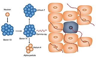
Neutron capture therapy (NCT) is a type of radiotherapy for treating locally invasive malignant tumors such as primary brain tumors, recurrent cancers of the head and neck region, and cutaneous and extracutaneous melanomas. It is a two-step process: first, the patient is injected with a tumor-localizing drug containing the stable isotope boron-10 (10B), which has a high propensity to capture low energy "thermal" neutrons. The neutron cross section of 10B is 1,000 times more than that of other elements, such as nitrogen, hydrogen, or oxygen, that occur in tissue. In the second step, the patient is radiated with epithermal neutrons, the sources of which in the past have been nuclear reactors and now are accelerators that produce higher energy epithermal neutrons. After losing energy as they penetrate tissue, the resultant low energy "thermal" neutrons are captured by the 10B atoms. The resulting decay reaction yields high-energy alpha particles that kill the cancer cells that have taken up enough 10B.

Docrates Cancer Center is the first and currently the only private hospital in the Nordic countries that comprehensively specialises in cancer treatment. It operates in Helsinki, Finland. It characterises its operations as those complementing the public sector. Docrates Oy was established in 2006 and the hospital started its operations at the premises of Eira Hospital in autumn 2007. It moved to its own premises in Jätkäsaari, Helsinki, in 2009, where it has hospital rights. There is a ward and Health and Recovery Center located at Docrates Cancer Center. Among other things, diagnostics, pharmacotherapy, radiation therapy and isotopic treatments are carried out at the hospital. Cancer surgeries are performed in partner hospitals. Docrates also participates in clinical trials and the testing and development of new treatments.

<span class="mw-page-title-main">Jayant S. Vaidya</span> British-Indian surgeon and oncologist

Jayant S. Vaidya is a British-Indian surgeon-oncologist and clinical academic who, together with Michael Baum and Jeffrey Tobias, developed the technique called targeted intra-operative radiotherapy (TARGIT). He is a professor of surgery and oncology at the University College London, London and the author of two books on breast cancer, one on tobacco eradication, and over 200 academic articles.

References

  1. Mirnezami, Reza; Chang, George J.; Das, Prajnan; Chandrakumaran, Kandiah; Tekkis, Paris; Darzi, Ara; Mirnezami, Alexander H. (March 2013). "Intraoperative radiotherapy in colorectal cancer: Systematic review and meta-analysis of techniques, long-term outcomes, and complications". Surgical Oncology. 22 (1): 22–35. doi:10.1016/j.suronc.2012.11.001. PMC   4663079 . PMID   23270946.
  2. Kraus-Tiefenbacher, Uta; Bauer, Lelia; Scheda, Antonella; Schoeber, Carola; Schaefer, Joerg; Steil, Volker; Wenz, Frederik (14 September 2007). "Intraoperative radiotherapy (IORT) is an option for patients with localized breast recurrences after previous external-beam radiotherapy". BMC Cancer. 7 (1): 178. doi: 10.1186/1471-2407-7-178 . PMC   2039741 . PMID   17854511.
  3. Hannoun-Levi, Jean-Michel; Ihrai, Tarik; Courdi, Adel (November 2013). "Local treatment options for ipsilateral breast tumour recurrence". Cancer Treatment Reviews. 39 (7): 737–741. doi:10.1016/j.ctrv.2013.02.003. PMID   23465859.
  4. Smyth, Chris (2014). "Single-dose radiotherapy eases breast cancer stress". The Times. Retrieved 26 July 2016.
  5. "Diagnosis and Treatment of Patients with Primary and Metastatic Breast Cancer" (PDF). AGO. Archived from the original (PDF) on 7 March 2017. Retrieved 26 July 2016.
  6. Belletti, B.; Vaidya, J. S.; D'Andrea, S.; Entschladen, F.; Roncadin, M.; Lovat, F.; Berton, S.; Perin, T.; Candiani, E.; Reccanello, S.; Veronesi, A.; Canzonieri, V.; Trovo, M. G.; Zaenker, K. S.; Colombatti, A.; Baldassarre, G.; Massarut, S. (3 March 2008). "Targeted Intraoperative Radiotherapy Impairs the Stimulation of Breast Cancer Cell Proliferation and Invasion Caused by Surgical Wounding". Clinical Cancer Research. 14 (5): 1325–1332. doi: 10.1158/1078-0432.CCR-07-4453 . PMID   18316551.
  7. Comas C., Prio A. Irradiation roentgen intra-abdominale, après intervention chirurgicale dans un cas de cancer de l'uterus, Congres International d'Electrologie. Imprenta Francesca Badia, Barcelona, pp 5-14, 1907
  8. Comas, C; Prio, A (1906). Comptes rendus des séances du 3e Congrès international d'électrologie et de radiologie médicales (Milan: 5-9 septembre 1906) (in French). Lille: C. Robbo. p.  321.
  9. Abe, Mitsuyuki; Takahashi, Masaji (July 1981). "Intraoperative radiotherapy: The japanese experience". International Journal of Radiation Oncology, Biology, Physics. 7 (7): 863–868. doi:10.1016/0360-3016(81)90001-8. PMID   7198109.
  10. Goldson A., Past, present and prospects of intraoperative radiotherapy (IOR). Semin Oncol 1981.
  11. Vaeth, J. M. (1996). Intraoperative Radiation Therapy in the Treatment of Cancer. pp. 65–70. doi:10.1159/000061147. ISBN   978-3-8055-6456-4. PMID   9263791.{{cite book}}: |journal= ignored (help)
  12. Abe M. History of Intraoperative radiation therapy. In:Debelbower RR, Abe M (eds) Intraoperative radiation therapy. CRC, Boca Raton; :1-10, 1989.
  13. Furhang, Eli E.; Sillanpaa, Jussi K.; Hu, Kenneth S.; Harrison, Louis B. (2011). "HDR-IORT: Physics and Techniques". In Gunderson, Leonard L.; Willett, Christopher G.; Calvo, Felipe A.; Harrison, Louis B. (eds.). Intraoperative Irradiation (2nd ed.). New York: Humana Press. pp. 73–84. doi:10.1007/978-1-61779-015-7_4. ISBN   978-1-61779-014-0.
  14. "INTRABEAM - breast cancer - Oncology - Medical Technology | ZEISS United States". Zeiss.com. Retrieved 2016-05-18.
  15. Hill, M. A. (15 December 2004). "The variation in biological effectiveness of X-rays and gamma rays with energy". Radiation Protection Dosimetry. 112 (4): 471–481. doi:10.1093/rpd/nch091. PMID   15623881.
  16. Correspondent, Maya Oppenheim, Health. "Breast cancer breakthrough as new treatment requires one shot of radiotherapy instead of multiple doses". Independent. ISSN   0951-9467 . Retrieved 2020-09-09.{{cite news}}: CS1 maint: multiple names: authors list (link)
  17. Vaidya, Jayant S.; Bulsara, Max; Baum, Michael; Wenz, Frederik; Massarut, Samuele; Pigorsch, Steffi; Alvarado, Michael; Douek, Michael; Saunders, Christobel; Flyger, Henrik L.; Eiermann, Wolfgang (2020-08-19). "Long term survival and local control outcomes from single dose targeted intraoperative radiotherapy during lumpectomy (TARGIT-IORT) for early breast cancer: TARGIT-A randomised clinical trial". BMJ. 370: m2836. doi: 10.1136/bmj.m2836 . ISSN   1756-1833. PMC   7500441 . PMID   32816842.