Invadopodia

Last updated
A microscope picture of a cell's invadopodium. Invadopodium.png
A microscope picture of a cell's invadopodium.

Invadopodia are actin-rich protrusions of the plasma membrane that are associated with degradation of the extracellular matrix in cancer invasiveness and metastasis. [1] [2] Very similar to podosomes, invadopodia are found in invasive cancer cells and are important for their ability to invade through the extracellular matrix, especially in cancer cell extravasation. [3] Invadopodia are generally visualized by the holes they create in ECM (fibronectin, collagen etc.)-coated plates, in combination with immunohistochemistry for the invadopodia localizing proteins such as cortactin, actin, Tks5 [1] [2] [4] etc. Invadopodia can also be used as a marker to quantify the invasiveness of cancer cell lines in vitro using a hyaluronic acid hydrogel assay. [5]

Contents

History and controversy

In the early 1980s, researchers noticed protrusions coming from the ventral membrane of cells that had been transformed by the Rous Sarcoma Virus and that they were at the sites of cell-to-extracellular matrix (ECM) adhesion. [1] They termed these structures podosomes, or cellular feet, but it was later noticed that degradation of the ECM was occurring at these sites and the name invadopodia was coined to highlight the invasive nature of these protrusions. [1] Since then, researchers have often used the two names interchangeably, but it is generally accepted that podosomes are the structures involved in normal biological processes (as when immune cells must cross tissue barriers or in bone remodeling [6] ) and invadopodia are the structures in invading cancer cells. [1] However, there remains controversy around this nomenclature, with some scientists arguing that the two are different enough to be considered distinct structures while others argue that invadopodia are simply disregulated podosomes and cancer cells don’t simply "invent" new mechanisms. Due to this confusion and the high similarity between the two structures, many have begun to group the two under the collective term invadosomes. [3]

Structure and formation

Invadopodia have an actin core, which is surrounded by a ring structure enriched in actin-binding proteins, adhesion molecules, integrins, and scaffold proteins. [1] [2] [3] [7] Invadopodia are generally longer than podosomes, with a width of 0.5- 2.0 um and a length greater than 2 um, and they last much longer than podosomes. [1] Invadopodia also penetrate deep into the ECM, while podosomes generally extend upward into the cytoplasm and do not cause as much ECM degradation. [3]

Invadopodia formation is a complex process that involves multiple signaling pathways and can be described as having three steps: initiation, stabilization, and maturation. [7] [8] Initiation of invadopodia involves the formation of buds in the plasma membrane and is initiated by growth factors like epidermal growth factor (EGF), transforming growth factor beta (TGFB) or platelet-derived growth factor (PDGF), which act through phosphoinositide 3-kinase (PI3K) to activate Src family kinases. [1] These kinases have key roles in the formation of invadopodia and when activated, phosphorylate multiple proteins involved in invadopodia formation including Tks5, synaptjanin-2, and the Abl-family kinase Arg4. The phosphorylation of these proteins leads to the recruitment of the Neural Wiskott-Aldrich syndrome protein (N-Wasp) to invadopodia, which requires Arp2/3, to activate actin polymerization and thus invadopodia elongation. [9] A key step during invadopodia formation is the stabilization of invadopodia, which involves the interaction of PX domain of Tks5 (a scaffold protein) with phospholipid, PI(3,4)P2 to anchor the invadopodia core to the plasma membrane. [7] Maturation of invadopodia requires sustained actin polymerization and there are several regulators of actin polymerization involved in this step, including cofilin, fascin, Arg kinase, and mDia2. [9] Invadopodia are considered mature when matrix metalloproteases (MMPs), specifically MMP2, 9, and 14, are recruited to the invadopodium to be released into the extracellular matrix. [9]

Role in cancer metastasis

Metastasis is the leading cause of mortality in cancer patients; it relies on the ability of cancer cells to degrade the surrounding extracellular matrix and invade other tissues. The mechanisms of this process are still not completely understood, and because of the invasive properties of invadopodia, they have been investigated in this context. Indeed, invadopodia have been implicated in many cancers and cancer cells. Increased invasiveness of cancer cells correlates with invadopodia presence, and cancer cells have been observed to project them into the endothelium of blood vessels during extravasation, an important step in metastasis. [10] Invadopodia have also been shown to correlate with a poorer prognosis in breast cancer patients. [11]

Tks5, a protein specific for invadopodia, has been implicated in cancer invasiveness. Increased levels of tks5 have been detected in prostate cancer and overexpression of Tks5 was sufficient to induce invadopodia formation and degradation of the extracellular matrix in an Src-dependent manner. [12] Increased Tks5 expression has been shown to correlate with poor patient prognosis in gliomas. [13] In a mouse model of lung adenocarcinoma, invasive tumors were shown to have an increased expression of a long isoform of tks5 while non-metastatic tumors had a short isoform. It was also shown that overexpression of the long isoform of tks5 was sufficient to cause non-metastatic tumors to become invasive. [14]

Therapeutic relevance

Due to the invasive nature of invadopodia in cancer cells, research has focused on targeting invadopodia as a potential therapeutic target to inhibit metastasis. Inhibiting invadopodia formation by targeting Src kinase with Saracatanib in a chicken model system showed a decreased incidence of invadopodia and decreased cancer extravasation. In mice, inhibiting invadopodia formation directly, through RNAi against tks4 or tks5, significantly reduced cancer extravasation. [10] Screening for drug activators and inhibitors of invadopodia revealed that Cdc5 can be a target for inhibiting invadopodia formation and also that, paradoxically, paclitaxel, a drug commonly used to treat cancer, induces invadopodia formation. [15] These results show potential for invadopodia as a therapeutic target, and research in this field continues.

See also

Related Research Articles

Integrin Instance of a defined set in Homo sapiens with Reactome ID (R-HSA-374573)

Integrins are transmembrane receptors that facilitate cell-cell and cell-extracellular matrix (ECM) adhesion. Upon ligand binding, integrins activate signal transduction pathways that mediate cellular signals such as regulation of the cell cycle, organization of the intracellular cytoskeleton, and movement of new receptors to the cell membrane. The presence of integrins allows rapid and flexible responses to events at the cell surface.

Pseudopodia False leg found on slime molds, archaea, protozoans, leukocytes and certain bacteria

A pseudopod or pseudopodium is a temporary arm-like projection of a eukaryotic cell membrane that is emerged in the direction of movement. Filled with cytoplasm, pseudopodia primarily consist of actin filaments and may also contain microtubules and intermediate filaments. Pseudopods are used for motility and ingestion. They are often found in amoebas.

Extracellular matrix Network of proteins and molecules outside cells that provides structural support for cells

In biology, the extracellular matrix (ECM) is a three-dimensional network consisting of extracellular macromolecules and minerals, such as collagen, enzymes, glycoproteins and hydroxyapatite that provide structural and biochemical support to surrounding cells. Because multicellularity evolved independently in different multicellular lineages, the composition of ECM varies between multicellular structures; however, cell adhesion, cell-to-cell communication and differentiation are common functions of the ECM.

Focal adhesion

In cell biology, focal adhesions are large macromolecular assemblies through which mechanical force and regulatory signals are transmitted between the extracellular matrix (ECM) and an interacting cell. More precisely, focal adhesions are the sub-cellular structures that mediate the regulatory effects of a cell in response to ECM adhesion.

Podosome

Podosomes are conical, actin-rich structures found on the outer surface of the plasma membrane of animal cells. Their size ranges from approximately 0.5 µm to 2.0 µm in diameter. While usually situated on the periphery of the cellular membrane, these unique structures display a polarized pattern of distribution in migrating cells, situating at the front border between the lamellipodium and lamellum. Their primary purpose is connected to cellular motility and invasion; therefore, they serve as both sites of attachment and degradation along the extracellular matrix. Many different specialized cells exhibit these dynamic structures such as invasive cancer cells, osteoclasts, vascular smooth muscle cells, endothelial cells, and certain immune cells like macrophages and dendritic cells.

Biological crosstalk refers to instances in which one or more components of one signal transduction pathway affects another. This can be achieved through a number of ways with the most common form being crosstalk between proteins of signaling cascades. In these signal transduction pathways, there are often shared components that can interact with either pathway. A more complex instance of crosstalk can be observed with transmembrane crosstalk between the extracellular matrix (ECM) and the cytoskeleton.

Anoikis is a form of programmed cell death that occurs in anchorage-dependent cells when they detach from the surrounding extracellular matrix (ECM). Usually cells stay close to the tissue to which they belong since the communication between proximal cells as well as between cells and ECM provide essential signals for growth or survival. When cells are detached from the ECM, there is a loss of normal cell–matrix interactions, and they may undergo anoikis. However, metastatic tumor cells may escape from anoikis and invade other organs.

Intravasation is the invasion of cancer cells through the basement membrane into a blood or lymphatic vessel. Intravasation is one of several carcinogenic events that initiate the escape of cancerous cells from their primary sites. Other mechanisms include invasion through basement membranes, extravasation, and colonization of distant metastatic sites. Cancer cell chemotaxis also relies on this migratory behavior to arrive at a secondary destination designated for cancer cell colonization.

Endoglin Protein-coding gene in the species Homo sapiens

Endoglin (ENG) is a type I membrane glycoprotein located on cell surfaces and is part of the TGF beta receptor complex. It is also commonly referred to as CD105, END, FLJ41744, HHT1, ORW and ORW1. It has a crucial role in angiogenesis, therefore, making it an important protein for tumor growth, survival and metastasis of cancer cells to other locations in the body.

Cortactin

Cortactin is a monomeric protein located in the cytoplasm of cells that can be activated by external stimuli to promote polymerization and rearrangement of the actin cytoskeleton, especially the actin cortex around the cellular periphery. It is present in all cell types. When activated, it will recruit Arp2/3 complex proteins to existing actin microfilaments, facilitating and stabilizing nucleation sites for actin branching. Cortactin is important in promoting lamellipodia formation, invadopodia formation, cell migration, and endocytosis.

Fascin Actin bundling protein

Fascin is an actin bundling protein.

MMP2

72 kDa type IV collagenase also known as matrix metalloproteinase-2 (MMP-2) and gelatinase A is an enzyme that in humans is encoded by the MMP2 gene. The MMP2 gene is located on chromosome 16 at position 12.2.

PTK2 Protein-coding gene in humans

PTK2 protein tyrosine kinase 2 (PTK2), also known as focal adhesion kinase (FAK), is a protein that, in humans, is encoded by the PTK2 gene. PTK2 is a focal adhesion-associated protein kinase involved in cellular adhesion and spreading processes. It has been shown that when FAK was blocked, breast cancer cells became less metastatic due to decreased mobility.

ADAM15 Protein-coding gene in the species Homo sapiens

Disintegrin and metalloproteinase domain-containing protein 15 is an enzyme that in humans is encoded by the ADAM15 gene.

RhoC

RhoC is a small signaling G protein, and is a member of the Rac subfamily of the family Rho family of GTPases. It is encoded by the gene RHOC.

The Akt signaling pathway or PI3K-Akt signaling pathway is a signal transduction pathway that promotes survival and growth in response to extracellular signals. Key proteins involved are PI3K and Akt.

Metastatic breast cancer Type of cancer

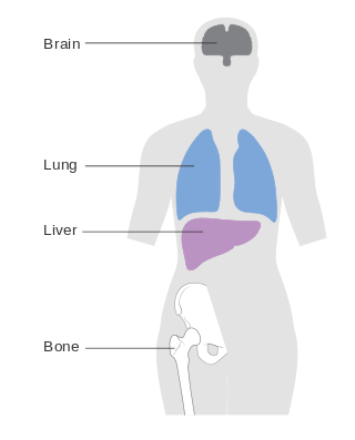
Metastatic breast cancer, also referred to as metastases, advanced breast cancer, secondary tumors, secondaries or stage IV breast cancer, is a stage of breast cancer where the breast cancer cells have spread to distant sites beyond the axillary lymph nodes. There is no cure for metastatic breast cancer; There is no stage after IV.

Rho-associated protein kinase

Rho-associated protein kinase (ROCK) is a kinase belonging to the AGC family of serine-threonine specific protein kinases. It is involved mainly in regulating the shape and movement of cells by acting on the cytoskeleton.

Role of cell adhesions in neural development

Cellular adhesions can be defined as proteins or protein aggregates that form mechanical and chemical linkages between the intracellular and extracellular space. Adhesions serve several critical processes including cell migration, signal transduction, tissue development and repair. Due to this functionality, adhesions and adhesion molecules have been a topic of study within the scientific community. Specifically, it has been found that adhesions are involved in tissue development, plasticity, and memory formation within the central nervous system (CNS), and may prove vital in the generation of CNS-specific therapeutics.
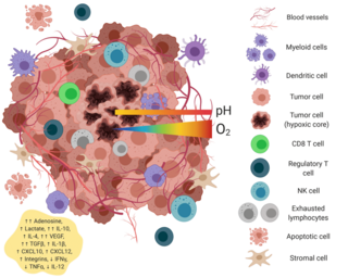
For cancer, invasion is the direct extension and penetration by cancer cells into neighboring tissues. It is generally distinguished from metastasis, which is the spread of cancer cells through the circulatory system or the lymphatic system to more distant locations. Yet, lymphovascular invasion is generally the first step of metastasis.

References

  1. 1 2 3 4 5 6 7 8 Murphy DA, Courtneidge SA (June 2011). "The 'ins' and 'outs' of podosomes and invadopodia: characteristics, formation and function". Nature Reviews Molecular Cell Biology. 12 (7): 413–26. doi:10.1038/nrm3141. PMC   3423958 . PMID   21697900.
  2. 1 2 3 Eddy RJ, Weidmann MD, Sharma VP, Condeelis JS (August 2017). "Tumor Cell Invadopodia: Invasive Protrusions that Orchestrate Metastasis". Trends in Cell Biology. 27 (8): 595–607. doi:10.1016/j.tcb.2017.03.003. PMC   5524604 . PMID   28412099.
  3. 1 2 3 4 Seano G, Primo L (2015). "Podosomes and invadopodia: tools to breach vascular basement membrane". Cell Cycle. 14 (9): 1370–4. doi:10.1080/15384101.2015.1026523. PMC   4614630 . PMID   25789660.
  4. Stylli SS, Stacey TT, Verhagen AM, Xu SS, Pass I, Courtneidge SA, Lock P (August 2009). "Nck adaptor proteins link Tks5 to invadopodia actin regulation and ECM degradation". Journal of Cell Science. 122 (Pt 15): 2727–40. doi:10.1242/jcs.046680. PMC   2909319 . PMID   19596797.
  5. Gurski LA, Xu X, Labrada LN, Nguyen NT, Xiao L, van Golen KL, Jia X, Farach-Carson MC (2009). "Hyaluronan (HA) interacting proteins RHAMM and hyaluronidase impact prostate cancer cell behavior and invadopodia formation in 3D HA-based hydrogels". PLOS ONE. 7 (11): e50075. doi: 10.1371/journal.pone.0050075 . PMC   3500332 . PMID   23166824.
  6. Weaver AM (May 2008). "Invadopodia". Current Biology. 18 (9): R362-4. doi: 10.1016/j.cub.2008.02.028 . PMID   18460310.
  7. 1 2 3 Sharma VP, Eddy R, Entenberg D, Kai M, Gertler FB, Condeelis J (November 2013). "Tks5 and SHIP2 regulate invadopodium maturation, but not initiation, in breast carcinoma cells". Current Biology. 23 (21): 2079–89. doi:10.1016/j.cub.2013.08.044. PMC   3882144 . PMID   24206842.
  8. Oser M, Yamaguchi H, Mader CC, Bravo-Cordero JJ, Arias M, Chen X, et al. (August 2009). "Cortactin regulates cofilin and N-WASp activities to control the stages of invadopodium assembly and maturation". The Journal of Cell Biology. 186 (4): 571–87. doi:10.1083/jcb.200812176. PMC   2733743 . PMID   19704022.
  9. 1 2 3 Jacob A, Prekeris R (February 2015). "The regulation of MMP targeting to invadopodia during cancer metastasis". Frontiers in Cell and Developmental Biology. 3: 4. doi: 10.3389/fcell.2015.00004 . PMC   4313772 . PMID   25699257.
  10. 1 2 Leong HS, Robertson AE, Stoletov K, Leith SJ, Chin CA, Chien AE, et al. (September 2014). "Invadopodia are required for cancer cell extravasation and are a therapeutic target for metastasis". Cell Reports. 8 (5): 1558–70. doi: 10.1016/j.celrep.2014.07.050 . PMID   25176655.
  11. Blouw B, Patel M, Iizuka S, Abdullah C, You WK, Huang X, et al. (2015). "The invadopodia scaffold protein Tks5 is required for the growth of human breast cancer cells in vitro and in vivo". PLOS ONE. 10 (3): e0121003. doi: 10.1371/journal.pone.0121003 . PMC   4380437 . PMID   25826475.
  12. Burger KL, Learman BS, Boucherle AK, Sirintrapun SJ, Isom S, Díaz B, et al. (February 2014). "Src-dependent Tks5 phosphorylation regulates invadopodia-associated invasion in prostate cancer cells". The Prostate. 74 (2): 134–48. doi:10.1002/pros.22735. PMC   4083496 . PMID   24174371.
  13. Stylli SS, I ST, Kaye AH, Lock P (March 2012). "Prognostic significance of Tks5 expression in gliomas". Journal of Clinical Neuroscience. 19 (3): 436–42. doi:10.1016/j.jocn.2011.11.013. PMID   22249020. S2CID   37199747.
  14. Li CM, Chen G, Dayton TL, Kim-Kiselak C, Hoersch S, Whittaker CA, et al. (July 2013). "Differential Tks5 isoform expression contributes to metastatic invasion of lung adenocarcinoma" (PDF). Genes & Development. 27 (14): 1557–67. doi:10.1101/gad.222745.113. PMC   3731545 . PMID   23873940.
  15. Courtneidge SA (February 2012). "Cell migration and invasion in human disease: the Tks adaptor proteins". Biochemical Society Transactions. 40 (1): 129–32. doi:10.1042/BST20110685. PMC   3425387 . PMID   22260678.