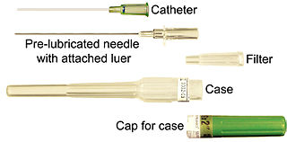
In urinary catheterization, a latex, polyurethane, or silicone tube known as a urinary catheter is inserted into the bladder through the urethra to allow urine to drain from the bladder for collection. It may also be used to inject liquids used for treatment or diagnosis of bladder conditions. A clinician, often a nurse, usually performs the procedure, but self-catheterization is also possible. A catheter may be in place for long periods of time or removed after each use.

In medicine, a catheter is a thin tube made from medical grade materials serving a broad range of functions. Catheters are medical devices that can be inserted in the body to treat diseases or perform a surgical procedure. Catheters are manufactured for specific applications, such as cardiovascular, urological, gastrointestinal, neurovascular and ophthalmic procedures. The process of inserting a catheter is called catheterization.

Pleurisy, also known as pleuritis, is inflammation of the membranes that surround the lungs and line the chest cavity (pleurae). This can result in a sharp chest pain while breathing. Occasionally the pain may be a constant dull ache. Other symptoms may include shortness of breath, cough, fever, or weight loss, depending on the underlying cause. Pleurisy can be caused by a variety of conditions, including viral or bacterial infections, autoimmune disorders, and pulmonary embolism.

Nasogastric intubation is a medical process involving the insertion of a plastic tube through the nose, down the esophagus, and down into the stomach. Orogastric intubation is a similar process involving the insertion of a plastic tube through the mouth. Abraham Louis Levin invented the NG tube. Nasogastric tube is also known as Ryle's tube in Commonwealth countries, after John Alfred Ryle.

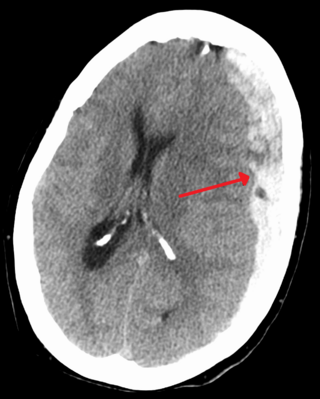
A subdural hematoma (SDH) is a type of bleeding in which a collection of blood—usually but not always associated with a traumatic brain injury—gathers between the inner layer of the dura mater and the arachnoid mater of the meninges surrounding the brain. It usually results from tears in bridging veins that cross the subdural space.

Epidural hematoma is when bleeding occurs between the tough outer membrane covering the brain and the skull. When this condition occurs in the spinal canal, it is known as a spinal epidural hematoma.

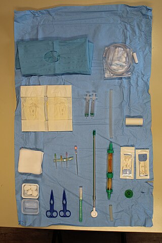
A chest tube is a surgical drain that is inserted through the chest wall and into the pleural space or the mediastinum. The insertion of the tube is sometimes a lifesaving procedure. The tube can be used to remove clinically undesired substances such as air (pneumothorax), excess fluid, blood (hemothorax), chyle (chylothorax) or pus (empyema) from the intrathoracic space. An intrapleural chest tube is also known as a Bülau drain or an intercostal catheter (ICC), and can either be a thin, flexible silicone tube, or a larger, semi-rigid, fenestrated plastic tube, which often involves a flutter valve or underwater seal.

Cauliflower ear is an irreversible condition that occurs when the external portion of the ear is hit and develops a blood clot or other collection of fluid under the perichondrium. This separates the cartilage from the overlying perichondrium that supplies its nutrients, causing it to die and resulting in the formation of fibrous tissue in the overlying skin. As a result, the outer ear becomes permanently swollen and deformed, resembling a cauliflower, hence the name.

An otoscope or auriscope is a medical device used by healthcare professionals to examine the ear canal and eardrum. This may be done as part of routine physical examinations, or for evaluating specific ear complaints, such as earaches, sense of fullness in the ear, or hearing loss.

A seroma is a pocket of clear serous fluid. They may sometimes develop in the body after surgery, particularly after breast surgery, abdominal surgery, and reconstructive surgery. They can be diagnosed by physical signs, and with a CT scan.

A surgical drain is a tube used to remove pus, blood or other fluids from a wound, body cavity, or organ. They are commonly placed by surgeons or interventional radiologists after procedures or some types of injuries, but they can also be used as an intervention for decompression. There are several types of drains, and selection of which to use often depends on the placement site and how long the drain is needed.

A trocar is a medical or veterinary device used in minimally invasive surgery. Trocars are typically made up of an awl, a cannula and often a seal. Some trocars also include a valve mechanism to allow for insufflation. Trocars are designed for placement through the chest and abdominal walls during thoracoscopic and laparoscopic surgery, and each trocar functions as a portal for the subsequent insertion of other endoscopic instruments such as grasper, scissors, stapler, electrocautery, suction tip, etc. — hence the more commonly used colloquial jargon "port". Trocars also allow passive evacuation of excess gas or fluid from organs within the body.

Nasal septal hematoma is a condition affecting the nasal septum. It can be associated with trauma.

In medicine, a port is a small appliance that is installed beneath the skin. A catheter connects the port to a vein. Under the skin, the port has a septum through which drugs can be injected and blood samples can be drawn many times, usually with less discomfort for the patient than a more typical "needle stick".
Autotransfusion is a process wherein a person receives their own blood for a transfusion, instead of banked allogenic (separate-donor) blood. There are two main kinds of autotransfusion: Blood can be autologously "pre-donated" before a surgery, or alternatively, it can be collected during and after the surgery using an intraoperative blood salvage device. The latter form of autotransfusion is utilized in surgeries where there is expected a large volume blood loss – e.g. aneurysm, total joint replacement, and spinal surgeries. The effectiveness, safety, and cost-savings of intraoperative cell salvage in people who are undergoing thoracic or abdominal surgery following trauma is not known.
Breast surgery is a form of surgery performed on the breast.

A breast mass, also known as a breast lump, is a localized swelling that feels different from the surrounding tissue. Breast pain, nipple discharge, or skin changes may be present. Concerning findings include masses that are hard, do not move easily, are of an irregular shape, or are firmly attached to surrounding tissue.
Pancreatic abscess is a late complication of acute necrotizing pancreatitis, occurring more than 4 weeks after the initial attack. A pancreatic abscess is a collection of pus resulting from tissue necrosis, liquefaction, and infection. It is estimated that approximately 3% of the patients with acute pancreatitis will develop an abscess.
Breast hematoma is a collection of blood within the breast. It arises from internal bleeding (hemorrhage) and may arise due to trauma or due to a non-traumatic cause.

Chest drains are surgical drains placed within the pleural space to facilitate removal of unwanted substances in order to preserve respiratory functions and hemodynamic stability. Some chest drains may utilize a flutter valve to prevent retrograde flow, but those that do not have physical valves employ a water trap seal design, often aided by continuous suction from a wall suction or a portable vacuum pump.


