Label-free quantification

Last updated

Label-free quantification is a method in mass spectrometry that aims to determine the relative amount of proteins in two or more biological samples. Unlike other methods for protein quantification, label-free quantification does not use a stable isotope containing compound to chemically bind to and thus label the protein. [1] [2]

Contents

Implementation

Label-free quantification experiment with 3 samples, 3 LC-MS files and 5 precursor ions/peptides. Intensities at the peak of the chromatographic peaks are used for quantification in this particular case. Peptides are identified via fragmentation mass spectra, and some of the precursor ions will be quantified, but not mapped to any peptide sequence. LCMS Features Label Free.png
Label-free quantification experiment with 3 samples, 3 LC-MS files and 5 precursor ions/peptides. Intensities at the peak of the chromatographic peaks are used for quantification in this particular case. Peptides are identified via fragmentation mass spectra, and some of the precursor ions will be quantified, but not mapped to any peptide sequence.

Label-free quantification may be based on precursor signal intensity or on spectral counting. The first method is useful when applied to high precision mass spectra, such as those obtained using the new generation of time-of-flight (ToF), fourier transform ion cyclotron resonance (FTICR), or Orbitrap mass analyzers. The high-resolution power facilitates the extraction of peptide signals on the MS1 level and thus uncouples the quantification from the identification process. In contrast, spectral counting simply counts the number of spectra identified for a given peptide in different biological samples and then integrates the results for all measured peptides of the protein(s) that are quantified.

The computational framework of label free approach includes detecting peptides, matching the corresponding peptides across multiple LC-MS data, selecting discriminatory peptides. [3] [4]

Intact protein expression spectrometry (IPEx) is a label-free quantification approach in mass spectrometry under development by the analytical chemistry group at the United States Food and Drug Administration Center for Food Safety and Applied Nutrition and elsewhere. Intact proteins are analyzed by an LCMS instrument, usually a quadrupole time-of-flight in profile mode, and the full protein profile is determined and quantified using data reduction software. Early results are very encouraging. In one study, two groups of treatment replicates from mammalian samples (different organisms with similar treatment histories, but not technical replicates) show dozens of low CV protein biomarkers, suggesting that IPEx is a viable technology for studying protein expression. [5]

Detecting peptides

Typically, peptide signals are detected at the MS1 level and distinguished from chemical noise through their characteristic isotopic pattern. These patterns are then tracked across the retention time dimension and used to reconstruct a chromatographic elution profile of the mono-isotopic peptide mass. The total ion current of the peptide signal is then integrated and used as a quantitative measurement of the original peptide concentration. For each detected peptide, all isotopic peaks are first found and the charge state is then assigned.

Label-free quantification may be based on precursor signal intensity and has problems due to isolation interference: in high-throughput studies, the identity of the peptide precursor ion being measured could easily be a completely different peptide with a similar m/z ratio and which elutes in a time frame overlapping with that of the former peptide. Spectral counting has problems due to the fact that the peptides are identified, thus making it necessary to run an additional MS/MS scan which takes time and therefore reduces the resolution of the experiment.

Matching corresponding peptides

In contrast to differential labelling, every biological specimen needs to be measured separately in a label-free experiment. The extracted peptide signals are then mapped across few or multiple LC-MS measurements using their coordinates on the mass-to-charge and retention-time dimensions. Data from high mass precision instruments greatly facilitate this process and increase the certainty of matching correct peptide signals across runs.

Clearly, differential processing of biological samples makes it necessary to have a standard which can be used to adjust the results. Peptides that are not expected to change in their expression levels in different biological samples may be used for this purpose. However, not all peptides ionize well and therefore the choice of candidates should be done after an initial study which should only characterize the protein content of the biological samples that will be investigated.

Selecting discriminatory peptides

Finally, sophisticated normalization methods are used to remove systematic artefacts in the peptide intensity values between LC-MS measurements. Then, discriminatory peptides are identified by selecting the peptides whose normalized intensities are different (e.g., p-value < 0.05) among multiple groups of samples.

In addition, newer hybrid mass spectrometers like LTQ OrbiTrap offer the possibility to acquire MS/MS peptide identifications in parallel to the high mass precision measurement of peptides on the MS1 level. This raises the computational challenge for the processing and integration of these two sources of information and has led to the development of novel promising quantification strategies.

Related Research Articles

<span class="mw-page-title-main">Inductively coupled plasma mass spectrometry</span> Type of mass spectrometry that uses an inductively coupled plasma to ionize the sample

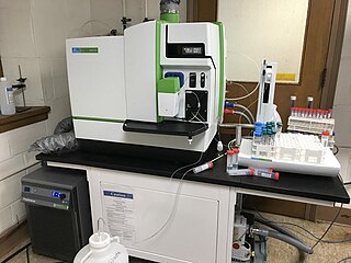
Inductively coupled plasma mass spectrometry (ICP-MS) is a type of mass spectrometry that uses an inductively coupled plasma to ionize the sample. It atomizes the sample and creates atomic and small polyatomic ions, which are then detected. It is known and used for its ability to detect metals and several non-metals in liquid samples at very low concentrations. It can detect different isotopes of the same element, which makes it a versatile tool in isotopic labeling.

<span class="mw-page-title-main">Proteomics</span> Large-scale study of proteins

Proteomics is the large-scale study of proteins. Proteins are vital parts of living organisms, with many functions such as the formation of structural fibers of muscle tissue, enzymatic digestion of food, or synthesis and replication of DNA. In addition, other kinds of proteins include antibodies that protect an organism from infection, and hormones that send important signals throughout the body.

<span class="mw-page-title-main">Tandem mass spectrometry</span> Type of mass spectrometry

Tandem mass spectrometry, also known as MS/MS or MS2, is a technique in instrumental analysis where two or more stages of analysis using one or more mass analyzer are performed with an additional reaction step in between these analyses to increase their abilities to analyse chemical samples. A common use of tandem MS is the analysis of biomolecules, such as proteins and peptides.

<span class="mw-page-title-main">Stable isotope labeling by amino acids in cell culture</span>

Stable Isotope Labeling by/with Amino acids in Cell culture (SILAC) is a technique based on mass spectrometry that detects differences in protein abundance among samples using non-radioactive isotopic labeling. It is a popular method for quantitative proteomics.

Surface-enhanced laser desorption/ionization (SELDI) is a soft ionization method in mass spectrometry (MS) used for the analysis of protein mixtures. It is a variation of matrix-assisted laser desorption/ionization (MALDI). In MALDI, the sample is mixed with a matrix material and applied to a metal plate before irradiation by a laser, whereas in SELDI, proteins of interest in a sample become bound to a surface before MS analysis. The sample surface is a key component in the purification, desorption, and ionization of the sample. SELDI is typically used with time-of-flight (TOF) mass spectrometers and is used to detect proteins in tissue samples, blood, urine, or other clinical samples, however, SELDI technology can potentially be used in any application by simply modifying the sample surface.

Phosphoproteomics is a branch of proteomics that identifies, catalogs, and characterizes proteins containing a phosphate group as a posttranslational modification. Phosphorylation is a key reversible modification that regulates protein function, subcellular localization, complex formation, degradation of proteins and therefore cell signaling networks. With all of these modification results, it is estimated that between 30–65% of all proteins may be phosphorylated, some multiple times. Based on statistical estimates from many datasets, 230,000, 156,000 and 40,000 phosphorylation sites should exist in human, mouse, and yeast, respectively.

A tandem mass tag (TMT) is a chemical label that facilitates sample multiplexing in mass spectrometry (MS)-based quantification and identification of biological macromolecules such as proteins, peptides and nucleic acids. TMT belongs to a family of reagents referred to as isobaric mass tags which are a set of molecules with the same mass, but yield reporter ions of differing mass after fragmentation. The relative ratio of the measured reporter ions represents the relative abundance of the tagged molecule, although ion suppression has a detrimental effect on accuracy. Despite these complications, TMT-based proteomics has been shown to afford higher precision than Label-free quantification. In addition to aiding in protein quantification, TMT tags can also increase the detection sensitivity of certain highly hydrophilic analytes, such as phosphopeptides, in RPLC-MS analyses.

<span class="mw-page-title-main">Protein mass spectrometry</span> Application of mass spectrometry

Protein mass spectrometry refers to the application of mass spectrometry to the study of proteins. Mass spectrometry is an important method for the accurate mass determination and characterization of proteins, and a variety of methods and instrumentations have been developed for its many uses. Its applications include the identification of proteins and their post-translational modifications, the elucidation of protein complexes, their subunits and functional interactions, as well as the global measurement of proteins in proteomics. It can also be used to localize proteins to the various organelles, and determine the interactions between different proteins as well as with membrane lipids.

<span class="mw-page-title-main">Quantitative proteomics</span> Analytical chemistry technique

Quantitative proteomics is an analytical chemistry technique for determining the amount of proteins in a sample. The methods for protein identification are identical to those used in general proteomics, but include quantification as an additional dimension. Rather than just providing lists of proteins identified in a certain sample, quantitative proteomics yields information about the physiological differences between two biological samples. For example, this approach can be used to compare samples from healthy and diseased patients. Quantitative proteomics is mainly performed by two-dimensional gel electrophoresis (2-DE), preparative native PAGE, or mass spectrometry (MS). However, a recent developed method of quantitative dot blot (QDB) analysis is able to measure both the absolute and relative quantity of an individual proteins in the sample in high throughput format, thus open a new direction for proteomic research. In contrast to 2-DE, which requires MS for the downstream protein identification, MS technology can identify and quantify the changes.

<span class="mw-page-title-main">Isobaric tag for relative and absolute quantitation</span>

Isobaric tags for relative and absolute quantitation (iTRAQ) is an isobaric labeling method used in quantitative proteomics by tandem mass spectrometry to determine the amount of proteins from different sources in a single experiment. It uses stable isotope labeled molecules that can be covalent bonded to the N-terminus and side chain amines of proteins.

An isotope-coded affinity tag (ICAT) is an in-vitro isotopic labeling method used for quantitative proteomics by mass spectrometry that uses chemical labeling reagents. These chemical probes consist of three elements: a reactive group for labeling an amino acid side chain, an isotopically coded linker, and a tag for the affinity isolation of labeled proteins/peptides. The samples are combined and then separated through chromatography, then sent through a mass spectrometer to determine the mass-to-charge ratio between the proteins. Only cysteine containing peptides can be analysed. Since only cysteine containing peptides are analysed, often the post translational modification is lost.

OpenMS is an open-source project for data analysis and processing in mass spectrometry and is released under the 3-clause BSD licence. It supports most common operating systems including Microsoft Windows, MacOS and Linux.

<span class="mw-page-title-main">Selected reaction monitoring</span> Tandem mass spectrometry method

Selected reaction monitoring (SRM), also called multiple reaction monitoring (MRM), is a method used in tandem mass spectrometry in which an ion of a particular mass is selected in the first stage of a tandem mass spectrometer and an ion product of a fragmentation reaction of the precursor ions is selected in the second mass spectrometer stage for detection.

Bioanalysis is a sub-discipline of analytical chemistry covering the quantitative measurement of xenobiotics and biotics in biological systems.

<span class="mw-page-title-main">Isobaric labeling</span>

Isobaric labeling is a mass spectrometry strategy used in quantitative proteomics. Peptides or proteins are labeled with chemical groups that have identical mass (isobaric), but vary in terms of distribution of heavy isotopes in their structure. These tags, commonly referred to as tandem mass tags, are designed so that the mass tag is cleaved at a specific linker region upon high-energy CID (HCD) during tandem mass spectrometry yielding reporter ions of different masses. The most common isobaric tags are amine-reactive tags. However, tags that react with cysteine residues and carbonyl groups have also been described. These amine-reactive groups go through N-hydroxysuccinimide (NHS) reactions, which are based around three types of functional groups. Isobaric labeling methods include tandem mass tags (TMT), isobaric tags for relative and absolute quantification (iTRAQ), mass differential tags for absolute and relative quantification, and dimethyl labeling. TMTs and iTRAQ methods are most common and developed of these methods. Tandem mass tags have a mass reporter region, a cleavable linker region, a mass normalization region, and a protein reactive group and have the same total mass.

A peptide spectral library is a curated, annotated and non-redundant collection/database of LC-MS/MS peptide spectra. One essential utility of a peptide spectral library is to serve as consensus templates supporting the identification of peptides and proteins based on the correlation between the templates with experimental spectra.

Secretomics is a type of proteomics which involves the analysis of the secretome—all the secreted proteins of a cell, tissue or organism. Secreted proteins are involved in a variety of physiological processes, including cell signaling and matrix remodeling, but are also integral to invasion and metastasis of malignant cells. Secretomics has thus been especially important in the discovery of biomarkers for cancer and understanding molecular basis of pathogenesis. The analysis of the insoluble fraction of the secretome has been termed matrisomics.

Terminal amine isotopic labeling of substrates (TAILS) is a method in quantitative proteomics that identifies the protein content of samples based on N-terminal fragments of each protein and detects differences in protein abundance among samples.

<span class="mw-page-title-main">Degradomics</span> Sub-discipline of biology

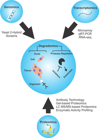
Degradomics is a sub-discipline of biology encompassing all the genomic and proteomic approaches devoted to the study of proteases, their inhibitors, and their substrates on a system-wide scale. This includes the analysis of the protease and protease-substrate repertoires, also called "protease degradomes". The scope of these degradomes can range from cell, tissue, and organism-wide scales.

Stable isotope standards and capture by anti-peptide antibodies (SISCAPA) is a mass spectrometry method for measuring the amount of a protein in a biological sample.

References

  1. Bantscheff M, Schirle M, Sweetman G, Rick J, Kuster B (October 2007). "Quantitative mass spectrometry in proteomics: a critical review". Analytical and Bioanalytical Chemistry. 389 (4): 1017–31. doi: 10.1007/s00216-007-1486-6 . PMID   17668192.
  2. Asara JM, Christofk HR, Freimark LM, Cantley LC (March 2008). "A label-free quantification method by MS/MS TIC compared to SILAC and spectral counting in a proteomics screen". Proteomics. 8 (5): 994–9. doi: 10.1002/pmic.200700426 . PMID   18324724.
  3. Bridges SM, Magee GB, Wang N, Williams WP, Burgess SC, Nanduri B (2007). "ProtQuant: a tool for the label-free quantification of MudPIT proteomics data". BMC Bioinformatics. 8 (Suppl 7): S24. doi: 10.1186/1471-2105-8-S7-S24 . PMC   2099493 . PMID   18047724.
  4. Lukas N. Mueller; et al. (2008). "An Assessment of Software Solutions for the Analysis of Mass Spectrometry Based Quantitative Proteomics Data". Journal of Proteome Research. 7 (1): 51–61. CiteSeerX   10.1.1.336.4416 . doi:10.1021/pr700758r. PMID   18173218.
  5. Scholl, PF, An intact protein LC/MS strategy for serum biomarker development: Biomarkers of hepatic responsiveness to chemopreventive treatment with the triterpenoid CDDO-Im, Abstract, TOA 8:35 a.m. ASMS Conference, 2007.