Lüder Deecke

Last updated • 8 min readFrom Wikipedia, The Free Encyclopedia
Lüder Deecke
Portrait of Luder Deecke.jpg
Born (1938-06-22) 22 June 1938 (age 86)
Lohe-Rickelshof, Germany
Nationality German-Austrian
Citizenship German-Austrian
Known for Bereitschaftspotential and Supplementary motor area
AwardsHans-Berger-Award, Prix Théophile Gluge, of the Royal Belgian Academy of Sciences, Brussels
Scientific career
Fields Neurology, Clinical neurology, Neurophysiology, Clinical neurophysiology, Neuroscience, Clinical neuroscience
Institutions University of Vienna, Medical University of Vienna

Lüder Deecke (German: [ˈdeːkə] ; born 22 June 1938) in Lohe-Rickelshof, Germany is a German Austrian neurologist, neuroscientist, teacher and physician whose scientific discoveries have influenced brain research and the treatment and rehabilitation of neurological disorders.

Contents

Full professor and head, Department of Clinical Neurology at the University of Vienna Medical University of Vienna, professor emeritus since October 2006, Deecke is also head of the Ludwig Boltzmann Institute for Functional Brain Topography and is the author of a number of books and more than 600 publications in the fields of neurology, clinical neurology, neurophysiology, clinical neurophysiology, neurosciences, brain research, movement disorders, etc.

His early research with Hans Helmut Kornhuber in the mid-1960s led to the discovery of the Bereitschaftspotential (or readiness potential), which is a measure of neural activity in the brain that precedes voluntary movements. This discovery set an important standard in research and rehabilitation of motor systems, and re-introduced the word will in key word registers.

Scientific contribution

In 1964 Deecke performed as doctoral student of Hans Helmut Kornhuber, EEG-recordings in man accompanying volitional movements and actions, and they discovered a slowly increasing activation (negative deflection) in the EEG, which they called Bereitschaftspotential [1]

The term Bereitschaftspotential (BP) can be found in the ‘List of German expressions in English‘. In order to record brain activity prior to an unforeseeable event – which a voluntary movement undoubtedly is – it needs a special method: the reverse averaging, which was invented by Kornhuber and Deecke in the same year (1964). The full paper appeared in 1965 [2] and was awarded a Citation Classic. [3]

In 1970 and 1971 Deecke was a research fellow in Toronto, Canada, under John M. Fredrickson. He performed experiments in the vestibular system (sense of balance) with rhesus monkeys and found the thalamic relay nucleus, nucleus ventralis posterior inferior (VPI) for the vestibular projection to the cortex. [4] In a second project, he investigated – with the rhesus monkey – normothermic perfusion as a therapeutic means with spinal cord compression, [5] and as a third project the alterations of the auditory evoked potentials under respiratory stress. [6]

In 1978 a further Citation Classic appeared with the discovery that the supplementary motor area (SMA) is active prior to voluntary actions and also prior to the activation of the primary motor cortex (M1, Brodmann-Area4). [7] This publication established the scientific knowledge that the early component of the Bereitschaftspotential (BP1 or BPearly) is generated by the activity of the SMA. BP1 is bilaterally symmetrical, because always – i.e. also with unilateral actions – the SMAs of both hemispheres are active, further substantiated by subsequent research. [8] The second component of the Bereitschaftspotential (BP2 oder BPlate) is generated by the primary motor cortex M1, and BP2 is asymmetrical with unilateral movements, namely dominant over the contralateral hemisphere. In Ulm, Deecke had projects with the DFG (German Research Foundation), and a productive team with research on the vestibular system and the motor system emerged including vestibular and neck interaction. [9] [10] In 1982 during Deecke's visiting professorship on invitation of Hal Weinberg in Vancouver, the Magnetoencephalographic-(MEG-) analogue of the Bereitschaftspotential, the Bereitschaftsmagnetfeld (Bereitschaftsfield, BF) was first recorded. [11]

From 1985 on in Vienna, Deecke has built his own MEG, the first generation with a five-channel MEG-System, and from 1995 on with the MEG Centre Vienna an MEG-whole head system with 143 channels (CTF Vancouver, Canada) has been established. Deecke and his team were successful to prove the participation of the SMA not only with the early Bereitschaftspotential but also with the Bereitschaftsmagnetfeld (Bereitschaftsfield in the MEG, solving the cancellation problem of the two SMAs opposing each other. [12] [13] In 1984 visual tracking movements were investigated. [14] [15] Evidence was found that the frontal cortex (SMA, prefrontal cortex) gives the starting command of the movement or action and supervises it, but the SMA does not execute the action. The frontal brain (including the SMA) is ‘delegating' this to the ‘expert systems for tracking in the brain‘, namely to the visual cortex and to the M1. [14]

In 2002 the term Bereitschafts-BOLD response was coined by Ross Cunnington et al. in event-related fMRI studies at the Department of Clinical Neurology and the Department of Radiodiagnostics Medical University of Vienna. [16] [17] [18] Thus, according to Deecke und Kornhuber [7],[15],[16] the early component of the BP (BP1 or BPearly) is generated by the following areas: the SMA proper, the pre-SMA and the cingulate motor area, CMA. This is now called anterior mid-cingulate cortex, aMCC. The second component (BP2 or BP late) is generated by the motor cortex (M1). Contrary to earlier views, the intentional activity according to Kornhuber and Deecke does not travel directly from the SMA to motor cortex M1 but is running via the cortico-basalganglio-thalamo-cortical loop in short motor loop. The motor loop has been discovered in patients with Parkinson's disease (PD). Deep brain stimulation improves frontal cortex function in PD patients. [19] This means that the formation of the will has already taken place in the frontal lobe and the preparation and planning of the action has been transferred initially to the unconscious routine processes of the basal ganglia, which do the groundwork for the motor cortex, M1. [20] [21] M1 finally generates the volley for the pyramidal tract, which then enters consciousness. [20] During the early BP, BP1, the action planning is not yet conscious, but during BP2 it is. From this observation Benjamin Libet, [22] postulated that we do not have free will (BP1) but with the control of the action (BP2) we do have free will. However, Kornhuber and Deecke, [20] [21] [23] [24] have shown that consciousness is not a sine qua non for free will. There are conscious and unconscious agendas in the brain, and both are important. The unconscious agendas far outweigh the conscious agendas, consciousness being only the ‘tip of an iceberg’. Therefore, free will is involved with both, the initiation of the action and for the control of the action. [20] [21] [23] [24]

The views of Kornhuber and Deecke upon the SMA and CMA [7] [20] [21] [23] [24] were confirmed in the meantime by Ross Cunnington and his team: The limbic system is always involved in the early planning for action – the matching with the inner needs, the emotional basic state, and our respective mood – has been postulated by Kornhuber and Deecke for quite some time [20] [21] [23] [24] and has been confirmed recently by the Cunnington group. [25] Kornhuber and Deecke have shown that freedom is given, a freedom in degrees of freedom, that humans can regulate up by their own efforts and learning in order to improve their free will, which is not a granted state but a dynamic process. [20] [21] [23] [24]

Awards and recognitions

On 2 October 2003 Deecke received the Dr. honoris causa (Dr. h.c.) from Simon Fraser University, Burnaby Greater Vancouver. 031205DeeckeCLR.L.dlBeschn.jpg
On 2 October 2003 Deecke received the Dr. honoris causa (Dr. h.c.) from Simon Fraser University, Burnaby Greater Vancouver.

Publications

Books

See also

Related Research Articles

<span class="mw-page-title-main">Apraxia</span> Loss of the ability to carry out learned purposeful movements

Apraxia is a motor disorder caused by damage to the brain, which causes difficulty with motor planning to perform tasks or movements. The nature of the damage determines the disorder's severity, and the absence of sensory loss or paralysis helps to explain the level of difficulty. Children may be born with apraxia; its cause is unknown, and symptoms are usually noticed in the early stages of development. Apraxia occurring later in life, known as acquired apraxia, is typically caused by traumatic brain injury, stroke, dementia, Alzheimer's disease, brain tumor, or other neurodegenerative disorders. The multiple types of apraxia are categorized by the specific ability and/or body part affected.

<span class="mw-page-title-main">Magnetoencephalography</span> Mapping brain activity by recording magnetic fields produced by currents in the brain

Magnetoencephalography (MEG) is a functional neuroimaging technique for mapping brain activity by recording magnetic fields produced by electrical currents occurring naturally in the brain, using very sensitive magnetometers. Arrays of SQUIDs are currently the most common magnetometer, while the SERF magnetometer is being investigated for future machines. Applications of MEG include basic research into perceptual and cognitive brain processes, localizing regions affected by pathology before surgical removal, determining the function of various parts of the brain, and neurofeedback. This can be applied in a clinical setting to find locations of abnormalities as well as in an experimental setting to simply measure brain activity.

Alien hand syndrome (AHS) or Dr. Strangelove syndrome is a category of conditions in which a person experiences their limbs acting seemingly on their own, without conscious control over the actions. There are a variety of clinical conditions that fall under this category, which most commonly affects the left hand. There are many similar terms for the various forms of the condition, but they are often used inappropriately. The affected person may sometimes reach for objects and manipulate them without wanting to do so, even to the point of having to use the controllable hand to restrain the alien hand. Under normal circumstances however, given that intent and action can be assumed to be deeply mutually entangled, the occurrence of alien hand syndrome can be usefully conceptualized as a phenomenon reflecting a functional "disentanglement" between thought and action.

<span class="mw-page-title-main">Frontal lobe</span> Part of the brain

The frontal lobe is the largest of the four major lobes of the brain in mammals, and is located at the front of each cerebral hemisphere. It is parted from the parietal lobe by a groove between tissues called the central sulcus and from the temporal lobe by a deeper groove called the lateral sulcus. The most anterior rounded part of the frontal lobe is known as the frontal pole, one of the three poles of the cerebrum.

<span class="mw-page-title-main">Motor cortex</span> Region of the cerebral cortex

The motor cortex is the region of the cerebral cortex involved in the planning, control, and execution of voluntary movements. The motor cortex is an area of the frontal lobe located in the posterior precentral gyrus immediately anterior to the central sulcus.

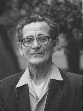
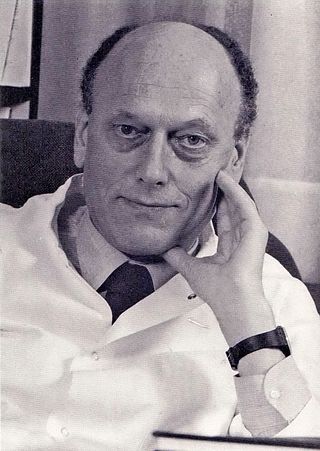
In neurology, the Bereitschaftspotential or BP, also called the pre-motor potential or readiness potential (RP), is a measure of activity in the motor cortex and supplementary motor area of the brain leading up to voluntary muscle movement. The BP is a manifestation of cortical contribution to the pre-motor planning of volitional movement. It was first recorded and reported in 1964 by Hans Helmut Kornhuber and Lüder Deecke at the University of Freiburg in Germany. In 1965 the full publication appeared after many control experiments.

<span class="mw-page-title-main">Benjamin Libet</span> American neuroscientist

Benjamin Libet was an American neuroscientist who was a pioneer in the field of human consciousness. Libet was a researcher in the physiology department of the University of California, San Francisco. In 2003, he was the first recipient of the Virtual Nobel Prize in Psychology from the University of Klagenfurt "for his pioneering achievements in the experimental investigation of consciousness, initiation of action, and free will".

<span class="mw-page-title-main">Pedunculopontine nucleus</span>

The pedunculopontine nucleus (PPN) or pedunculopontine tegmental nucleus is a collection of neurons located in the upper pons in the brainstem. It is involved in voluntary movements, arousal, and provides sensory feedback to the cerebral cortex and one of the main components of the reticular activating system. It is a potential target for deep brain stimulation treatment for Parkinson's disease. It was first described in 1909 by Louis Jacobsohn-Lask, a German neuroanatomist.

<span class="mw-page-title-main">Supplementary eye field</span> Region of the frontal cortex of the brain

Supplementary eye field (SEF) is the name for the anatomical area of the dorsal medial frontal lobe of the primate cerebral cortex that is indirectly involved in the control of saccadic eye movements. Evidence for a supplementary eye field was first shown by Schlag, and Schlag-Rey. Current research strives to explore the SEF's contribution to visual search and its role in visual salience. The SEF constitutes together with the frontal eye fields (FEF), the intraparietal sulcus (IPS), and the superior colliculus (SC) one of the most important brain areas involved in the generation and control of eye movements, particularly in the direction contralateral to their location. Its precise function is not yet fully known. Neural recordings in the SEF show signals related to both vision and saccades somewhat like the frontal eye fields and superior colliculus, but currently most investigators think that the SEF has a special role in high level aspects of saccade control, like complex spatial transformations, learned transformations, and executive cognitive functions.

<span class="mw-page-title-main">Premotor cortex</span> Part of the human brain

The premotor cortex is an area of the motor cortex lying within the frontal lobe of the brain just anterior to the primary motor cortex. It occupies part of Brodmann's area 6. It has been studied mainly in primates, including monkeys and humans. The functions of the premotor cortex are diverse and not fully understood. It projects directly to the spinal cord and therefore may play a role in the direct control of behavior, with a relative emphasis on the trunk muscles of the body. It may also play a role in planning movement, in the spatial guidance of movement, in the sensory guidance of movement, in understanding the actions of others, and in using abstract rules to perform specific tasks. Different subregions of the premotor cortex have different properties and presumably emphasize different functions. Nerve signals generated in the premotor cortex cause much more complex patterns of movement than the discrete patterns generated in the primary motor cortex.

<span class="mw-page-title-main">Supplementary motor area</span> Midline region in front of the motor cortex of the brain

The supplementary motor area (SMA) is a part of the motor cortex of primates that contributes to the control of movement. It is located on the midline surface of the hemisphere just in front of the primary motor cortex leg representation. In monkeys, the SMA contains a rough map of the body. In humans, the body map is not apparent. Neurons in the SMA project directly to the spinal cord and may play a role in the direct control of movement. Possible functions attributed to the SMA include the postural stabilization of the body, the coordination of both sides of the body such as during bimanual action, the control of movements that are internally generated rather than triggered by sensory events, and the control of sequences of movements. All of these proposed functions remain hypotheses. The precise role or roles of the SMA is not yet known.

Beevor's Axiom is the idea that the brain does not know muscles, only movements. In other words, the brain registers the movements that muscles combine to make, not the individual muscles that are making the movements. Hence, this is why one can sign their name with their foot. Beevor's Axiom was coined by Dr. Charles Edward Beevor, an English neurologist.

Premovement neuronal activity in neurophysiological literature refers to neuronal modulations that alter the rate at which neurons fire before a subject produces movement. Through experimentation with multiple animals, predominantly monkeys, it has been shown that several regions of the brain are particularly active and involved in initiation and preparation of movement. Two specific membrane potentials, the bereitschaftspotential, or the BP, and contingent negative variation, or the CNV, play a pivotal role in premovement neuronal activity. Both have been shown to be directly involved in planning and initiating movement. Multiple factors are involved with premovement neuronal activity including motor preparation, inhibition of motor response, programming of the target of movement, closed-looped and open-looped tasks, instructed delay periods, short-lead and long-lead changes, and mirror motor neurons.

<span class="mw-page-title-main">Blocq's disease</span> Loss of memory of specialized movements causing the inability to maintain an upright posture

Blocq's disease was first considered by Paul Blocq (1860–1896), who described this phenomenon as the loss of memory of specialized movements causing the inability to maintain an upright posture, despite normal function of the legs in the bed. The patient is able to stand up, but as soon as the feet are on the ground, the patient cannot hold himself upright nor walk; however when lying down, the subject conserved the integrity of muscular force and the precision of movements of the lower limbs. The motivation of this study came when a fellow student Georges Marinesco (1864) and Paul published a case of parkinsonian tremor (1893) due to a tumor located in the substantia nigra.

In neuroscience, the lateralized readiness potential (LRP) is an event-related brain potential, or increase in electrical activity at the surface of the brain, that is thought to reflect the preparation of motor activity on a certain side of the body; in other words, it is a spike in the electrical activity of the brain that happens when a person gets ready to move one arm, leg, or foot. It is a special form of bereitschaftspotential. LRPs are recorded using electroencephalography (EEG) and have numerous applications in cognitive neuroscience.

<span class="mw-page-title-main">Primary motor cortex</span> Brain region

The primary motor cortex is a brain region that in humans is located in the dorsal portion of the frontal lobe. It is the primary region of the motor system and works in association with other motor areas including premotor cortex, the supplementary motor area, posterior parietal cortex, and several subcortical brain regions, to plan and execute voluntary movements. Primary motor cortex is defined anatomically as the region of cortex that contains large neurons known as Betz cells, which, along with other cortical neurons, send long axons down the spinal cord to synapse onto the interneuron circuitry of the spinal cord and also directly onto the alpha motor neurons in the spinal cord which connect to the muscles.

<span class="mw-page-title-main">Neuroscience of free will</span> Neurophilosophical study of topics related to free will

The neuroscience of free will, a part of neurophilosophy, is the study of topics related to free will using neuroscience and the analysis of how findings from such studies may impact the free will debate.

The anti-saccade (AS) task is a way of measuring how well the frontal lobe of the brain can control the reflexive saccade, or eye movement. Saccadic eye movement is primarily controlled by the frontal cortex.

<span class="mw-page-title-main">Hans Helmut Kornhuber</span> German neurologist (1928–2009)

Hans Helmut Kornhuber was a German neurologist and neurophysiologist.

The Ludwig-Boltzmann-Institute for functional Brain Topography was a research institute for the investigation of the function of brain areas. It was founded in 1993 in Vienna, Austria by Lüder Deecke. With his retirement in 2006 the institute was closed.

References

  1. H. H. Kornhuber, L. Deecke: Hirnpotentialänderungen beim Menschen vor und nach Willkürbewegungen, dargestellt mit Magnetbandspeicherung und Rückwärtsanalyse. In: Pflügers Arch. 281, 1964, S. 52.
  2. H. H. Kornhuber, L. Deecke: Hirnpotentialänderungen bei Willkürbewegungen und passiven Bewegungen des Menschen: Bereitschaftspotential und reafferente Potentiale. In: Pflügers Arch. 284, 1965, S. 1–17; Englisch translation: Kornhuber HH, Deecke L (2016). "Brain potential changes in voluntary and passive movements in humans: readiness potential and reafferent potentials". Pflügers Archiv: European Journal of Physiology. 468 (7): 1115–24. doi: 10.1007/s00424-016-1852-3 . PMID   27392465., PDF (accessed October 21, 2016).
  3. H. H. Kornhuber, L. Deecke: Readiness for movement - the Bereitschaftspotential story. In: Current Contents Life Sciences. 33 (4): 14 (1990) and Current Contents Clinical Medicine. 18 (4): 14 (1990)
  4. L. Deecke, DWF Schwarz, JM Fredrickson: Nucleus ventroposterior inferior (VPI) as the vestibular thalamic relay in the rhesus monkey. I. Field potential investigation. Exp Brain Res 20: 88-100 (1974)
  5. CH Tator, L. Deecke: Value of normothermic perfusion, hypothermic perfusion, and durotomy in the treatment of experimental acute spinal cord trauma. J Neurosurg 39: 52-64 (1973)
  6. Deecke L, Goode RC, Whitehead G, Johnson WH, Bryce DP: Hearing under respiratory stress: Latency changes of the human auditory evoked response during hyperventilation, hypoxia, asphyxia, and hypercapnia. Aerospace Med 44: 1106-1111 (1973)
  7. 1 2 L. Deecke, H. H. Kornhuber: "An electrical sign of participation of the mesial “supplementary” motor cortex in human voluntary finger movements." In: Brain Res. 159, 1978, S. 473–476, (Citation Classic).
  8. Deecke L, Lang W (1996) Generation of movement-related potentials and fields in the supplementary sensorimotor area and the primary motor area. Advances in Neurology, Vol. 70: Supplementary Sensorimotor Area, HO Lüders (Ed) pp 127-146
  9. Mergner T, Deecke L, Becker W (1981) Patterns of vestibular and neck responses and their interaction: A comparison between cat cortical neurons and human psychophysics. Ann NY Acad Sci 374: 361–372
  10. Deecke L (1996) Planning, preparation, execution, and imagery of volitional action, (Introduction/Editorial) in: Deecke L, Lang W, Berthoz A (Eds) Mental representations of motor acts (Special Issue) Cogn Brain Res 3 (2): 59-64
  11. L. Deecke, H. Weinberg, P. Brickett: Magnetic fields of the human brain accompanying voluntary movement. Bereitschaftsmagnetfeld. In: Exp Brain Res. 48, 1982, S. 144–148.
  12. W. Lang, D. Cheyne, R. Kristeva, R. Beisteiner, G. Lindinger, L. Deecke: Three-dimensional localization of SMA activity preceding voluntary movement. A study of electric and magnetic fields in a patient with infarction of the right supplementary motor area. Exp Brain Res 87: 688-695 (1991)
  13. M. Erdler, R. Beisteiner, D. Mayer, T. Kaindl, V. Edward, C. Windischberger, G. Lindinger, L. Deecke: Supplementary motor area activation preceding voluntary movement is detectable with a whole scalp magnetoencephalography system. NeuroImage 11: 697-707 (2000)
  14. 1 2 M. Lang, W. Lang, B. Heise, L. Deecke, H. H. Kornhuber: Brain potentials related to voluntary hand tracking, motivation and attention. In: Hum Neurobiol. 3, 1984, S. 235–240.
  15. Deecke L, Heise B, Kornhuber HH, Lang M, Lang W (1984) Brain potentials associated with voluntary manual tracking: Bereitschaftspotential, conditioned pre-motion positivity, directed attention potential, and relaxation potential. Anticipatory activity of the limbic and frontal cortex. In: Karrer R, Cohen J, Tueting P (Eds): Brain and information: Event-related potentials. Ann NY Acad Sci, Vol 425: 450-464
  16. R. Cunnington, C. Windischberger, L. Deecke, E. Moser: The use of single event fMRI and fuzzy clustering analysis to examine haemodynamic response time courses in supplementary motor and primary motor cortical areas. Biomed Technik 44 (Suppl 2): 116-119 (1999)
  17. R. Cunnington, C. Windischberger, L. Deecke, E. Moser: The preparation and execution of self-initiated and externally-triggered movement: A study of event-related fMRI. NeuroImage 15: 373-385 (2002)
  18. R. Cunnington, C. Windischberger, L. Deecke, E. Moser: The preparation and readiness for voluntary movement: a high-field event-related fMRI study of the Bereitschafts-BOLD response. NeuroImage 20: 404–412 (2003)
  19. Gerschlager W, Alesch F, Cunnington R, Deecke L, Dirnberger G, Endl W, Lindinger G, Lang W (1999) Bilateral subthalamic nucleus stimulation improves frontal cortex function in Parkinson's disease. An electrophysiological study of the contingent negative variation. Brain 122: 2365-2373
  20. 1 2 3 4 5 6 7 H. H. Kornhuber, L. Deecke: Wille und Gehirn. 2. überarb. Auflage. Edition Sirius/ Aisthesis-Verlag, Bielefeld/ Basel 2009, ISBN   978-3-89528-628-5.
  21. 1 2 3 4 5 6 H. H. Kornhuber, L. Deecke: The will and its brain – an appraisal of reasoned free will. University Press of America, Lanham MD, USA 2012, ISBN   978-0-7618-5862-1.
  22. B. Libet, C. A. Gleason, E. W. Wright, D. K. Pearl: Time of conscious intention to act in relation to onset of cerebral activity (readiness potential): The unconscious initiation of a freely voluntary act. In: Brain. 106, 1983, S. 623–642.
  23. 1 2 3 4 5 Kornhuber HH, Deecke L, Lang W, Lang M, Kornhuber A (1989) Will, volitional action, attention and cerebral potentials in man: Bereitschaftspotential, performance-related potentials, directed attention potential, EEG spectrum changes. Chapter 6 in: Hershberger WA (Ed) Volitional action. Amsterdam, Elsevier (North Holland), pp 107-168
  24. 1 2 3 4 5 Deecke L, Kornhuber HH (2003) Human freedom, reasoned will, and the brain: The Bereitschaftspotential story. In: M Jahanshahi, M Hallett(Eds) The Bereitschaftspotential, movement-related cortical potentials. Kluwer Academic / Plenum Publishers New York, pp 283-320 ISBN   0-306-47407-7
  25. V. T. Nguyen, M. Breakspear, R. Cunnington: Reciprocal interactions of the SMA and cingulate cortex sustain pre-movement activity for voluntary actions. In: J Neurosci. 34, 2014, S. 16397–16407.
  26. "Lüder Deecke Homepage". Archived from the original on 2015-05-30. Retrieved 2015-06-13.