Related Research Articles

The cervix or cervix uteri is the lower part of the uterus (womb) in the human female reproductive system. The cervix is usually 2 to 3 cm long and roughly cylindrical in shape, which changes during pregnancy. The narrow, central cervical canal runs along its entire length, connecting the uterine cavity and the lumen of the vagina. The opening into the uterus is called the internal os, and the opening into the vagina is called the external os. The lower part of the cervix, known as the vaginal portion of the cervix, bulges into the top of the vagina. The cervix has been documented anatomically since at least the time of Hippocrates, over 2,000 years ago.
Dilationand curettage (D&C) refers to the dilation (widening/opening) of the cervix and surgical removal of part of the lining of the uterus and/or contents of the uterus by scraping and scooping (curettage). It is a gynecologic procedure used for diagnostic and therapeutic purposes, and is the most commonly used method for first-trimester miscarriage or abortion.

The uterus or womb is the organ in the reproductive system of most female mammals, including humans, that accommodates the embryonic and fetal development of one or more embryos until birth. The uterus is a hormone-responsive sex organ that contains glands in its lining that secrete uterine milk for embryonic nourishment.
Gynaecology or gynecology is the area of medicine that involves the treatment of women's diseases, especially those of the reproductive organs. It is often paired with the field of obstetrics, forming the combined area of obstetrics and gynecology (OB-GYN).
In medicine, prolapse is a condition in which organs fall down or slip out of place. It is used for organs protruding through the vagina, rectum, or for the misalignment of the valves of the heart. A spinal disc herniation is also sometimes called "disc prolapse". Prolapse means "to fall out of place", from the Latin prolabi meaning "to fall out".

Hysterectomy is the partial or total surgical removal of the uterus. It may also involve removal of the cervix, ovaries (oophorectomy), Fallopian tubes (salpingectomy), and other surrounding structures. Partial hysterectomies allow for hormone regulation while total hysterectomies do not.
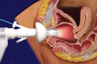
A pessary is a prosthetic device inserted into the vagina for structural and pharmaceutical purposes. It is most commonly used to treat stress urinary incontinence to stop urinary leakage and to treat pelvic organ prolapse to maintain the location of organs in the pelvic region. It can also be used to administer medications locally in the vagina or as a method of contraception.

Umbilical cord prolapse is when the umbilical cord comes out of the uterus with or before the presenting part of the baby. The concern with cord prolapse is that pressure on the cord from the baby will compromise blood flow to the baby. It usually occurs during labor but can occur anytime after the rupture of membranes.

A cystocele, also known as a prolapsed bladder, is a medical condition in which a woman's bladder bulges into her vagina. Some may have no symptoms. Others may have trouble starting urination, urinary incontinence, or frequent urination. Complications may include recurrent urinary tract infections and urinary retention. Cystocele and a prolapsed urethra often occur together and is called a cystourethrocele. Cystocele can negatively affect quality of life.
Endometrial ablation is a surgical procedure that is used to remove (ablate) or destroy the endometrial lining of the uterus. The goal of the procedure is to decrease the amount of blood loss during menstrual periods. Endometrial ablation is most often employed in people with excessive menstrual bleeding, who do not wish to undergo a hysterectomy, following unsuccessful medical therapy.
Sacrohysteropexy is a surgical procedure to correct uterine prolapse. It involves a resuspension of the prolapsed uterus using a strip of synthetic mesh to lift the uterus and hold it in place. It allows for normal sexual function and preserves childbearing function.

Uterine prolapse is a form of pelvic organ prolapse in which the uterus and a portion of the upper vagina protrude into the vaginal canal and, in severe cases, through the opening of the vagina. It is most often caused by injury or damage to structures that hold the uterus in place within the pelvic cavity. Symptoms may include vaginal fullness, pain with sexual intercourse, difficulty urinating, and urinary incontinence. Risk factors include older age, pregnancy, vaginal childbirth, obesity, chronic constipation, and chronic cough. Prevalence, based on physical exam alone, is estimated to be approximately 14%.

Uterus didelphys represents a uterine malformation where the uterus is present as a paired organ when the embryogenetic fusion of the Müllerian ducts fails to occur. As a result, there is a double uterus with two separate cervices, and possibly a double vagina as well. Each uterus has a single horn linked to the ipsilateral fallopian tube that faces its ovary.

The cardinal ligament is a major ligament of the uterus formed as a thickening of connective tissue of the base of the broad ligament of the uterus. It extends laterally from the cervix and vaginal fornix to attach onto the lateral wall of the pelvis. The female ureter, uterine artery, and inferior hypogastric (nervous) plexus course within the cardinal ligament. The cardinal ligament supports the uterus.

Uterine inversion is when the uterus turns inside out, usually following childbirth. Symptoms include postpartum bleeding, abdominal pain, a mass in the vagina, and low blood pressure. Rarely inversion may occur not in association with pregnancy.
An obstetric labor complication is a difficulty or abnormality that arises during the process of labor or delivery.

Bovine uterine prolapse occurs when the bovine uterus protrudes after calving. It is most common in dairy cattle and can occur in beef cows occasionally with hypocalcaemia. It is not as commonly seen in heifers, but occasionally can be seen in dairy heifers and most commonly Herefords.
Müllerian duct anomalies are those structural anomalies caused by errors in müllerian duct development during embryonic morphogenesis. Factors that precipitate include genetics, and maternal exposure to teratogens.
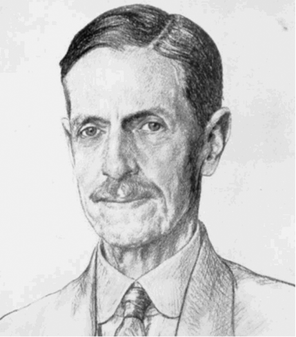
Archibald Donald was consulting gynaecological surgeon at Manchester Royal Infirmary and professor of obstetrics and gynaecology at the Victoria University of Manchester. Donald was notable for routinely sterilising catgut sutures and for a surgical repair technique for Uterine prolapse that later became known as the Fothergills Repair and later still became known as the Manchester operation

Bovine vaginal prolapse is a medical condition in cattle, characterised by an abnormally positioned (prolapsed) vagina. In most cases the bovine vaginal prolapse occurs near the time of calving, yet there are some examples of the vaginal prolapse in younger and non-pregnant animals. Another, but less common and more severe reproductive prolapse in cattle is so-called bovine uterine prolapse, where a uterus is the one being abnormally positioned.
References
- ↑ C., Dutta, D. (2014-04-30). DC Dutta's textbook of gynecology : including contraception (Enlarged & revised reprint of sixth ed.). New Delhi. ISBN 9789351520689. OCLC 872736102.
- ↑ "Abstract by Dr. A. Ayhan, Dr. S. Esin, Dr. Calhoun. Salman and Dr. O. Ozcu". Archived from the original on 2020-03-13. Retrieved 2011-01-21.