Melanosis coli

Last updated
Melanosis coli
Other namesPseudomelanosis coli,
Melanosis coli.jpg
Melanosis coli identified on colonoscopy as a brownish moiré pattern on the wall of the colon.
Micrograph of melanosis coli, with the characteristic mucosal lipofuscin-laden macrophages (brown). Melanosis coli high mag.jpg
Micrograph of melanosis coli, with the characteristic mucosal lipofuscin-laden macrophages (brown).

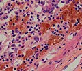
Melanosis coli, also pseudomelanosis coli, is a disorder of pigmentation of the wall of the colon, often identified at the time of colonoscopy. It is benign and may have no significant correlation with disease. The brown pigment is lipofuscin in macrophages, not melanin.[ citation needed ] It is most commonly associated with the use of certain laxatives.

Contents

Cause

The most common cause of melanosis coli is the extended use of laxatives, and commonly anthraquinone containing laxatives such as senna, aloe vera, and other plant glycosides. [1] The anthranoid laxatives pass through the gastrointestinal tract unabsorbed until they reach the large intestine, where they are changed into their active forms. The resulting active compounds cause damage to the cells in the lining of the intestine and leads to apoptosis (a form of cell death). The damaged (apoptotic) cells appear as darkly pigmented bodies that may be taken up by scavenger cells known as macrophages. When enough cells have been damaged, the characteristic pigmentation of the bowel wall develops. The condition can develop after just a few months of laxative use. [2]

However, other causes are identified, including an increase in colonic epithelial apoptosis. [3] Endoscopically, the mucosa may show a brownish discoloration in a moiré pattern.[ citation needed ]

Histologic appearance

On biopsy, melanosis coli shows characteristic pigment-laden macrophages within the mucosa on PAS staining. [4]

The histologic differential diagnosis of mucosal pigmentation is: lipofuscin (melanosis coli), hemosiderin-laden macrophages, and melanin (rare).

Prognosis

No adverse effects or consequences of melanosis coli have been identified. [4]

Relation to true melanoses

The condition is unrelated to true melanoses, such as Peutz–Jeghers syndrome and smoker's melanosis. [5]

Peutz–Jeghers syndrome causes pigmentation of the skin and mucous surfaces with melanin, and polyps in the digestive tract.

Non-colonic pseudomelanoses

Pseudomelanoses of other parts of the gastrointestinal tract have also been reported, and are of unclear relevance. [6]

Patients with colostomies can have melanosis involving the stoma, which is also of no significance. [7]

Related Research Articles

<span class="mw-page-title-main">Duodenum</span> First section of the small intestine

The duodenum is the first section of the small intestine in most higher vertebrates, including mammals, reptiles, and birds. In mammals it may be the principal site for iron absorption. The duodenum precedes the jejunum and ileum and is the shortest part of the small intestine.

<span class="mw-page-title-main">Lamina propria</span> Thin connective layer forming part of the mucous membranes

The lamina propria is a thin layer of connective tissue that forms part of the moist linings known as mucous membranes or mucosae, which line various tubes in the body, such as the respiratory tract, the gastrointestinal tract, and the urogenital tract.

<span class="mw-page-title-main">Melanism</span> Congenital excess of melanin in an organism resulting in dark pigment

Melanism is the congenital excess of melanin in an organism resulting in dark pigment.

<span class="mw-page-title-main">Nevus</span> Mole or birthmark; visible, circumscribed, chronic skin lesion

Nevus is a nonspecific medical term for a visible, circumscribed, chronic lesion of the skin or mucosa. The term originates from nævus, which is Latin for "birthmark"; however, a nevus can be either congenital or acquired. Common terms, including mole, birthmark, and beauty mark, are used to describe nevi, but these terms do not distinguish specific types of nevi from one another.

<span class="mw-page-title-main">Hyperpigmentation</span> Darkening of an area of skin or nails caused by increased melanin

Hyperpigmentation is the darkening of an area of skin or nails caused by increased melanin.

<span class="mw-page-title-main">Peutz–Jeghers syndrome</span> Medical condition

Peutz–Jeghers syndrome is an autosomal dominant genetic disorder characterized by the development of benign hamartomatous polyps in the gastrointestinal tract and hyperpigmented macules on the lips and oral mucosa (melanosis). This syndrome can be classed as one of various hereditary intestinal polyposis syndromes and one of various hamartomatous polyposis syndromes. It has an incidence of approximately 1 in 25,000 to 300,000 births.

<span class="mw-page-title-main">Sex cord–gonadal stromal tumour</span> Medical condition

Sex cord–gonadal stromal tumour is a group of tumors derived from the stromal component of the ovary and testis, which comprises the granulosa, thecal cells and fibrocytes. In contrast, the epithelial cells originate from the outer epithelial lining surrounding the gonad while the germ cell tumors arise from the precursor cells of the gametes, hence the name germ cell. In humans, this group accounts for 8% of ovarian cancers and under 5% of testicular cancers. Their diagnosis is histological: only a biopsy of the tumour can make an exact diagnosis. They are often suspected of being malignant prior to operation, being solid ovarian tumours that tend to occur most commonly in post menopausal women.

Gastrointestinal cancer refers to malignant conditions of the gastrointestinal tract and accessory organs of digestion, including the esophagus, stomach, biliary system, pancreas, small intestine, large intestine, rectum and anus. The symptoms relate to the organ affected and can include obstruction, abnormal bleeding or other associated problems. The diagnosis often requires endoscopy, followed by biopsy of suspicious tissue. The treatment depends on the location of the tumor, as well as the type of cancer cell and whether it has invaded other tissues or spread elsewhere. These factors also determine the prognosis.

<span class="mw-page-title-main">Blood in stool</span> Medical condition

Blood in stool or rectal bleeding looks different depending on how early it enters the digestive tract—and thus how much digestive action it has been exposed to—and how much there is. The term can refer either to melena, with a black appearance, typically originating from upper gastrointestinal bleeding; or to hematochezia, with a red color, typically originating from lower gastrointestinal bleeding. Evaluation of the blood found in stool depends on its characteristics, in terms of color, quantity and other features, which can point to its source, however, more serious conditions can present with a mixed picture, or with the form of bleeding that is found in another section of the tract. The term "blood in stool" is usually only used to describe visible blood, and not fecal occult blood, which is found only after physical examination and chemical laboratory testing.

Gut-associated lymphoid tissue (GALT) is a component of the mucosa-associated lymphoid tissue (MALT) which works in the immune system to protect the body from invasion in the gut.

The mucosa-associated lymphoid tissue (MALT), also called mucosa-associated lymphatic tissue, is a diffuse system of small concentrations of lymphoid tissue found in various submucosal membrane sites of the body, such as the gastrointestinal tract, nasopharynx, thyroid, breast, lung, salivary glands, eye, and skin. MALT is populated by lymphocytes such as T cells and B cells, as well as plasma cells, dendritic cells and macrophages, each of which is well situated to encounter antigens passing through the mucosal epithelium. In the case of intestinal MALT, M cells are also present, which sample antigen from the lumen and deliver it to the lymphoid tissue. MALT constitute about 50% of the lymphoid tissue in human body. Immune responses that occur at mucous membranes are studied by mucosal immunology.

<span class="mw-page-title-main">Small intestine cancer</span> Medical condition

Small intestine cancer starts when cells in the small intestine start to grow out of control. The small intestine is the largest part of the gastrointestinal tract, which processes food for energy and rids the body of solid waste. The small intestine is not as common as other cancers of the GI tract such as colon, rectal, stomach, and esophageal cancers in the United States. They account for fewer than 1 in 10 cancers in the GI tract, and fewer than 1 in 100 cancers overall.

<span class="mw-page-title-main">Intestinal gland</span> Gland between the intestinal villi that produces new cells

In histology, an intestinal gland is a gland found in between villi in the intestinal epithelium lining of the small intestine and large intestine. The glands and intestinal villi are covered by epithelium, which contains multiple types of cells: enterocytes, goblet cells, enteroendocrine cells, cup cells, tuft cells, and at the base of the gland, Paneth cells and stem cells.

Microfold cells are found in the gut-associated lymphoid tissue (GALT) of the Peyer's patches in the small intestine, and in the mucosa-associated lymphoid tissue (MALT) of other parts of the gastrointestinal tract. These cells are known to initiate mucosal immunity responses on the apical membrane of the M cells and allow for transport of microbes and particles across the epithelial cell layer from the gut lumen to the lamina propria where interactions with immune cells can take place.

<span class="mw-page-title-main">Cronkhite–Canada syndrome</span> Medical condition

Cronkhite–Canada syndrome is a rare syndrome characterized by multiple polyps of the digestive tract. It is sporadic, and it is currently considered acquired and idiopathic.

<span class="mw-page-title-main">Smoker's melanosis</span> Medical condition

Smoker's melanosis is seen with the naked eye as a brown to black pigmentation of the oral tissue i.e. the gums, cheeks or palate as well as in larynx. It is most often seen in the lower labial gingiva of tobacco users. Most easily it is found in Caucasians, due to their lack of a genetically caused melanin pigmentation.

<span class="mw-page-title-main">Colorectal polyp</span> Growth found in bowel wall

A colorectal polyp is a polyp occurring on the lining of the colon or rectum. Untreated colorectal polyps can develop into colorectal cancer.

<span class="mw-page-title-main">STK11</span> Protein-coding gene in the species Homo sapiens

Serine/threonine kinase 11 (STK11) also known as liver kinase B1 (LKB1) or renal carcinoma antigen NY-REN-19 is a protein kinase that in humans is encoded by the STK11 gene.

Gum depigmentation, also known as gum bleaching, is a procedure used in cosmetic dentistry to lighten or remove black spots or patches on the gums consisting of melanin. Melanin in skin is very common in inhabitants in many parts of the world due to genetic factors. Melanin pigmentation in skin, oral mucosa, inner ear and other organs is a detoxification mechanism. Some toxic agents bind to melanin and will move out of the tissue with the ageing cells and are expelled to the tissue surfaces. Also in the gums and oral mucosa a visible pigmentation is most often caused by genetic factors, but also by tobacco smoking or in a few cases by long-term use of certain medications. If stopping smoking or change of medication do not solve the problem with a disfigurating melanin pigmentation, a surgical operation may be performed. The procedure itself can involve laser ablation techniques.

Oral pigmentation is asymptomatic and does not usually cause any alteration to the texture or thickness of the affected area. The colour can be uniform or speckled and can appear solitary or as multiple lesions. Depending on the site, depth, and quantity of pigment, the appearance can vary considerably.

References

  1. Müller-Lissner, SA. (Oct 1993). "Adverse effects of laxatives: fact and fiction". Pharmacology. 47 (Suppl 1): 138–45. doi:10.1159/000139853. PMID   8234421.
  2. "What is Melanosis Coli? Symptoms, Causes & Treatment".
  3. Byers, R.J.; Marsh, P.; Parkinson, D.; Haboubi, N.Y (October 2003). "Melanosis coli is associated with an increase in colonic epithelial apoptosis and not with laxative use". Histopathology. 30 (2): 160–164. doi:10.1046/j.1365-2559.1997.d01-574.x. PMID   9067741. S2CID   3175917.
  4. 1 2 Wittoesch, JH.; Jackman, RJ.; McDonald, JR. (1958). "Melanosis coli: general review and a study of 887 cases". Dis Colon Rectum. 1 (3): 172–80. doi:10.1007/bf02616828. PMID   13537819. S2CID   46183745.
  5. Vellappally, S.; Fiala, Z.; Smejkalová, J.; Jacob, V.; Somanathan, R. (2007). "Smoking related systemic and oral diseases". Acta Medica. 50 (3): 161–6. doi: 10.14712/18059694.2017.76 . PMID   18254267.
  6. Ghadially, FN.; Walley, VM. (Sep 1994). "Melanoses of the gastrointestinal tract". Histopathology. 25 (3): 197–207. doi:10.1111/j.1365-2559.1994.tb01319.x. PMID   7821887. S2CID   22088870.
  7. Fleischer, I.; Bryant, D. (May 1995). "Melanosis coli or mucosa ischemia? A case report". Ostomy Wound Manage. 41 (4): 44, 46–7. PMID   7598783.