Microtubule-associated protein 2

Last updated
MAP2
Identifiers
Aliases MAP2 , MAP2A, MAP2B, MAP2C, microtubule associated protein 2, MAP-2
External IDs OMIM: 157130; MGI: 97175; HomoloGene: 1779; GeneCards: MAP2; OMA:MAP2 - orthologs
Orthologs
SpeciesHumanMouse
Entrez
Ensembl
UniProt
RefSeq (mRNA)

NM_001039934
NM_008632
NM_001310634

RefSeq (protein)

NP_001035023
NP_001297563
NP_032658

Location (UCSC) Chr 2: 209.42 – 209.73 Mb Chr 1: 66.21 – 66.48 Mb
PubMed search [3] [4]
Wikidata
View/Edit Human View/Edit Mouse

Microtubule-associated protein 2 is a protein in humans that is encoded by the MAP2 gene. [5] [6]

Function

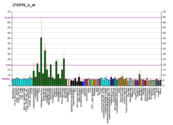
This gene encodes a protein that belongs to the microtubule-associated protein family. The proteins of this family were originally isolated since they copurify with tubulin in polymerization experiments: tubulin in cell extracts can be made to polymerize to produce microtubules (MT) under the influence of heat and the addition of GTP, and the MT can then be collected by centrifugation. When this is done a series of microtubule associated proteins are collected along with the MT and can be detected by SDS-PAGE and other methods. Brain extracts are rich in several of these proteins, MAP2 being one of these. The single MAP2 gene produces four major transcripts producing four proteins, MAP2A, MAP2B, MAP2C and MAP2D. MAP2A and MAP2B are very high molecular weight proteins, with apparent molecular weight on SDS-PAGE about 250 kDa, while MAP2C and MAP2D are much lower molecular weight forms with apparent SDS-PAGE size about 70 kDa. [7] All forms of MAP2 share a common core sequence which includes MT binding domains, 18 amino acid sequences which are found in other MT associated proteins such as MAP Tau and MAP1B. The MAP2 isoforms are thought to be involved in MT assembly, which is an essential step in neuritogenesis. MAP2 serves to stabilize MT growth by crosslinking MT with intermediate filaments and other MTs. MAP2 isoforms are neuron-specific cytoskeletal proteins enriched in dendrites and perikarya, implicating a role in determining and stabilizing neuronal morphology during neuron development. As a result antibodies to MAP2 are widely used to identify neuronal cells and trace dendritic processes in experimental contexts.

Interactions

MAP2 has been shown to interact with Grb2, [8] [9] NEFL [10] and MYO7A. [11] All MAP2 isoforms bind to microtubules.

Neurons were grown in tissue culture and stained with antibody to MAP2 protein in green and antibody to another microtubule associated protein MAP-tau in red using the immunofluorescence technique. MAP2 is found only in dendrites and perikarya, while MAP-tau is found not only in the dendrites and perikarya but also in axons. As a result, axons appear red since they contain only MAP-tau while the dendrites and perikarya appear yellow, since they contain both proteins and the red and green signals superimpose to produce a yellow signal. DNA is shown in blue using the DAPI stain which highlights the nuclei. Image courtesy EnCor Biotechnology Inc. MAP2-tau in neurons.jpg
Neurons were grown in tissue culture and stained with antibody to MAP2 protein in green and antibody to another microtubule associated protein MAP-tau in red using the immunofluorescence technique. MAP2 is found only in dendrites and perikarya, while MAP-tau is found not only in the dendrites and perikarya but also in axons. As a result, axons appear red since they contain only MAP-tau while the dendrites and perikarya appear yellow, since they contain both proteins and the red and green signals superimpose to produce a yellow signal. DNA is shown in blue using the DAPI stain which highlights the nuclei. Image courtesy EnCor Biotechnology Inc.

References

  1. 1 2 3 GRCh38: Ensembl release 89: ENSG00000078018 Ensembl, May 2017
  2. 1 2 3 GRCm38: Ensembl release 89: ENSMUSG00000015222 Ensembl, May 2017
  3. "Human PubMed Reference:". National Center for Biotechnology Information, U.S. National Library of Medicine.
  4. "Mouse PubMed Reference:". National Center for Biotechnology Information, U.S. National Library of Medicine.
  5. Neve RL, Harris P, Kosik KS, Kurnit DM, Donlon TA (May 1987). "Identification of cDNA clones for the human microtubule-associated protein tau and chromosomal localization of the genes for tau and microtubule-associated protein 2". Brain Res. 387 (3): 271–80. doi:10.1016/0169-328x(86)90033-1. PMID   3103857.
  6. Kalcheva N, Albala J, O'Guin K, Rubino H, Garner C, Shafit-Zagardo B (December 1995). "Genomic structure of human microtubule-associated protein 2 (MAP-2) and characterization of additional MAP-2 isoforms". Proc Natl Acad Sci U S A. 92 (24): 10894–8. Bibcode:1995PNAS...9210894K. doi: 10.1073/pnas.92.24.10894 . PMC   40537 . PMID   7479905.
  7. "Entrez Gene: MAP2 microtubule-associated protein 2".
  8. Lim RW, Halpain S (July 2000). "Regulated association of microtubule-associated protein 2 (MAP2) with Src and Grb2: evidence for MAP2 as a scaffolding protein". J. Biol. Chem. 275 (27): 20578–87. doi: 10.1074/jbc.M001887200 . PMID   10781592.
  9. Zamora-Leon SP, Lee G, Davies P, Shafit-Zagardo B (October 2001). "Binding of Fyn to MAP-2c through an SH3 binding domain. Regulation of the interaction by ERK2". J. Biol. Chem. 276 (43): 39950–8. doi: 10.1074/jbc.M107807200 . PMID   11546790.
  10. Frappier T, Stetzkowski-Marden F, Pradel LA (April 1991). "Interaction domains of neurofilament light chain and brain spectrin". Biochem. J. 275 (Pt 2): 521–7. doi:10.1042/bj2750521. PMC   1150082 . PMID   1902666.
  11. Todorov PT, Hardisty RE, Brown SD (March 2001). "Myosin VIIA is specifically associated with calmodulin and microtubule-associated protein-2B (MAP-2B)". Biochem. J. 354 (Pt 2): 267–74. doi:10.1042/0264-6021:3540267. PMC   1221652 . PMID   11171103.

Further reading