Mitochondrial antiviral-signaling protein

Last updated

MAVS
Available structures
PDB Ortholog search: PDBe RCSB
Identifiers
Aliases MAVS , CARDIF, IPS-1, IPS1, VISA, mitochondrial antiviral signaling protein
External IDs OMIM: 609676; MGI: 2444773; HomoloGene: 17004; GeneCards: MAVS; OMA:MAVS - orthologs
Orthologs
SpeciesHumanMouse
Entrez
Ensembl
UniProt
RefSeq (mRNA)

NM_001206491
NM_020746
NM_001385663

NM_001206382
NM_001206383
NM_001206385
NM_144888

RefSeq (protein)

NP_001193420
NP_065797

NP_001193311
NP_001193312
NP_001193314
NP_659137

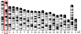
Location (UCSC) Chr 20: 3.85 – 3.88 Mb Chr 2: 131.08 – 131.09 Mb
PubMed search [3] [4]
Wikidata
View/Edit Human View/Edit Mouse

Mitochondrial antiviral-signaling protein (MAVS) is a protein that is essential for antiviral innate immunity. MAVS is located in the outer membrane of the mitochondria, peroxisomes, and mitochondrial-associated endoplasmic reticulum membrane (MAM). [5] [6] Upon viral infection, a group of cytosolic proteins will detect the presence of the virus and bind to MAVS, thereby activating MAVS. The activation of MAVS leads the virally infected cell to secrete cytokines. This induces an immune response which kills the host's virally infected cells, resulting in clearance of the virus.

Contents

Structure

Schematic representation of MAVS gene. Ribosome recognition sites for translation are located at position 1 and position 142 on the MAVS gene, highlighted by the yellow band. Schematic representation of MAVS gene.png
Schematic representation of MAVS gene. Ribosome recognition sites for translation are located at position 1 and position 142 on the MAVS gene, highlighted by the yellow band.

MAVS is also known as IFN-β promoter stimulator I (IPS-1), caspase activation recruitment domain adaptor inducing IFN-β(CARDIF), or virus induced signaling adaptor (VISA). [7] MAVS is encoded by a MAVS gene. [7] [8] MAVS is a 540 amino acid protein that consists of three components, a N terminal caspase activation recruitment domain (CARD), a proline rich domain, and a transmembrane C terminal domain (TM). [7]

After the MAVS gene has been transcribed into RNA, ribosomes can translate the MAVS protein from two different sites. [7] The initial translation site generates the full-length MAVS protein. The alternative translation site generates a shorter protein, termed as “miniMAVS” or short-MAVS (sMAVS). [7] sMAVS is a 398 amino acid MAVS protein that lacks the CARD domain. This is significant because the CARD domain is where two cytosolic proteins bind to activate MAVS, signaling that there is a virus present in the cell. [7]

Function

Double stranded RNA viruses are recognized by either the transmembrane toll-like receptor 3 (TLR3) or by one of two cytosolic proteins, retinoic acid-inducible gene I (RIG-I)-like receptors and melanoma differentiation-associated gene 5 (MDA5). [7] [8] [9] [10] RIG-I and MDA5 differ in the viral RNA that they recognize, but they share many structural features, including the N-terminal CARD that allows them to bind to MAVS. [7] MAVS activation leads to the increased levels of pro-inflammatory cytokines via activation of transcription factors, nuclear factor kB (NF-κB), interferon regulatory factor 1 (IRF1), and interferon regulatory factor 3 (IRF3). [7] [8] [9] NF-κB, IRF1, and IRF3 are transcription factors and play critical roles in the production of cytokines.

Cellular mechanisms of MAVS pathway Cellular mechanisms of MAVS pathway.pdf
Cellular mechanisms of MAVS pathway

At a resting state for the cell, a protein called mitofusin 2 (MFN2) is known to interact with MAVS, preventing MAVS from binding to the cytosolic proteins, such as RIG-I and MDA5. [7] [8] Upon recognition of the virus in the cytosol, mitochondria-associated ER membranes (MAM) and mitochondria will become physically tethered by MFN2 and RIG-I binds to a second RIG-I protein to form a protein complex. [7] [8] [9] This complex binds to TRIM25 and molecular chaperone 14-3-3e to form a complex termed “translocon”. [7] [8] [9] [10] The translocon travels to the mitochondria where it binds to the CARD region on MAVS, leading to activation of MAVS. [7] [8] [9] [10] Subsequently, MAVS proteins bind to each other through the CARD and TM domain to recruit several downstream signaling factors to form the MAVS signaling complex. [7] [8] The formation of this MAVS signaling complex is aided by augmented levels of mitochondrial reactive oxygen species (mROS), independent of the RNA sensing. [8] [9] The MAVS signaling complex interacts with TANK binding kinase 1 and/or protein kinases IKKA (CHUK) and IKKB (IKBKB), which leads to the phosphorylation and nuclear translocation of IRF3. [7] Although MAVS signal transduction and regulation is not fully understood, activated MAVS proteins in the mitochondria, ER, and peroxisome are needed to maximize the antiviral innate immune response.

MAVS protein induces apoptosis in host virally infected cells by interacting with a protease called caspase 8. [7] Activation of apoptosis by caspase 8 is independent of the Bax/Bak apoptotic pathway, the main pathway of apoptosis in cells. [7]

Viral evasion

Certain viruses, such as human cytomegalovirus (HCMV) and hepatitis C (HCV), have adapted to suppress the function of MAVS in the antiviral innate immune response, aiding in viral replication. [7] [11] HCMV impairs MAVS through the viral mitochondria-localized inhibitor of apoptosis protein (vMIA), thus reducing the pro-inflammatory cytokine response. [11] vMIA also localizes to the peroxisome where it interacts with cytoplasmic chaperone protein Pex19, disabling the transport machinery of peroxisomal membrane proteins. [11] The HCV NS3-NS4A strain inactivates MAVS signaling by cleaving the MAVS protein directly upstream of MAVS membrane-targeting domain in the MAM and peroxisome, preventing MAVS downstream signaling. [7]

Regulation

The expression and function of MAVS are regulated at the transcriptional, posttranscriptional, and posttranslational level. At the transcriptional level, the reactive oxygen species (ROS) generated during antiviral response acts as a negative regulator. [7] [8] [9] MAVS, additionally, encodes a number of splice variants that have been proposed to regulate MAVS. At the post-transcriptional level, there are two translational sites present on MAVS that can generate two proteins of MAVS. The alternative translation site resides upstream, resulting in expression of sMAVS. [7] [8] [9] At the translational level, proteins such as a family of ubiquitin E3 ligase regulate MAVS activity. [7] [8] [9]

Related Research Articles

<span class="mw-page-title-main">Interferon</span> Signaling proteins released by host cells in response to the presence of pathogens

Interferons are a group of signaling proteins made and released by host cells in response to the presence of several viruses. In a typical scenario, a virus-infected cell will release interferons causing nearby cells to heighten their anti-viral defenses.

<i>Dengue virus</i> Species of virus

Dengue virus (DENV) is the cause of dengue fever. It is a mosquito-borne, single positive-stranded RNA virus of the family Flaviviridae; genus Flavivirus. Four serotypes of the virus have been found, and a reported fifth has yet to be confirmed, all of which can cause the full spectrum of disease. Nevertheless, the mainstream scientific community's understanding of dengue virus may be simplistic as, rather than distinct antigenic groups, a continuum appears to exist. This same study identified 47 strains of dengue virus. Additionally, coinfection with and lack of rapid tests for Zika virus and chikungunya complicate matters in real-world infections.

<span class="mw-page-title-main">Innate immune system</span> Immunity strategy in living beings

The innate immune system or nonspecific immune system is one of the two main immunity strategies in vertebrates. The innate immune system is an alternate defense strategy and is the dominant immune system response found in plants, fungi, prokaryotes, and invertebrates.

<span class="mw-page-title-main">CARD (domain)</span> Interaction motifs found in a wide array of proteins

Caspase recruitment domains, or caspase activation and recruitment domains (CARDs), are interaction motifs found in a wide array of proteins, typically those involved in processes relating to inflammation and apoptosis. These domains mediate the formation of larger protein complexes via direct interactions between individual CARDs. CARDs are found on a strikingly wide range of proteins, including helicases, kinases, mitochondrial proteins, caspases, and other cytoplasmic factors.

<span class="mw-page-title-main">IRF3</span> Protein-coding gene in the species Homo sapiens

Interferon regulatory factor 3, also known as IRF3, is an interferon regulatory factor.

<span class="mw-page-title-main">Toll-like receptor 6</span> Protein found in humans

Toll-like receptor 6 is a protein that in humans is encoded by the TLR6 gene. TLR6 is a transmembrane protein, member of toll-like receptor family, which belongs to the pattern recognition receptor (PRR) family. TLR6 acts in a heterodimer form with toll-like receptor 2 (TLR2). Its ligands include multiple diacyl lipopeptides derived from gram-positive bacteria and mycoplasma and several fungal cell wall saccharides. After dimerizing with TLR2, the NF-κB intracellular signalling pathway is activated, leading to a pro-inflammatory cytokine production and activation of innate immune response. TLR6 has also been designated as CD286.

<span class="mw-page-title-main">RIG-I</span> Mammalian protein found in humans

RIG-I is a cytosolic pattern recognition receptor (PRR) that can mediate induction of a type-I interferon (IFN1) response. RIG-I is an essential molecule in the innate immune system for recognizing cells that have been infected with a virus. These viruses can include West Nile virus, Japanese Encephalitis virus, influenza A, Sendai virus, flavivirus, and coronaviruses.

<span class="mw-page-title-main">TANK-binding kinase 1</span> Protein-coding gene in the species Homo sapiens

TBK1 is an enzyme with kinase activity. Specifically, it is a serine / threonine protein kinase. It is encoded by the TBK1 gene in humans. This kinase is mainly known for its role in innate immunity antiviral response. However, TBK1 also regulates cell proliferation, apoptosis, autophagy, and anti-tumor immunity. Insufficient regulation of TBK1 activity leads to autoimmune, neurodegenerative diseases or tumorigenesis.

<span class="mw-page-title-main">IRF5</span> Protein-coding gene in the species Homo sapiens

Interferon regulatory factor 5 is a protein that in humans is encoded by the IRF5 gene. The IRF family is a group of transcription factors that are involved in signaling for virus responses in mammals along with regulation of certain cellular functions.

<span class="mw-page-title-main">MDA5</span> Mammalian protein found in Homo sapiens

MDA5 is a RIG-I-like receptor dsRNA helicase enzyme that is encoded by the IFIH1 gene in humans. MDA5 is part of the RIG-I-like receptor (RLR) family, which also includes RIG-I and LGP2, and functions as a pattern recognition receptor capable of detecting viruses. It is generally believed that MDA5 recognizes double stranded RNA (dsRNA) over 2000nts in length, however it has been shown that whilst MDA5 can detect and bind to cytoplasmic dsRNA, it is also activated by a high molecular weight RNA complex composed of ssRNA and dsRNA. For many viruses, effective MDA5-mediated antiviral responses are dependent on functionally active LGP2. The signaling cascades in MDA5 is initiated via CARD domain. Some observations made in cancer cells show that MDA5 also interacts with cellular RNA is able to induce an autoinflammatory response.

Mitophagy is the selective degradation of mitochondria by autophagy. It often occurs to defective mitochondria following damage or stress. The process of mitophagy was first described in 1915 by Margaret Reed Lewis and Warren Harmon Lewis. Ashford and Porter used electron microscopy to observe mitochondrial fragments in liver lysosomes by 1962, and a 1977 report suggested that "mitochondria develop functional alterations which would activate autophagy." The term "mitophagy" was in use by 1998.

RIG-I-like receptors are a type of intracellular pattern recognition receptor involved in the recognition of viruses by the innate immune system. RIG-I is the best characterized receptor within the RIG-I like receptor (RLR) family. Together with MDA5 and LGP2, this family of cytoplasmic pattern recognition receptors (PRRs) are sentinels for intracellular viral RNA that is a product of viral infection. The RLR receptors provide frontline defence against viral infections in most tissues.

<span class="mw-page-title-main">Stimulator of interferon genes</span> Protein-coding gene in the species Homo sapiens

Stimulator of interferon genes (STING), also known as transmembrane protein 173 (TMEM173) and MPYS/MITA/ERIS is a protein that in humans is encoded by the STING1 gene.

Shu Hongbing is a Chinese cytologist and immunologist. He became a member of the Chinese Academy of Sciences in 2011 and TWAS in 2012. Shu is mainly known for his work on cell signal transduction related to immunity.

<span class="mw-page-title-main">MUL1</span> Protein-coding gene in the species Homo sapiens

Mitochondrial E3 ubiquitin protein ligase 1 (MUL1) is an enzyme that in humans is encoded by the MUL1 gene on chromosome 1. This enzyme localizes to the outer mitochondrial membrane, where it regulates mitochondrial morphology and apoptosis through multiple pathways, including the Akt, JNK, and NF-κB. Its proapoptotic function thus implicates it in cancer and Parkinson's disease.

The cGAS–STING pathway is a component of the innate immune system that functions to detect the presence of cytosolic DNA and, in response, trigger expression of inflammatory genes that can lead to senescence or to the activation of defense mechanisms. DNA is normally found in the nucleus of the cell. Localization of DNA to the cytosol is associated with tumorigenesis, viral infection, and invasion by some intracellular bacteria. The cGAS – STING pathway acts to detect cytosolic DNA and induce an immune response.

<span class="mw-page-title-main">ECSIT</span> Protein-coding gene in the species Homo sapiens

Evolutionarily conserved signaling intermediate in Toll pathway, mitochondrial (ECSIT), also known as SITPEC, is a protein that in humans is encoded by the ECSIT gene. ECSIT is a cytosolic adaptor protein involved in inflammatory responses, embryonic development, and the assembly and stabilization of mitochondrial NADH:ubiquinone oxidoreductase.

<span class="mw-page-title-main">SNX8</span> Protein

The SNX8 is a sorting nexin protein involved in intracellular molecular traffic from the early endosomes to the trans-golgi network. It is suggested that it acts as an adaptor protein in events related to immune response and cholesterol regulation, for example. As a protein of the SNXs family, the SNX8 is formed of 465 aminoacids and presents a BAR domain and a PX domain which are very relevant in relation to its functions. Furthermore, SNX8 study is motivated by its medical significance in relation to diseases such as Alzheimer's Disease, cancer, neurodevelopmental malformations and to its role in fighting against viral infections.

<span class="mw-page-title-main">Viral strategies for immune response evasion</span>

The mammalian immune system has evolved complex methods for addressing and adapting to foreign antigens. At the same time, viruses have co-evolved evasion machinery to address the many ways that host organisms attempt to eradicate them. DNA and RNA viruses use complex methods to evade immune cell detection through disruption of the Interferon Signaling Pathway, remodeling of cellular architecture, targeted gene silencing, and recognition protein cleavage.

Jonathan C. Kagan is an American immunologist and the Marian R. Neutra, Ph.D. Professor of Pediatrics at Harvard Medical School. He is also the director of Basic Research and Shwachman Chair in Gastroenterology at Boston Children's Hospital. Kagan is a world leader in defining the molecular basis of innate immunity and inflammation.

References

  1. 1 2 3 GRCh38: Ensembl release 89: ENSG00000088888 Ensembl, May 2017
  2. 1 2 3 GRCm38: Ensembl release 89: ENSMUSG00000037523 Ensembl, May 2017
  3. "Human PubMed Reference:". National Center for Biotechnology Information, U.S. National Library of Medicine.
  4. "Mouse PubMed Reference:". National Center for Biotechnology Information, U.S. National Library of Medicine.
  5. Vazquez C, Beachboard DC, Horner SM (2017). "Methods to Visualize MAVS Subcellular Localization". Innate Antiviral Immunity. Methods in Molecular Biology. Vol. 1656. pp. 131–142. doi:10.1007/978-1-4939-7237-1_7. ISBN   978-1-4939-7236-4. PMC   6103534 . PMID   28808966.
  6. Seth RB, Sun L, Ea CK, Chen ZJ (September 2005). "Identification and characterization of MAVS, a mitochondrial antiviral signaling protein that activates NF-kappaB and IRF 3". Cell. 122 (5): 669–682. doi: 10.1016/j.cell.2005.08.012 . PMID   16125763. S2CID   11104354.
  7. 1 2 3 4 5 6 7 8 9 10 11 12 13 14 15 16 17 18 19 20 21 22 Vazquez C, Horner SM (July 2015). "MAVS Coordination of Antiviral Innate Immunity". Journal of Virology. 89 (14): 6974–6977. doi:10.1128/JVI.01918-14. PMC   4473567 . PMID   25948741.
  8. 1 2 3 4 5 6 7 8 9 10 11 12 Mohanty A, Tiwari-Pandey R, Pandey NR (September 2019). "Mitochondria: the indispensable players in innate immunity and guardians of the inflammatory response". Journal of Cell Communication and Signaling. 13 (3): 303–318. doi:10.1007/s12079-019-00507-9. PMC   6732146 . PMID   30719617.
  9. 1 2 3 4 5 6 7 8 9 Jacobs JL, Coyne CB (December 2013). "Mechanisms of MAVS regulation at the mitochondrial membrane". Journal of Molecular Biology. 425 (24): 5009–5019. doi:10.1016/j.jmb.2013.10.007. PMC   4562275 . PMID   24120683.
  10. 1 2 3 Jiang QX (2019). "Structural Variability in the RLR-MAVS Pathway and Sensitive Detection of Viral RNAs". Medicinal Chemistry. 15 (5): 443–458. doi:10.2174/1573406415666181219101613. PMC   6858087 . PMID   30569868.
  11. 1 2 3 Ashley CL, Abendroth A, McSharry BP, Slobedman B (2019). "Interferon-Independent Innate Responses to Cytomegalovirus". Frontiers in Immunology. 10: 2751. doi: 10.3389/fimmu.2019.02751 . PMC   6917592 . PMID   31921100.

Further reading

This article incorporates text from the United States National Library of Medicine, which is in the public domain.