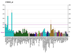
TBK1 (TANK-binding kinase 1) is an enzyme with kinase activity. Specifically, it is a serine / threonine protein kinase. [5] It is encoded by the TBK1 gene in humans. [6] This kinase is mainly known for its role in innate immunity antiviral response. However, TBK1 also regulates cell proliferation, apoptosis, autophagy, and anti-tumor immunity. [5] Insufficient regulation of TBK1 activity leads to autoimmune, neurodegenerative diseases or tumorigenesis. [7] [8]
TBK1 is a non-canonical IKK kinase that phosphorylates the nuclear factor kB (NFkB). It shares sequence homology with canonical IKK. [5]
The N-terminus of the protein contains the kinase domain (region 9-309) and the ubiquitin-like domain (region 310-385). The C-terminus is formed by two coiled-coil structures (region 407-713) that provide a surface for homodimerization. [5] [6]
The autophosphorylation of serine 172, which requires homodimerization and ubiquitinylation of lysines 30 and 401, is necessary for kinase activity. [9]
TBK1 is involved in many signaling pathways and forms a node between them. For this reason, regulation of its involvement in individual signaling pathways is necessary. This is provided by adaptor proteins that interact with the dimerization domain of TBK1 to determine its location and access to substrates. Binding to TANK leads to localization to the perinuclear region and phosphorylation of substrates which is required for subsequent production of type I interferons (IFN-I). In contrast, binding to NAP1 and SINTBAD leads to localization in the cytoplasm and involvement in autophagy. Another adaptor protein that determines the location of TBK1 is TAPE. TAPE targets TBK1 to endolysosomes. [5]
A key interest in TBK1 is due to its role in innate immunity, especially in antiviral responses. TBK1 is redundant with IKK , but TBK1 seems to play a more important role. After triggering antiviral signaling through PRRs (pattern recognition receptors), TBK1 is activated. Subsequently, it phosphorylates the transcription factor IRF3, which is translocated to the nucleus, and promotes production of IFN-I. [7]
As a non-canonical IKK, TBK1 is also involved in the non-canonical NFkB pathway. It phosphorylates p100/NF-κB2, which is subsequently processed in the proteasome and released as a p52 subunit. This subunit dimerizes with RelB and mediates gene expression. [10]
In the canonical NFkB pathway, the NF-kappa-B (NFKB) complex of proteins is inhibited by I-kappa-B (IKB) proteins, which inactivate NFKB by trapping it in the cytoplasm. Phosphorylation of serine residues on the IKB proteins by IKB kinases marks them for destruction via the ubiquitination pathway, thereby allowing activation and nuclear translocation of the NFKB complex. The protein encoded by this gene is similar to IKB kinases and can mediate NFkB activation in response to certain growth factors. [6]
TBK1 promotes autophagy involved in pathogen and mitochondrial clearance. [11] TBK1 phosphorylates autophagy receptors [12] [13] and components of the autophagy apparatus. [14] [15] Furthermore, TBK1 is also involved in the regulation of cell proliferation, apoptosis and glucose metabolism. [10]
TANK-binding kinase 1 has been shown to interact with:
Transcription factors activated upon TBK1 activation include IRF3, IRF7 [21] and ZEB1. [22]
Deregulation of TBK1 activity and mutations in this protein are associated with many diseases. Due to the role of TBK1 in cell survival, deregulation of its activity is associated with tumorogenesis. [8] There are also many autoimmune (e.g., rheumatoid arthritis, sympathetic lupus), neurodegenerative (e.g., amyotrophic lateral sclerosis), and infantile (e.g., herpesviral encephalitis) diseases. [9] [7]
The loss of TBK1 cause embryonic lethality in mice. [21]
Inhibition of IκB kinase (IKK) and IKK-related kinases, IKBKE (IKKε) and TANK-binding kinase 1 (TBK1), has been investigated as a therapeutic option for the treatment of inflammatory diseases and cancer, [23] and a way to overcome resistance to cancer immunotherapy. [24]
Nuclear factor kappa-light-chain-enhancer of activated B cells (NF-κB) is a protein complex that controls transcription of DNA, cytokine production and cell survival. NF-κB is found in almost all animal cell types and is involved in cellular responses to stimuli such as stress, cytokines, free radicals, heavy metals, ultraviolet irradiation, oxidized LDL, and bacterial or viral antigens. NF-κB plays a key role in regulating the immune response to infection. Incorrect regulation of NF-κB has been linked to cancer, inflammatory and autoimmune diseases, septic shock, viral infection, and improper immune development. NF-κB has also been implicated in processes of synaptic plasticity and memory.
Caspase recruitment domains, or caspase activation and recruitment domains (CARDs), are interaction motifs found in a wide array of proteins, typically those involved in processes relating to inflammation and apoptosis. These domains mediate the formation of larger protein complexes via direct interactions between individual CARDs. CARDs are found on a strikingly wide range of proteins, including helicases, kinases, mitochondrial proteins, caspases, and other cytoplasmic factors.
NF-kappa-B essential modulator (NEMO) also known as inhibitor of nuclear factor kappa-B kinase subunit gamma (IKK-γ) is a protein that in humans is encoded by the IKBKG gene. NEMO is a subunit of the IκB kinase complex that activates NF-κB. The human gene for IKBKG is located on the chromosome band Xq28. Multiple transcript variants encoding different isoforms have been found for this gene.
IKK-β also known as inhibitor of nuclear factor kappa-B kinase subunit beta is a protein that in humans is encoded by the IKBKB gene.
The IκB kinase is an enzyme complex that is involved in propagating the cellular response to inflammation, specifically the regulation of lymphocytes.
Transcription factor p65 also known as nuclear factor NF-kappa-B p65 subunit is a protein that in humans is encoded by the RELA gene.
Inhibitor of nuclear factor kappa-B kinase subunit alpha (IKK-α) also known as IKK1 or conserved helix-loop-helix ubiquitous kinase (CHUK) is a protein kinase that in humans is encoded by the CHUK gene. IKK-α is part of the IκB kinase complex that plays an important role in regulating the NF-κB transcription factor. However, IKK-α has many additional cellular targets, and is thought to function independently of the NF-κB pathway to regulate epidermal differentiation.
Mitogen-activated protein kinase kinase kinase 7 (MAP3K7), also known as TAK1, is an enzyme that in humans is encoded by the MAP3K7 gene.
Interleukin-1 receptor-associated kinase 1 (IRAK-1) is an enzyme in humans encoded by the IRAK1 gene. IRAK-1 plays an important role in the regulation of the expression of inflammatory genes by immune cells, such as monocytes and macrophages, which in turn help the immune system in eliminating bacteria, viruses, and other pathogens. IRAK-1 is part of the IRAK family consisting of IRAK-1, IRAK-2, IRAK-3, and IRAK-4, and is activated by inflammatory molecules released by signaling pathways during pathogenic attack. IRAK-1 is classified as a kinase enzyme, which regulates pathways in both innate and adaptive immune systems.
Receptor-interacting serine/threonine-protein kinase 1 (RIPK1) functions in a variety of cellular pathways related to both cell survival and death. In terms of cell death, RIPK1 plays a role in apoptosis and necroptosis. Some of the cell survival pathways RIPK1 participates in include NF-κB, Akt, and JNK.
Inhibitor of nuclear factor kappa-B kinase subunit epsilon also known as I-kappa-B kinase epsilon or IKK-epsilon is an enzyme that in humans is encoded by the IKBKE gene.
Serine/threonine-protein kinase D3 (PKD3) or PKC-nu is an enzyme that in humans is encoded by the PRKD3 gene.
Caspase recruitment domain-containing protein 11 also known as CARD-containing MAGUK protein 1 is a protein in the CARD-CC protein family that in humans is encoded by the CARD11 gene. CARD 11 is a membrane associated protein that is found in various human tissues, including the thymus, spleen, liver, and peripheral blood leukocytes. Similarly, CARD 11 is also found in abundance in various lines of cancer cells.
Mitochondrial antiviral-signaling protein (MAVS) is a protein that is essential for antiviral innate immunity. MAVS is located in the outer membrane of the mitochondria, peroxisomes, and mitochondrial-associated endoplasmic reticulum membrane (MAM). Upon viral infection, a group of cytosolic proteins will detect the presence of the virus and bind to MAVS, thereby activating MAVS. The activation of MAVS leads the virally infected cell to secrete cytokines. This induces an immune response which kills the host's virally infected cells, resulting in clearance of the virus.
TRAF family member-associated NF-kappa-B activator is a protein that in humans is encoded by the TANK gene.
Adapter protein CIKS is a protein that in humans is encoded by the TRAF3IP2 gene.
5-azacytidine-induced protein 2 is a protein that in humans is encoded by the AZI2 gene.
The Akt signaling pathway or PI3K-Akt signaling pathway is a signal transduction pathway that promotes survival and growth in response to extracellular signals. Key proteins involved are PI3K and Akt.
Shu Hongbing is a Chinese cytologist and immunologist. He became a member of the Chinese Academy of Sciences in 2011 and TWAS in 2012. Shu is mainly known for his work on cell signal transduction related to immunity.
The interleukin-1 receptor (IL-1R) associated kinase (IRAK) family plays a crucial role in the protective response to pathogens introduced into the human body by inducing acute inflammation followed by additional adaptive immune responses. IRAKs are essential components of the Interleukin-1 receptor signaling pathway and some Toll-like receptor signaling pathways. Toll-like receptors (TLRs) detect microorganisms by recognizing specific pathogen-associated molecular patterns (PAMPs) and IL-1R family members respond the interleukin-1 (IL-1) family cytokines. These receptors initiate an intracellular signaling cascade through adaptor proteins, primarily, MyD88. This is followed by the activation of IRAKs. TLRs and IL-1R members have a highly conserved amino acid sequence in their cytoplasmic domain called the Toll/Interleukin-1 (TIR) domain. The elicitation of different TLRs/IL-1Rs results in similar signaling cascades due to their homologous TIR motif leading to the activation of mitogen-activated protein kinases (MAPKs) and the IκB kinase (IKK) complex, which initiates a nuclear factor-κB (NF-κB) and AP-1-dependent transcriptional response of pro-inflammatory genes. Understanding the key players and their roles in the TLR/IL-1R pathway is important because the presence of mutations causing the abnormal regulation of Toll/IL-1R signaling leading to a variety of acute inflammatory and autoimmune diseases.