Muscular hydrostat

Last updated
The tongue is a muscular hydrostat. Que lengua.jpg
The tongue is a muscular hydrostat.

A muscular hydrostat is a biological structure found in animals. It is used to manipulate items (including food) or to move its host about and consists mainly of muscles with no skeletal support. It performs its hydraulic movement without fluid in a separate compartment, as in a hydrostatic skeleton.

Contents

A muscular hydrostat, like a hydrostatic skeleton, relies on the fact that water is effectively incompressible at physiological pressures. In contrast to a hydrostatic skeleton, where muscle surrounds a fluid-filled cavity, a muscular hydrostat is composed mainly of muscle tissue. Since muscle tissue itself is mainly made of water and is also effectively incompressible, similar principles apply.

Muscular anatomy

Closeup of the trunk of an Asian elephant Elephas Maximus Trunk Closeup.jpg
Closeup of the trunk of an Asian elephant

Muscles provide the force to move a muscular hydrostat. Since muscles are only able to produce force by contracting and becoming shorter, different groups of muscles have to work against each other, with one group relaxing and lengthening as the other group provides the force by contracting. Such complementary muscle groups are termed antagonistic pairs.

The muscle fibers in a muscular hydrostat are oriented in three directions: parallel to the long axis, perpendicular to the long axis, and wrapped obliquely around the long axis. [1] [2]

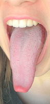
The muscles parallel to the long axis are arranged in longitudinal bundles. The more peripherally these are located, the more elaborate bending movements are possible. A more peripheral distribution is found in tetrapod tongues, octopus arms, nautilus tentacles, and elephant trunks. Tongues that are adapted for protrusion typically have centrally located longitudinal fibers. These are found in snake tongues, many lizard tongues, and the mammalian anteaters.

The muscles perpendicular to the long axis may be arranged in a transverse, circular, or radial pattern. A transverse arrangement involves sheets of muscle fibers running perpendicular to the long axis, usually alternating between horizontal and vertical orientations. This arrangement is found in the arms and tentacles of squid, octopuses, and in most mammalian tongues. A radial arrangement involves fibers radiating out in all directions from the center of the organ. This is found in the tentacles of the chambered nautilus and in the elephant proboscis (trunk). A circular arrangement has rings of contractive fibers around the long axis. This is found in many mammalian and lizard tongues along with squid tentacles.

Helical or oblique fibers around the long axis are generally present in two layers with opposite chirality and wrap around the central core of musculature.

Mechanism of operation

In a muscular hydrostat, the musculature itself both creates movement and provides skeletal support for that movement. It can provide this support because it is composed primarily of an incompressible “liquid" and is thus constant in volume. The most important biomechanical feature of a muscular hydrostat is its constant volume. Muscle is composed primarily of an aqueous liquid that is essentially incompressible at physiological pressures. In a muscular hydrostat or any other structure of constant volume, a decrease in one dimension will cause a compensatory increase in at least one other dimension. [3] The mechanisms of elongation, bending and torsion in muscular hydrostats all depend on constancy of volume to effect shape changes in the absence of stiff skeletal attachments. [4] Since muscular hydrostats are under constant volume when the diameter increases or decreases, the length must also decrease or increase, respectively. When looking at a cylinder the volume is: V=πr²l. When the radius is differentiated with respect to the length: dr/dl=-r/(2l). From this, if a diameter decreases by 25%, the length will increase by approximately 80% which may produce a large amount of force depending on what the animal is trying to do. [5]

Elongation and shortening

Arms and tentacles of the squid Abralia veranyi Abralia veranyi tentacles.jpg
Arms and tentacles of the squid Abralia veranyi

Elongation in hydrostats is caused by the contraction of transverse or helical musculature arrangements. Given the constant volume of muscular hydrostats, these contractions cause an elongation of the longitudinal muscles. Change in length is proportional to the square of the decrease in diameter. [3] Therefore, contractions of muscles perpendicular to the long axis will cause a decrease in diameter while keeping a constant volume will elongate the organ length-wise. Shortening, on the other hand, can be caused by contraction of the muscles parallel to the long axis resulting in the organ increasing in diameter as well as shortening in length.

The muscles used in elongation and shortening maintain support through the constant volume principle and their antagonistic relationships with each other. These mechanisms are seen often in prey capture of shovelnose frogs and chameleons, as well as in the human tongue and many other examples. In some frogs, the tongue elongates up to 180% of its resting length. [6] Extra-oral tongues show higher length/width ratios than intra-oral tongues, allowing for a greater increase in length (more than 100% of resting length, as compared to intra-oral tongues at only about 50% of resting length increase). Greater elongation lengths trade off with the force produced by the organ; as the length/width ratio is increased elongation increases while force is decreased. [1] Squids have been shown to use muscular hydrostat elongation in prey capture and feeding as well. [7]

Bending

The bending of a muscular hydrostat can occur in two ways, both of which require the use of antagonistic muscles. [1] The unilateral contraction of a longitudinal muscle will produce little or no bending and will serve to increase the diameter of the muscular hydrostat because of the constant volume principle that must be met. To bend the hydrostat structure, the unilateral contraction of longitudinal muscle must be accompanied by contractile activity of transverse, radial, or circular muscles to maintain a constant diameter. Bending of a muscular hydrostat can also occur by the contraction of transverse, radial, or circular muscles which decreases the diameter. Bending is produced by longitudinal muscle activity which maintains a constant length on one side of the structure.

The bending of a muscular hydrostat is particularly important in animal tongues. This motion provides the mechanism by which a snake flicks the air with its tongue to sense its surroundings, and it is also responsible for the complexities of human speech. [2]

Stiffening

The stiffening of a muscular hydrostat is accomplished by the muscle or connective tissue of the hydrostat resisting dimensional changes. [3]

Torsion

Torsion is the twisting of a muscular hydrostat along its long axis and is produced by a helical or oblique arrangement of musculature [3] which have varying direction. For a counter-clockwise torsion it is necessary for a right-hand helix to contract. Contraction of a left-hand helix causes clockwise torsion. The simultaneous contraction of both right and left-hand helixes results in an increase in resistance to torsional forces. The oblique or helical muscle arrays in the muscular hydrostats are located in the periphery of the structure, wrapping the inner core of musculature, and this peripheral location provides a larger moment through which the torque is applied than a more central location. The effect of helically arranged muscle fibers, which may also contribute to changes in length of a muscular hydrostat, depends on fiber angle—the angle that the helical muscle fibers make with the long axis of the structure.

The length of the helical fiber is at a minimum when the fiber angle equals 54°44′ and is at maximum length when the fiber angle approaches 0° and 90°. [3] Summed up, this means that helically arranged muscle fibers with a fiber angle greater than 54°44′ will create force for both torsion and elongation while helically arranged muscle fibers with a fiber angle less than 54°44′ will create force for both torsion and shortening. [8] The fiber angle of the oblique or helical muscle layers must increase during shortening and decrease during lengthening. In addition to creating a torsional force, the oblique muscle layers will therefore create a force for elongation that may aid the transverse musculature in resisting longitudinal compression.

Examples

West Indian manatee Manatee Florida.jpg
West Indian manatee

Technological applications

A group of engineers and biologists have collaborated[ when? ] to develop robotic arms that are able to manipulate and handle various objects of different size, mass, surface texture and mechanical properties. These robotic arms have many advantages over previous robotic arms that were not based on muscular hydrostats. [13]

Related Research Articles

<span class="mw-page-title-main">Cephalopod</span> Class of mollusks

A cephalopod is any member of the molluscan class Cephalopoda such as a squid, octopus, cuttlefish, or nautilus. These exclusively marine animals are characterized by bilateral body symmetry, a prominent head, and a set of arms or tentacles modified from the primitive molluscan foot. Fishers sometimes call cephalopods "inkfish", referring to their common ability to squirt ink. The study of cephalopods is a branch of malacology known as teuthology.

<span class="mw-page-title-main">Skeletal muscle</span> One of three major skeletal system types that connect to bones

Skeletal muscles are organs of the vertebrate muscular system and typically are attached by tendons to bones of a skeleton. The muscle cells of skeletal muscles are much longer than in the other types of muscle tissue, and are often known as muscle fibers. The muscle tissue of a skeletal muscle is striated – having a striped appearance due to the arrangement of the sarcomeres.

A hydrostatic skeleton or hydroskeleton is a type of skeleton supported by hydrostatic fluid pressure, common among soft-bodied invertebrate animals colloquially referred to as "worms". While more advanced organisms can be considered hydrostatic, they are sometimes referred to as hydrostatic for their possession of a hydrostatic organ instead of a hydrostatic skeleton, where the two may have the same capabilities but are not the same. As the prefix hydro- meaning "water", being hydrostatic means being fluid-filled.

<span class="mw-page-title-main">Tentacle</span> Varied organ found in many animals and used for palpation and manipulation

In zoology, a tentacle is a flexible, mobile, and elongated organ present in some species of animals, most of them invertebrates. In animal anatomy, tentacles usually occur in one or more pairs. Anatomically, the tentacles of animals work mainly like muscular hydrostats. Most forms of tentacles are used for grasping and feeding. Many are sensory organs, variously receptive to touch, vision, or to the smell or taste of particular foods or threats. Examples of such tentacles are the eyestalks of various kinds of snails. Some kinds of tentacles have both sensory and manipulatory functions.

<span class="mw-page-title-main">Muscle contraction</span> Activation of tension-generating sites in muscle

Muscle contraction is the activation of tension-generating sites within muscle cells. In physiology, muscle contraction does not necessarily mean muscle shortening because muscle tension can be produced without changes in muscle length, such as when holding something heavy in the same position. The termination of muscle contraction is followed by muscle relaxation, which is a return of the muscle fibers to their low tension-generating state.

<span class="mw-page-title-main">Transversus thoracis muscle</span>

The transversus thoracis muscle, also known as triangularis sterni, lies internal to the thoracic cage, anteriorly. It is usually a thin plane of muscular and tendinous fibers, however on athletic individuals it can be a thick 'slab of meat', situated upon the inner surface of the front wall of the chest. It is in the same layer as the subcostal muscles and the innermost intercostal muscles.

<span class="mw-page-title-main">Abdominal internal oblique muscle</span> Muscle in the abdominal wall

The abdominal internal oblique muscle, also internal oblique muscle or interior oblique, is an abdominal muscle in the abdominal wall that lies below the external oblique muscle and just above the transverse abdominal muscle.

The apex beat, also called the apical impulse, is the pulse felt at the point of maximum impulse (PMI), which is the point on the precordium farthest outwards (laterally) and downwards (inferiorly) from the sternum at which the cardiac impulse can be felt. The cardiac impulse is the vibration resulting from the heart rotating, moving forward, and striking against the chest wall during systole. The PMI is not the apex of the heart but is on the precordium not far from it. Another theory for the occurrence of the PMI is the early systolic contraction of the longitudinal fibers of the left ventricle located on the endocardial surface of this chamber. This period of the cardiac cycle is called isovolumic contraction. Because the contraction starts near the base of the left ventricle and spreads toward the apex most of the longitudinal fibers of the left ventricle have shortened before the apex. The rapidly increasing pressure developed by the shortening of these fibers causes the aortic valve to open and the apex to move outward causing the PMI. Anatomical dissection of the musculature of the apex reveals that muscle fibers are no longer longitudinal oriented but form a spiral mass of muscular tissues which may also have an effect on the ability of the apex to contract longitudinally. After the longitudinal fibers contract, the ejection of blood out of the left ventricle is accomplished by the torsional action of the circumferential muscle fibers of the left ventricle that are in the mid-portion of the ventricle and contract after the longitudinal fibers. During the longitudinal fiber contraction, the volume of the left ventricle has not changed keeping the apex in intimate contact with the chest wall allowing the ability to feel the apex move outward before the heart empties greater than 55% of its volume and the apex falling away from the chest wall.

<span class="mw-page-title-main">Quadratus lumborum muscle</span> Muscle in the lower back

The quadratus lumborum muscle, informally called the QL, is a paired muscle of the left and right posterior abdominal wall. It is the deepest abdominal muscle, and commonly referred to as a back muscle. Each is irregular and quadrilateral in shape.

<span class="mw-page-title-main">Myofilament</span> The two protein filaments of myofibrils in muscle cells

Myofilaments are the three protein filaments of myofibrils in muscle cells. The main proteins involved are myosin, actin, and titin. Myosin and actin are the contractile proteins and titin is an elastic protein. The myofilaments act together in muscle contraction, and in order of size are a thick one of mostly myosin, a thin one of mostly actin, and a very thin one of mostly titin.

A pennate or pinnate muscle is a type of skeletal muscle with fascicles that attach obliquely to its tendon. This type of muscle generally allows higher force production but a smaller range of motion. When a muscle contracts and shortens, the pennation angle increases.

<span class="mw-page-title-main">Cephalopod limb</span> Limbs of cephalopod molluscs

All cephalopods possess flexible limbs extending from their heads and surrounding their beaks. These appendages, which function as muscular hydrostats, have been variously termed arms, legs or tentacles.

<span class="mw-page-title-main">Undulatory locomotion</span>

Undulatory locomotion is the type of motion characterized by wave-like movement patterns that act to propel an animal forward. Examples of this type of gait include crawling in snakes, or swimming in the lamprey. Although this is typically the type of gait utilized by limbless animals, some creatures with limbs, such as the salamander, forgo use of their legs in certain environments and exhibit undulatory locomotion. In robotics this movement strategy is studied in order to create novel robotic devices capable of traversing a variety of environments.

Role of skin in locomotion describes how the integumentary system is involved in locomotion. Typically the integumentary system can be thought of as skin, however the integumentary system also includes the segmented exoskeleton in arthropods and feathers of birds. The primary role of the integumentary system is to provide protection for the body. However, the structure of the skin has evolved to aid animals in their different modes of locomotion. Soft bodied animals such as starfish rely on the arrangement of the fibers in their tube feet for movement. Eels, snakes, and fish use their skin like an external tendon to generate the propulsive forces need for undulatory locomotion. Vertebrates that fly, glide, and parachute also have a characteristic fiber arrangements of their flight membranes that allows for the skin to maintain its structural integrity during the stress and strain experienced during flight.

Ballistic movement can be defined as muscle contractions that exhibit maximum velocities and accelerations over a very short period of time. They exhibit high firing rates, high force production, and very brief contraction times.

<span class="mw-page-title-main">Architectural gear ratio</span> Ratio between muscle-shortening velocity and fiber-shortening velocity

Architectural gear ratio, also called anatomical gear ratio (AGR) is a feature of pennate muscle defined by the ratio between the longitudinal strain of the muscle and muscle fiber strain. It is sometimes also defined as the ratio between muscle-shortening velocity and fiber-shortening velocity.

<i>Euapta lappa</i> Species of sea cucumber

Euapta lappa, the beaded sea cucumber, is a species of sea cucumbers in the family Synaptidae in the phylum Echinodermata. It is found on coral reefs in the Caribbean region.

Muscle architecture is the physical arrangement of muscle fibers at the macroscopic level that determines a muscle's mechanical function. There are several different muscle architecture types including: parallel, pennate and hydrostats. Force production and gearing vary depending on the different muscle parameters such as muscle length, fiber length, pennation angle, and the physiological cross-sectional area (PCSA).

<span class="mw-page-title-main">Anatomical terms of muscle</span> Muscles terminology

Anatomical terminology is used to uniquely describe aspects of skeletal muscle, cardiac muscle, and smooth muscle such as their actions, structure, size, and location.

<span class="mw-page-title-main">Gastrointestinal wall</span> Digestive system structure

The gastrointestinal wall of the gastrointestinal tract is made up of four layers of specialised tissue. From the inner cavity of the gut outwards, these are the mucosa, the submucosa, the muscular layer and the serosa or adventitia.

References

  1. 1 2 3 4 5 6 Kier, W. M.; Smith, K. K. (1985). "Tongues, tentacles and trunks: The biomechanics of movement in muscular-hydrostats". Zoological Journal of the Linnean Society. 83 (4): 307–324. doi:10.1111/j.1096-3642.1985.tb01178.x.
  2. 1 2 Smith, Kathleen K.; William M. Kier (Jan–Feb 1989). "Trunks, tongues, and tentacles: Moving with skeletons of muscle". American Scientist. 77 (1): 28–35.
  3. 1 2 3 4 5 Kier, W. M. (1985). "The musculature of squid arms and tentacles: Ultrastructural evidence for functional differences". Journal of Morphology. 185 (2): 223–239. doi:10.1002/jmor.1051850208. PMID   30011972. S2CID   51631466.
  4. Wainwright, P. C.; Bennett, A. F. (1992). "The mechanism of tongue projection in chameleons: II. Role of shape change in a muscular hydrostat". The Journal of Experimental Biology168: 23–40.
  5. Alexander, R. McN. (2003). Principles of Animal Locomotion. Princeton, NJ: Princeton University Press.
  6. Nishikawa, K. C.; Kier, W. M.; Smith, K. K. (1999). "Morphology and mechanics of tongue movement in the African pig-nosed frog Hemisus marmoratum: a muscular hydrostatic model http://jeb.biologists.org/content/202/7/771.short". The Journal of Experimental Biology202: 771–780.
  7. 1 2 Kier, W. M. (1982). "The functional morphology of the musculature of squid (Loliginidae) arms and tentacles". Journal of Morphology. 172 (2): 179–192. doi:10.1002/jmor.1051720205. PMID   30103569. S2CID   51980244.
  8. Meyers, J. J.; O'Reilly, J. C.; Monroy, J. A.; Nishikawa, K. C. (2004). "Mechanism of tongue protraction in microhylid frogs. The Journal of Experimental Biology207: 21–31.
  9. Price, Rebecca (Dec 2003). "Columellar muscle of neogastropods: Muscle attachment and the function of columellar folds". Biological Bulletin. 205 (3): 351–366. doi:10.2307/1543298. JSTOR   1543298. PMID   14672989. S2CID   12277966.
  10. Matzner, H.; Gutfreund, Y.; Hochner, B. (2000). "Neuromuscular system of the flexible arm of the octopus: Physiological characterization". Journal of Neurophysiology. 83 (3): 1315–1328. doi:10.1152/jn.2000.83.3.1315. PMID   10712459. S2CID   14402766.
  11. Yekutieli, Y.; Sumbre, G.; Flash, T.; Hochner, B. (2002). "How to move with no rigid skeleton? The octopus has the answers". Biologist (London, England). 49 (6): 250–254. PMID   12486300.
  12. Marshall, C. D.; Clark, L. A.; Reep, R. L. (1998). "The muscular hydrostat of the Florida manatee (Trichechus manatus latirostris): A functional morphological model of perioral bristle use". Marine Mammal Science. 14 (2): 290–303. doi:10.1111/j.1748-7692.1998.tb00717.x.
  13. Walker, I.D.; Dawson, D.M.; Flash, T.; Grasso, F.W.; Hanlon, R.T.; Hochner, B.; Kier, W.M.; Pagano, C.C.; Rahn, C.D.; Zhang, Q.M. (2005). "Continuum robot arms inspired by cephalopods". Proceedings of SPIE5804: 303–314.