Patulous Eustachian tube

Last updated
Patulous Eustachian tube
Other namesPatent Eustachian tube
Specialty ENT surgery

Patulous Eustachian tube is the name of a physical disorder where the Eustachian tube, which is normally closed, instead stays intermittently open. When this occurs, the person experiences autophony, the hearing of self-generated sounds. [1] These sounds, such as one's own breathing, voice, and heartbeat, vibrate directly onto the ear drum and can create a "bucket on the head" effect, making it difficult for the patient to attend to environmental sounds. Patulous Eustachian tube is a form of Eustachian tube dysfunction, which is said to be present in about 1 percent of the general population. [2]

Contents

Signs and symptoms

With patulous Eustachian tube, variations in upper airway pressure associated with respiration are transmitted to the middle ear through the Eustachian tube. This causes an unpleasant fullness feeling in the middle ear and alters the auditory perception. Complaints seem to include muffled hearing and autophony. In addition, patulous Eustachian tube generally feels dry with no clogged feeling or sinus pressure.

Patients hear their own voice or its echo from inside. [3] [4] They describe it as being amplified and unpleasant. Lying head down may help since it increases venous blood pressure and congestion of the mucosa.

Causes

Patulous Eustachian tube is a physical disorder. The exact causes may vary depending on the person but the causes or often unknown. [5] Weight loss is a commonly cited cause of the disorder due to the nature of the Eustachian tube itself and is associated with approximately one-third of reported cases. [6] Fatty tissues hold the tube closed most of the time in healthy individuals. When circumstances cause overall body fat to diminish, the tissue surrounding the Eustachian tube shrinks and this function is disrupted. [7]

Activities and substances which dehydrate the body have the same effect and are also possible causes of patulous Eustachian tube. Examples are stimulants (including caffeine) and exercise. Exercise may have a more short-term effect than caffeine or weight loss in this regard.

Pregnancy can also be a cause of patulous Eustachian tube due to the effects of pregnancy hormones on surface tension and mucus in the respiratory system. [8]

Granulomatosis with polyangiitis can also be a cause of this disorder. It is yet unknown why.

Patulous Eustachian tube can occur as a result of liquid residue in the Eustachian tube, after suffering a middle ear infection (otitis media).

Potential causes list

It's worth noting that there hasn't been much comprehensive scientific research conducted to establish a clear correlation between most of the claimed potential causes and patulous eustachian tube disorder.

Diagnosis

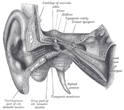
Upon examination of a suspected case of patulous Eustachian tube, a doctor can directly view the tympanic membrane with a light and observe that it vibrates with every breath taken by the patient. A tympanogram may also help with the diagnosis. Patulous Eustachian tube is likely if brisk inspiration causes a significant pressure shift.

Patulous Eustachian tube is frequently misdiagnosed as standard congestion due to the similarity in symptoms and rarity of the disorder. Audiologists are more likely to recognize the disorder, usually with tympanometry or nasally delivered masking noise during a hearing assessment, which is highly sensitive to this condition. [11]

When misdiagnosis occurs, a decongestant medication is sometimes prescribed. This type of medication aggravates the condition, as the Eustachian tube relies on sticky fluids to keep closed and the drying effect of a decongestant would make it even more likely to remain open and cause symptoms.

Incidentally, patients who instead suffer from the even rarer condition of superior canal dehiscence are at risk for misdiagnosis of patulous Eustachian tube due to the similar autophony in both conditions.

Treatment

Estrogen nasal drops or saturated potassium iodide have been used to induce edema of the eustachian tube opening. Nasal medications containing diluted hydrochloric acid, chlorobutanol, and benzyl alcohol have been reported to be effective in some patients, with few side effects. Food and Drug Administration approval is still pending, however. [12] Nasal sprays have also been a very effective temporary treatment for this disease, as well. [13]

In extreme cases surgical intervention may attempt to restore the Eustachian tube tissues with fat, gel foam, or cartilage or scar it closed with cautery. These methods are not always successful. For example, there is the case of the early attempts at surgical correction involving injections of tetrafluoroetheylene (Teflon) paste but although this treatment was able to give transient relief, it was discontinued due to several deaths that resulted from inadvertent intracarotid injections. [14]

Although a temporary solution, surgical ventilation tube placement in the ear drum has also proven to be an effective treatment option. This treatment is known as either a unilateral or bilateral myringotomy. 50% of patients reported relief of patulous Eustachian tube symptoms when given this treatment. [15]

Related Research Articles

<span class="mw-page-title-main">Otorhinolaryngology</span> Medical specialty

Otorhinolaryngology is a surgical subspeciality within medicine that deals with the surgical and medical management of conditions of the head and neck. Doctors who specialize in this area are called otorhinolaryngologists, otolaryngologists, head and neck surgeons, or ENT surgeons or physicians. Patients seek treatment from an otorhinolaryngologist for diseases of the ear, nose, throat, base of the skull, head, and neck. These commonly include functional diseases that affect the senses and activities of eating, drinking, speaking, breathing, swallowing, and hearing. In addition, ENT surgery encompasses the surgical management of cancers and benign tumors and reconstruction of the head and neck as well as plastic surgery of the face, scalp, and neck.

<span class="mw-page-title-main">Ménière's disease</span> Disorder of the inner ear

Ménière's disease (MD) is a disease of the inner ear that is characterized by potentially severe and incapacitating episodes of vertigo, tinnitus, hearing loss, and a feeling of fullness in the ear. Typically, only one ear is affected initially, but over time, both ears may become involved. Episodes generally last from 20 minutes to a few hours. The time between episodes varies. The hearing loss and ringing in the ears can become constant over time.

<span class="mw-page-title-main">Cholesteatoma</span> Medical condition

Cholesteatoma is a destructive and expanding growth consisting of keratinizing squamous epithelium in the middle ear and/or mastoid process. Cholesteatomas are not cancerous as the name may suggest, but can cause significant problems because of their erosive and expansile properties. This can result in the destruction of the bones of the middle ear (ossicles), as well as growth through the base of the skull into the brain. They often become infected and can result in chronically draining ears. Treatment almost always consists of surgical removal.

<span class="mw-page-title-main">Otitis media</span> Inflammation of the middle ear

Otitis media is a group of inflammatory diseases of the middle ear. One of the two main types is acute otitis media (AOM), an infection of rapid onset that usually presents with ear pain. In young children this may result in pulling at the ear, increased crying, and poor sleep. Decreased eating and a fever may also be present. The other main type is otitis media with effusion (OME), typically not associated with symptoms, although occasionally a feeling of fullness is described; it is defined as the presence of non-infectious fluid in the middle ear which may persist for weeks or months often after an episode of acute otitis media. Chronic suppurative otitis media (CSOM) is middle ear inflammation that results in a perforated tympanic membrane with discharge from the ear for more than six weeks. It may be a complication of acute otitis media. Pain is rarely present. All three types of otitis media may be associated with hearing loss. If children with hearing loss due to OME do not learn sign language, it may affect their ability to learn.

<span class="mw-page-title-main">Eustachian tube</span> Tube connecting middle ear to throat

The Eustachian tube, also called the auditory tube or pharyngotympanic tube, is a tube that links the nasopharynx to the middle ear, of which it is also a part. In adult humans, the Eustachian tube is approximately 35 mm (1.4 in) long and 3 mm (0.12 in) in diameter. It is named after the sixteenth-century Italian anatomist Bartolomeo Eustachi.

<span class="mw-page-title-main">Nosebleed</span> Bleeding from the nose

A nosebleed, also known as epistaxis, is an instance of bleeding from the nose. Blood can flow down into the stomach, and cause nausea and vomiting. In more severe cases, blood may come out of both nostrils. Rarely, bleeding may be so significant that low blood pressure occurs. Blood may also come up the nasolacrimal duct and out from the eye.

<span class="mw-page-title-main">Nasal polyp</span> Noncancerous growths within the nose or sinuses

Nasal polyps (NP) are noncancerous growths within the nose or sinuses. Symptoms include trouble breathing through the nose, loss of smell, decreased taste, post nasal drip, and a runny nose. The growths are sac-like, movable, and nontender, though face pain may occasionally occur. They typically occur in both nostrils in those who are affected. Complications may include sinusitis and broadening of the nose.

<span class="mw-page-title-main">Conductive hearing loss</span> Medical condition

Conductive hearing loss (CHL) occurs when there is a problem transferring sound waves anywhere along the pathway through the outer ear, tympanic membrane (eardrum), or middle ear (ossicles). If a conductive hearing loss occurs in conjunction with a sensorineural hearing loss, it is referred to as a mixed hearing loss. Depending upon the severity and nature of the conductive loss, this type of hearing impairment can often be treated with surgical intervention or pharmaceuticals to partially or, in some cases, fully restore hearing acuity to within normal range. However, cases of permanent or chronic conductive hearing loss may require other treatment modalities such as hearing aid devices to improve detection of sound and speech perception.

<span class="mw-page-title-main">Ear pain</span> Pain in the ear

Ear pain, also known as earache or otalgia, is pain in the ear. Primary ear pain is pain that originates from the ear. Secondary ear pain is a type of referred pain, meaning that the source of the pain differs from the location where the pain is felt.

<span class="mw-page-title-main">Otology</span> Branch of medicine for the ear

Otology is a branch of medicine which studies normal and pathological anatomy and physiology of the ear as well as their diseases, diagnosis and treatment. Otologic surgery generally refers to surgery of the middle ear and mastoid related to chronic otitis media, such as tympanoplasty, or ear drum surgery, ossiculoplasty, or surgery of the hearing bones, and mastoidectomy. Otology also includes surgical treatment of conductive hearing loss, such as stapedectomy surgery for otosclerosis.

<span class="mw-page-title-main">Tympanostomy tube</span> Medical device inserted into the eardrum

Tympanostomy tube, also known as a grommet,myringotomy tube, or pressure equalizing tube, is a small tube inserted into the eardrum via a surgical procedure called myringotomy to keep the middle ear aerated for a prolonged period of time, typically to prevent accumulation of fluid in the middle ear. The tube itself is made in a variety of designs, most often shaped like a grommet for short-term use, or with long flanges and sometimes resembling a T-shape for long-term use. Materials used to manufacture the tubes are often made from fluoroplastic or silicone, which have largely replaced the use of metal tubes made from stainless steel, titanium, or gold.

<span class="mw-page-title-main">Perforated eardrum</span> Injury leading to a hole in the eardrum

A perforated eardrum is a prick in the eardrum. It can be caused by infection, trauma, overpressure, inappropriate ear clearing, and changes in middle ear pressure. An otoscope can be used to view the eardrum to diagnose a perforation. Perforations may heal naturally or require surgery.

<span class="mw-page-title-main">Contact granuloma</span> Medical condition

Contact granuloma is a condition that develops due to persistent tissue irritation in the posterior larynx. Benign granulomas, not to be confused with other types of granulomas, occur on the vocal process of the vocal folds, where the vocal ligament attaches. Signs and symptoms may include hoarseness of the voice, or a sensation of having a lump in the throat, but contact granulomas may also be without symptoms. There are two common causes associated with contact granulomas; the first common cause is sustained periods of increased pressure on the vocal folds, and is commonly seen in people who use their voice excessively, such as singers. Treatment typically includes voice therapy and changes to lifestyle factors. The second common cause of granulomas is gastroesophageal reflux and is controlled primarily through the use of anti-reflux medication. Other associated causes are discussed below.

Chronic atrophic rhinitis, or simply atrophic rhinitis, is a chronic inflammation of the nose characterised by atrophy of nasal mucosa, including the glands, turbinate bones and the nerve elements supplying the nose. Chronic atrophic rhinitis may be primary and secondary. Special forms of chronic atrophic rhinitis are rhinitis sicca anterior and ozaena. It can also be described as the empty nose syndrome.

The semicircular canal dehiscence (SCD) is a category of rare neurotological diseases/disorders affecting the inner ears, which gathers the superior SCD, lateral SCD and posterior SCD. These SCDs induce SCD syndromes (SCDSs), which define specific sets of hearing and balance symptoms. This entry mainly deals with the superior SCDS.

<span class="mw-page-title-main">Tympanosclerosis</span> Medical condition

Tympanosclerosis is a condition caused by hyalinization and subsequent calcification of subepithelial connective tissue of the tympanic membrane and middle ear, sometimes resulting in a detrimental effect to hearing.

<span class="mw-page-title-main">Hypernasal speech</span> Medical condition

Hypernasal speech is a disorder that causes abnormal resonance in a human's voice due to increased airflow through the nose during speech. It is caused by an open nasal cavity resulting from an incomplete closure of the soft palate and/or velopharyngeal sphincter. In normal speech, nasality is referred to as nasalization and is a linguistic category that can apply to vowels or consonants in a specific language. The primary underlying physical variable determining the degree of nasality in normal speech is the opening and closing of a velopharyngeal passage way between the oral vocal tract and the nasal vocal tract. In the normal vocal tract anatomy, this opening is controlled by lowering and raising the velum or soft palate, to open or close, respectively, the velopharyngeal passageway.

<span class="mw-page-title-main">Tympanic membrane retraction</span> Medical condition

Tympanic membrane retraction describes a condition in which a part of the eardrum lies deeper within the ear than its normal position.

Eustachian tube dysfunction (ETD) is a disorder where pressure abnormalities in the middle ear result in symptoms.

Middle ear barotrauma (MEBT), also known to underwater divers as ear squeeze and reverse ear squeeze, is an injury caused by a difference in pressure between the external ear canal and the middle ear. It is common in underwater divers and usually occurs when the diver does not equalise sufficiently during descent or, less commonly, on ascent. Failure to equalise may be due to inexperience or eustachian tube dysfunction, which can have many possible causes. Unequalised ambient pressure increase during descent causes a pressure imbalance between the middle ear air space and the external auditory canal over the eardrum, referred to by divers as ear squeeze, causing inward stretching, serous effusion and haemorrhage, and eventual rupture. During ascent internal over-pressure is normally passively released through the eustachian tube, but if this does not happen the volume expansion of middle ear gas will cause outward bulging, stretching and eventual rupture of the eardrum known to divers as reverse ear squeeze. This damage causes local pain and hearing loss. Tympanic rupture during a dive can allow water into the middle ear, which can cause severe vertigo from caloric stimulation. This may cause nausea and vomiting underwater, which has a high risk of aspiration of vomit or water, with possibly fatal consequences.

References

  1. Ikeda R, Kikuchi T, Oshima H, Kobayashi T (2020). "Management of Patulous Eustachian Tube". Japan Medical Association Journal . 3 (2): 101–108. doi:10.31662/jmaj.2020-0007. PMC   7590401 . PMID   33150241.
  2. Dornhoffer, John; Gluth, Michael (2016). The Chronic Ear. Stuttgart: Thieme. ISBN   9781604068658.
  3. Gopen, Quinton (2013). Fundamental Otology: Pediatric and Adult Practice. New Delhi: Jaypee Brothers Medical Publishers Pvt. Ltd. p. 181. ISBN   9789350902691.
  4. Sataloff, Joseph; Sataloff, Robert (2005). Hearing Loss, 4th ed. Boca Raton, FL: CRC Press. p. 186. ISBN   9780824754358.
  5. Bance, Manohar; Tysome, James R.; Smith, Matthew E. (2019-09-01). "Patulous Eustachian tube (PET), a practical overview". World Journal of Otorhinolaryngology - Head and Neck Surgery. Focused Issue on Diagnosis and Management of the Eustachian Tube Disorders. 5 (3): 137–142. doi:10.1016/j.wjorl.2019.08.003. ISSN   2095-8811. PMC   6849362 .
  6. Gulya, Aina; Minor, Lloyd; Poe, Dennis (2010). Glasscock-Shambaugh Surgery of the Ear 6th ed. Shelton, CT: PMPH-USA. p. 252. ISBN   9781607950264.
  7. Patulous Eustachian Tube Overview at eMedicine
  8. Hillman, Edawrd J. (1995). Otolaryngologic Manifestations Of Pregnancy. BCM
  9. Poe, Dennis S. (August 2007). "Diagnosis and Management of the Patulous Eustachian Tube". Otology & Neurotology. 28 (5): 668. doi:10.1097/mao.0b013e31804d4998. ISSN   1531-7129.
  10. Muñoz, Daniel; Aedo, Cristian; Der, Carolina (October 2010). "Patulous eustachian tube in bariatric surgery patients". Otolaryngology–Head and Neck Surgery. 143 (4): 521–524. doi:10.1016/j.otohns.2010.07.004. ISSN   0194-5998.
  11. Hori, Y., Kawase, T., Hasegawa, J., Sato, T., Yoshida, N., et al. (2006). Audiometry with nasally presented masking noise: Novel diagnostic method for patulous eustachian tube. Otol Neurotol, 27, 596-599.
  12. Patel, K; Levine, S (July 31, 2010). "Emedicine - Patulous Eustachian Tube".
  13. "Patulous Eustachian Tube | Genetic and Rare Diseases Information Center (GARD) – an NCATS Program". rarediseases.info.nih.gov. Retrieved 2018-12-12.
  14. Snow, James; Ballenger, John Jacob (2009). Ballenger's Otorhinolaryngology: Head and Neck Surgery. Sheldon, CT: PMPH-USA. p. 207. ISBN   9781550093377.
  15. Chen, DA; Luxford, WM (1990). "Myringotomy and tube for relief of patulous eustachian tube symptoms". The American Journal of Otology. 11 (4): 272–3. ISSN   0192-9763. PMID   2399947.