Persistent stapedial artery

Last updated

Persistent stapedial artery
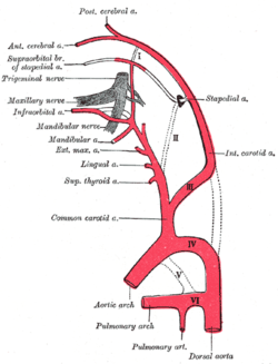
Gray474.png
The main branches of the carotid arteries, the stapedial artery is visible in the top right in grey.
Specialty Otolaryngology, angiology
Symptoms Pulsatile tinnitus, conductive hearing loss
Usual onsetFrom birth
Diagnostic method CT scan, angiography, radiography
Treatment Coagulation, hearing aids
FrequencyExtremely rare

A persistent stapedial artery (PSA) is a rare anomaly in human anatomy where the stapedial branch of posterior auricular artery, or simply stapedial artery, remains within the ear of a fetus after the first ten weeks of pregnancy. Whilst not problematic for the majority of people with the anomaly, it can cause difficulties with hearing.

Contents

Signs and symptoms

Most cases of PSA cases remain asymptomatic for a person's life; [1] however, symptoms can include vertigo, [2] pulsatile tinnitus, conductive hearing loss or sensorineural hearing loss via bone erosion of the otic capsule in rare cases. [3] Hearing loss in children can also cause developmental delays. [4]

Cause

During pregnancy, the stapedial artery originates as a branch of the hyoid artery in the dorsal branch of the second aortic arch, which is in turn connected to the internal carotid artery. The stapedial artery after passing through the stapes splits into two vessels, the supraorbital branch and the maxillomandibular branch, the latter of which has a smaller mandibular branch. The former later anastomoses with the opthalmic artery and becomes the middle meningeal artery, the latter anastomoses with the internal maxillary branch of the external carotid artery, becoming the inferior alveolar artery and infraorbital artery, respectively. [3]

Diagnosis

Since most cases of PSA do not present with symptoms, it is usually discovered incidentally upon middle ear surgery, or during postmortem temporal bone dissections. [5] The absence of the foramen spinosum is sometimes associated with a PSA, [6] although the prevalence of a missing foramen spinosum is much higher than the prevalence of a PSA. [7] On the other hand, a foramen spinosum may be present if the maxillomandibular artery originates from the stapedial artery. [8] A differential diagnosis can help eliminate other possible conditions such as glomus tumour of the tympanicum, facial nerve schwannoma, and aberrant internal carotid artery (ICA) based on results of high-resolution computed tomograms, angiograms, or magnetic resonance angiograms. [9] An aberrant ICA refers to when agenesis of the cervical portion of the ICA occurs, causing the inferior tympanic artery to anastomose with the caroticotympanic artery. This causes the internal carotid artery to enter the middle ear through the same canal as the tympanic nerve, rather than the normal carotid canal. [10] Aberrant internal carotid arteries are often found alongside a PSA, [1] [11] although both anomalies can occur independent of each other. [10]

Treatment

An electrocoagulation machine Apparaat voor electrocoagulatie 2.jpg
An electrocoagulation machine

Treatment of PSA usually involves some form of coagulation, either laser coagulation or electrocoagulation. [12] Palliative care such as the installation of hearing aids can help resolve hearing loss caused by a PSA; [13] conversely, the PSA may prevent the insertion of cochlear implants meant to alleviate the hearing loss. [14] Ablation of the PSA is also an option, although this treatment carries the risk of potential facial nerve damage and haemorrhage. [13]

The central hospital of the University of Porto reported in 2016 the first successful implementation of a bone-anchored hearing aid in an 8-year-old patient without complications, successfully resolving their hearing loss. [4] Further successful insertions of cochlear implants to resolve hearing loss have also been reported. [15]

Epidemiology

The prevalence of persistent stapedial arteries is thought to be somewhere between 0.02 to 0.48% of the general population; [16] [N 1] a study in the American Journal of Radiology in 2000 stated that only fifty-six cases of PSA had been reported since the first report of PSA was published in 1836 by Austrian anatomist Josef Hyrtl. [1]

Notes

  1. 0.02% by incidence during surgery, 0.48% histopathologically. [16] [17]

Related Research Articles

<i>Stapes</i> Bone in the middle ear

The stapes or stirrup is a bone in the middle ear of humans and other animals which is involved in the conduction of sound vibrations to the inner ear. This bone is connected to the oval window by its annular ligament, which allows the footplate to transmit sound energy through the oval window into the inner ear. The stapes is the smallest and lightest bone in the human body, and is so-called because of its resemblance to a stirrup.

Tinnitus is a variety of sound that is heard when no corresponding external sound is present. Nearly everyone experiences faint "normal tinnitus" in a completely quiet room; but it is of concern only if it is bothersome, interferes with normal hearing, or is associated with other problems. The word tinnitus comes from the Latin tinnire, "to ring". In some people, it interferes with concentration, and can be associated with anxiety and depression.

<span class="mw-page-title-main">Brachiocephalic artery</span> Artery of the mediastinum

The brachiocephalic artery is an artery of the mediastinum that supplies blood to the right arm and the head and neck. It was previously known as the innominate artery, meaning unnamed artery.

<span class="mw-page-title-main">Cochlear implant</span> Prosthesis

A cochlear implant (CI) is a surgically implanted neuroprosthesis that provides a person who has moderate-to-profound sensorineural hearing loss with sound perception. With the help of therapy, cochlear implants may allow for improved speech understanding in both quiet and noisy environments. A CI bypasses acoustic hearing by direct electrical stimulation of the auditory nerve. Through everyday listening and auditory training, cochlear implants allow both children and adults to learn to interpret those signals as speech and sound.

<span class="mw-page-title-main">Ear</span> Organ of hearing and balance

An ear is the organ that enables hearing and body balance using the vestibular system. In mammals the ear is usually described as having three parts: the outer ear, the middle ear and the inner ear. The outer ear consists of the pinna and the ear canal. Since the outer ear is the only visible portion of the ear in most animals, the word "ear" often refers to the external part alone. The middle ear includes the tympanic cavity and the three ossicles. The inner ear sits in the bony labyrinth, and contains structures which are key to several senses: the semicircular canals, which enable balance and eye tracking when moving; the utricle and saccule, which enable balance when stationary; and the cochlea, which enables hearing. The ear is a self cleaning organ through its relationship with earwax and the ear canals. The ears of vertebrates are placed somewhat symmetrically on either side of the head, an arrangement that aids sound localization.

<span class="mw-page-title-main">Cerebral angiography</span> Angiography that produces images of blood vessels in and around the brain

Cerebral angiography is a form of angiography which provides images of blood vessels in and around the brain, thereby allowing detection of abnormalities such as arteriovenous malformations and aneurysms. It was pioneered in 1927 by the Portuguese neurologist Egas Moniz at the University of Lisbon, who also helped develop thorotrast for use in the procedure.

Unilateral hearing loss (UHL) is a type of hearing impairment where there is normal hearing in one ear and impaired hearing in the other ear.

<span class="mw-page-title-main">Otology</span> Branch of medicine for the ear

Otology is a branch of medicine which studies normal and pathological anatomy and physiology of the ear as well as their diseases, diagnosis and treatment. Otologic surgery generally refers to surgery of the middle ear and mastoid related to chronic otitis media, such as tympanoplasty, or ear drum surgery, ossiculoplasty, or surgery of the hearing bones, and mastoidectomy. Otology also includes surgical treatment of conductive hearing loss, such as stapedectomy surgery for otosclerosis.

<span class="mw-page-title-main">The House Institute Foundation</span> Californian non-profit to advance hearing science

The House Institute Foundation (HIF), formerly the House Ear Institute, is a non-profit 501(c)(3) organization, based in Los Angeles, California, and dedicated to advancing hearing science through research, education, and global hearing health to improve quality of life.

<span class="mw-page-title-main">Anterior inferior cerebellar artery</span> Major blood supply to the cerebellum

The anterior inferior cerebellar artery (AICA) is one of three pairs of arteries that supplies blood to the cerebellum.

<span class="mw-page-title-main">Aortic arches</span>

The aortic arches or pharyngeal arch arteries are a series of six paired embryological vascular structures which give rise to the great arteries of the neck and head. They are ventral to the dorsal aorta and arise from the aortic sac.

<span class="mw-page-title-main">Aberrant subclavian artery</span> Medical condition

Aberrant subclavian artery, or aberrant subclavian artery syndrome, is a rare anatomical variant of the origin of the right or left subclavian artery. This abnormality is the most common congenital vascular anomaly of the aortic arch, occurring in approximately 1% of individuals.

<span class="mw-page-title-main">Deep temporal arteries</span> Artery of the head

The deep temporal arteries are two arteries of the head. They ascend between the temporalis muscle and the pericranium. They anastomose with the middle temporal artery, among other vessels. They supply the temporalis muscle.

The caroticotympanic artery is a small, sometimes doubled artery which arises from the internal carotid artery. It leaves the carotid canal through a foramen to reach the tympanic cavity. It contributes arterial supply to the osseous part of the pharyngotympanic tube.

In human anatomy, the stapedial branch of posterior auricular artery, or stapedial artery for short, is a small artery supplying the stapedius muscle in the inner ear.

<span class="mw-page-title-main">Leptomeningeal collateral circulation</span>

The leptomeningeal collateral circulation is a network of small blood vessels in the brain that connects branches of the middle, anterior and posterior cerebral arteries, with variation in its precise anatomy between individuals. During a stroke, leptomeningeal collateral vessels allow limited blood flow when other, larger blood vessels provide inadequate blood supply to a part of the brain.

<span class="mw-page-title-main">Tympanosclerosis</span> Medical condition

Tympanosclerosis is a condition caused by hyalinization and subsequent calcification of subepithelial connective tissue of the tympanic membrane and middle ear, sometimes resulting in a detrimental effect to hearing.

<span class="mw-page-title-main">Panoramic radiograph</span> Type of X-ray

A panoramic radiograph is a panoramic scanning dental X-ray of the upper and lower jaw. It shows a two-dimensional view of a half-circle from ear to ear. Panoramic radiography is a form of focal plane tomography; thus, images of multiple planes are taken to make up the composite panoramic image, where the maxilla and mandible are in the focal trough and the structures that are superficial and deep to the trough are blurred.

<span class="mw-page-title-main">Trigeminal artery</span>

The trigeminal artery is an artery that supplies the basilar artery with blood during human embryonic development. Normally, the trigeminal artery involutes after the formation of the posterior communicating artery. However, in some cases, the artery persists into adulthood and can cause medical complications, including intracranial aneurysms.

<span class="mw-page-title-main">Michel aplasia</span> Medical condition

Michel aplasia, also known as complete labyrinthine aplasia (CLA), is a congenital abnormality of the inner ear. It is characterized by the bilateral absence of differentiated inner ear structures and results in complete deafness (anacusis). Michel aplasia should not be confused with michel dysplasia. It may affect one or both ears.

References

  1. 1 2 3 Silbergleit R, Quint DJ, Mehta BA, et al. (March 2000). "The Persistent Stapedial Artery". American Journal of Radiology . 21 (3): 572–577. PMC   8174972 . PMID   10730654.
  2. Sanjuan M, Chapon F, Magnan J (August 2020). "An atypical stapedial artery". The Journal of International Advanced Otology . 16 (2): 274–277. doi: 10.5152/iao.2019.4002 . PMC   7419084 . PMID   32510458.
  3. 1 2 Tien HC, Linthicum FH (November 2001). "Persistent Stapedial Artery". Otology & Neurotology . 22 (6): 975–976. doi:10.1097/00129492-200111000-00044. PMID   11698828.
  4. 1 2 Santos M, Esteves SS, Pinto A, et al. (15 December 2016). "Clinical Experience of bone-anchored hearing aid in a patient with Persistent Stapedial Artery". Acta Otorrinolaringológica . 9 (1): 140–145. ISSN   2340-3438.
  5. Bonasia S, Smajda S, Ciccio G, et al. (September 2020). "Stapedial Artery: From Embryology to Different Possible Adult Configurations". American Journal of Neuroradiology . 41 (10): 1768–1776. doi: 10.3174/ajnr.A6738 . PMC   7661070 . PMID   32883664.
  6. Guinto FC, Garrabrant EC, Radcliffe WB (November 1972). "Radiology of the Persistent Stapedial Artery". Radiology . 105 (2): 365–369. doi:10.1148/105.2.365. PMID   5079662.
  7. Klostranec JM, Krings T (2022). "Cerebral neurovascular embryology, anatomic variations, and congenital brain arteriovenous lesions". Journal of NeuroInterventional Surgery . 14 (9): 910–919. doi: 10.1136/neurintsurg-2021-018607 . PMID   35169032. S2CID   246829491.
  8. Hitier M, Zhang M, Labrousse M, et al. (2013). "Persistent stapedial arteries in human: from phylogeny to surgical consequences". Surgical and Radiologic Anatomy . 35 (10): 883–891. doi:10.1007/s00276-013-1127-z. PMID   23640742. S2CID   21065191.
  9. Yilmaz T, Bilgen C, Savas R, Alper H (June 2003). "Persistent Stapedial Artery: MR Angiographic and CT Findings". American Journal of Neuroradiology . 24 (6): 1133–1135. PMC   8148996 . PMID   12812939.
  10. 1 2 Hatipoglu HG, Cetin MA, Yuksel E, et al. (May 2011). "A case of a coexisting aberrant internal carotid artery and persistent stapedial artery: The role of MR angiography in the diagnosis". Ear, Nose & Throat Journal . 90 (5): 17–20. doi: 10.1177/014556131109000513 . PMID   21563075.
  11. Sullivan AM, Curtin HD, Moonis G (February 2019). "Arterial Anomalies of the Middle Ear: A Pictorial Review with Clinical-Embryologic and Imaging Correlation". Neuroimaging Clinics of North America . 29 (1): 93–102. doi:10.1016/j.nic.2018.09.010. PMID   30466646. S2CID   53716332.
  12. Quarte R, Manipoud P, Schmerber S (June 2019). "Persistent stapedial artery in PHACE syndrome". European Annals of Otorhinolaryngology, Head and Neck Diseases . 136 (3): 215–217. doi: 10.1016/j.anorl.2019.02.015 . PMID   30876851.
  13. 1 2 Hill FC, Teh B, Tykocinski M (2018). "Persistent Stapedial Artery with Ankylosis of the Stapes Footplate". Ear, Nose & Throat Journal. 97 (7): 227–228. doi: 10.1177/014556131809700702 . PMID   30036438.
  14. Wardrop P, Kerr AI, Moussa SA (September 1995). "Persistent stapedial artery preventing successful cochlear implantation: a case report". The Annals of Otology, Rhinology, and Laryngology . 166: 443–445. PMID   7668745.
  15. Jones H, Hintze J, Gendre A, et al. (2022). "Persistent Stapedial Artery Encountered during Cochlear Implantation". Case Reports in Otolaryngology . 2022: 1–3. doi: 10.1155/2022/8179062 . PMC   8888055 . PMID   35242393.
  16. 1 2 Moreano EH, Paparella MM, Zelterman D, et al. (2 March 1993). "Prevalence of facial canal dehiscence and of persistent stapedial artery in the human middle ear: A report of 1000 temporal bones". The Laryngoscope . 104 (3 Pt 1): 309–320. doi:10.1288/00005537-199403000-00012. PMID   8127188. S2CID   10575546.
  17. Jehl J, Jeunet L, Berraiah M, et al. (2006). "Bilateral Persistent Pharyngo-Stapedial Arteries Revealed during Evaluation of a Carotid-Cavernous Fistula". Interventional Neuroradiology . 12 (4): 327–334. doi:10.1177/159101990601200406. PMC   3354603 . PMID   20569590.