Photostimulation

Last updated

Photostimulation is the use of light to artificially activate biological compounds, cells, tissues, or even whole organisms. Photostimulation can be used to noninvasively probe various relationships between different biological processes, using only light. In the long run, photostimulation has the potential for use in different types of therapy, such as migraine headache. Additionally, photostimulation may be used for the mapping of neuronal connections between different areas of the brain by “uncaging” signaling biomolecules with light. [1] Therapy with photostimulation has been called light therapy, phototherapy, or photobiomodulation.

Contents

ATP(1) can be inactivated until photolysis by the addition of a caging group(2). Likewise, the active site of cAMP(3) can be inactivated by the addition of a caging group(4). Photostimulation of cAMP.png
ATP(1) can be inactivated until photolysis by the addition of a caging group(2). Likewise, the active site of cAMP(3) can be inactivated by the addition of a caging group(4).

Photostimulation methods fall into two general categories: one set of methods uses light to uncage a compound that then becomes biochemically active, binding to a downstream effector. For example, uncaging glutamate is useful for finding excitatory connections between neurons, since the uncaged glutamate mimics the natural synaptic activity of one neuron impinging upon another. The other major photostimulation method is the use of light to activate a light-sensitive protein such as rhodopsin, which can then excite the cell expressing the opsin.

Scientists have long postulated the need to control one type of cell while leaving those surrounding it untouched and unstimulated. Well-known scientific advancements such as the use of electrical stimuli and electrodes have succeeded in neural activation but fail to achieve the aforementioned goal because of their imprecision and inability to distinguish between different cell types. [2] The use of optogenetics (artificial cell activation via the use of light stimuli) is unique in its ability to deliver light pulses in a precise and timely fashion. Optogenetics is somewhat bidirectional in its ability to control neurons. Channels can be either depolarized or hyperpolarized depending on the wavelength of light that targets them. [3] For instance, the technique can be applied to channelrhodopsin cation channels to initiate neuronal depolarization and eventually activation upon illumination. Conversely, activity inhibition of a neuron can be triggered via the use of optogenetics as in the case of the chloride pump halorhodopsin which functions to hyperpolarize neurons. [3]

Before optogenetics can be performed, however, the subject at hand must express the targeted channels. Natural and abundant in microbials, rhodopsins—including bacteriorhodopsin, halorhodopsin and channelrhodopsin—each have a different characteristic action spectrum which describes the set of colors and wavelengths that they respond to and are driven to function by. [4]

It has been shown that channelrhodopsin-2, a monolithic protein containing a light sensor and a cation channel, provides electrical stimulation of appropriate speed and magnitude to activate neuronal spike firing. Recently, photoinhibition, the inhibition of neural activity with light, has become feasible with the application of molecules such as the light-activated chloride pump halorhodopsin to neural control. Together, blue-light activated channelrhodopsin-2 and the yellow light-activated chloride pump halorhodopsin enable multiple-color, optical activation and silencing of neural activity. (See also Photobiomodulation)

Methods

A caged protein is a protein that is activated in the presence of a stimulating light source. In most cases, photo-uncaging is the technique revealing the active region of a compound by the process of photolysis of the shielding molecule (‘cage’). However, uncaging the protein requires an appropriate wavelength, intensity, and timing of the light. Achieving this is possible due to the fact that the optical fiber may be modified to deliver specific amounts of light. In addition, short bursts of stimulation allow results similar to the physiological norm. The steps of photostimulation are time independent in that protein delivery and light activation can be done at different times. This is because the two steps are dependent on each other for activation of the protein. [5]

Some proteins are innately photosensitive and function in the presence of light. Proteins known as opsins form the crux of the photosensitive proteins. These proteins are often found in the eye. In addition, many of these proteins function as ion channels and receptors. One example is when a certain wavelength of light is put onto certain channels, the blockage in the pore is relieved and allows ion transduction. [6]

To uncage molecules, a photolysis system is required to cleave the covalent bond. An example system can consist of a light source (generally a laser or a lamp), a controller for the amount of light that enters, a guide for the light, and a delivery system. Often, the design function in such a way that a medium is met between the diffusing light that may cause additional, unwanted photolysis and light attenuation; both being significant problems with a photolysis system. [5]

History

The idea of photostimulation as a method of controlling biomolecule function was developed in the 1970s. Two researchers, Walther Stoeckenius and Dieter Oesterhelt discovered an ion pump known as bacteriorhodopsin which functions in the presence of light in 1971. [7] In 1978, J.F. Hoffman invented the term “caging”. Unfortunately, this term caused some confusion among scientists due to the fact that the term is often used to describe a molecule which is trapped within another molecule. It could also be confused with the “caged effect” in the recombination of radicals. Therefore, some authors decided to use the term “light-activated” instead of “caging”. Both terms are currently in use. The first “caged molecule” synthesized by Hoffman et al. at Yale was the caged precursor to ATP derivative 1. [8]

Applications

Photostimulation is notable for its temporal precision, which may be used to obtain an accurate starting time of activation of caged effectors. In conjunction with caged inhibitors, the role of biomolecules at specific timepoints in an organism's lifecycle may be studied. A caged inhibitor of N-ethylmaleimide sensitive fusion protein (NSF), a key mediator of synaptic transmission, has been used to study the time dependency of NSF. [9] Several other studies have effected action potential firing through use of caged neurotransmitters such as glutamate. [10] [11] Caged neurotransmitters, including photolable precursors of glutamate, dopamine, serotonin, and GABA, are commercially available. [12]

Signaling during mitosis has been studied using reporter molecules with a caged fluorophore, which is not phosphorylated if photolysis has not occurred. [13] The advantage of this technique is that it provides a “snapshot” of kinase activity at specific timepoints rather than recording all activity since the reporter's introduction.

Calcium ions play an important signaling role, and controlling their release with caged channels has been extensively studied. [14] [15] [16]

Unfortunately, not all organisms produce or hold sufficient amounts of opsins. Thus, the opsin gene must be introduced to target neurons if they are not already present in the organism of study. The addition and expression of this gene is sufficient for the use of optogenetics. Possible means of achieving this include the construction of transgenic lines containing the gene or acute gene transfer to a specific area or region within an individual. These methods are known as germline transgenesis and somatic gene delivery, respectively. [17]

Optogenetics has shown significant promise in the treatment of a series of neurological disorders such as Parkinson's disease and epilepsy. Optogenetics has the potential to facilitate the manipulation and targeting of specific cell types or neural circuits, characteristics that are lacking in current brain stimulation techniques like DBS. At this point, the use of optogenetics in treating neural diseases has only been practically implemented in the field of neurobiology to reveal more about the mechanisms of specific disorders. Before the technique can be implemented to directly treat these disorders developments in other related fields such as gene therapy, opsin engineering, and optoelectronics must also make certain developments. [18]

Related Research Articles

<span class="mw-page-title-main">Neuron</span> Electrically excitable cell found in the nervous system of animals

Within a nervous system, a neuron, neurone, or nerve cell is an electrically excitable cell that fires electric signals called action potentials across a neural network. Neurons communicate with other cells via synapses - specialized connections that commonly use minute amounts of chemical neurotransmitters to pass the electric signal from the presynaptic neuron to the target cell through the synaptic gap. The neuron is the main component of nervous tissue in all animals except sponges and placozoa. Non-animals like plants and fungi do not have nerve cells.

<span class="mw-page-title-main">Photoreceptor cell</span> Type of neuroepithelial cell

A photoreceptor cell is a specialized type of neuroepithelial cell found in the retina that is capable of visual phototransduction. The great biological importance of photoreceptors is that they convert light into signals that can stimulate biological processes. To be more specific, photoreceptor proteins in the cell absorb photons, triggering a change in the cell's membrane potential.

<span class="mw-page-title-main">Guanylate cyclase</span> Lyase enzyme that synthesizes cGMP from GTP

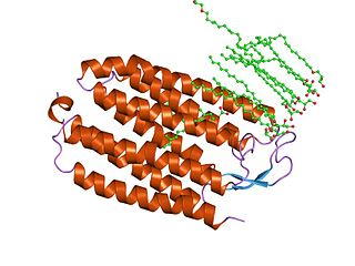
Guanylate cyclase is a lyase enzyme that converts guanosine triphosphate (GTP) to cyclic guanosine monophosphate (cGMP) and pyrophosphate:

<span class="mw-page-title-main">Behavioral neuroscience</span> Field of study

Behavioral neuroscience, also known as biological psychology, biopsychology, or psychobiology, is the application of the principles of biology to the study of physiological, genetic, and developmental mechanisms of behavior in humans and other animals.

Channelrhodopsins are a subfamily of retinylidene proteins (rhodopsins) that function as light-gated ion channels. They serve as sensory photoreceptors in unicellular green algae, controlling phototaxis: movement in response to light. Expressed in cells of other organisms, they enable light to control electrical excitability, intracellular acidity, calcium influx, and other cellular processes. Channelrhodopsin-1 (ChR1) and Channelrhodopsin-2 (ChR2) from the model organism Chlamydomonas reinhardtii are the first discovered channelrhodopsins. Variants that are sensitive to different colors of light or selective for specific ions have been cloned from other species of algae and protists.

<span class="mw-page-title-main">Halorhodopsin</span> Family of transmembrane proteins


Halorhodopsin is a seven-transmembrane retinylidene protein from microbial rhodopsin family. It is a chloride-specific light-gated ion pump found in archaea known as halobacteria. It is activated by green light wavelengths of approximately 578nm. Halorhodopsin also shares sequence similarity to channelrhodopsin, another light-driven ion channel.

Retinylidene proteins, or rhodopsins in a broad sense, are proteins that use retinal as a chromophore for light reception. They are the molecular basis for a variety of light-sensing systems from phototaxis in flagellates to eyesight in animals. Retinylidene proteins include all forms of opsin and rhodopsin. While rhodopsin in the narrow sense refers to a dim-light visual pigment found in vertebrates, usually on rod cells, rhodopsin in the broad sense refers to any molecule consisting of an opsin and a retinal chromophore in the ground state. When activated by light, the chromophore is isomerized, at which point the molecule as a whole is no longer rhodopsin, but a related molecule such as metarhodopsin. However, it remains a retinylidene protein. The chromophore then separates from the opsin, at which point the bare opsin is a retinylidene protein. Thus, the molecule remains a retinylidene protein throughout the phototransduction cycle.

Light-gated ion channels are a family of ion channels regulated by electromagnetic radiation. Other gating mechanisms for ion channels include voltage-gated ion channels, ligand-gated ion channels, mechanosensitive ion channels, and temperature-gated ion channels. Most light-gated ion channels have been synthesized in the laboratory for study, although two naturally occurring examples, channelrhodopsin and anion-conducting channelrhodopsin, are currently known. Photoreceptor proteins, which act in a similar manner to light-gated ion channels, are generally classified instead as G protein-coupled receptors.

<span class="mw-page-title-main">Gero Miesenböck</span>

Gero Andreas Miesenböck is an Austrian scientist. He is currently Waynflete Professor of Physiology and Director of the Centre for Neural Circuits and Behaviour (CNCB) at the University of Oxford and a fellow of Magdalen College, Oxford.

<span class="mw-page-title-main">OPN3</span> Protein-coding gene in the species Homo sapiens

Opsin-3 also known as encephalopsin or panopsin is a protein that, in humans, is encoded by the OPN3 gene. Alternative splicing of this gene results in multiple transcript variants encoding different protein isoforms.

Optogenetics is a biological technique to control the activity of neurons or other cell types with light. This is achieved by expression of light-sensitive ion channels, pumps or enzymes specifically in the target cells. On the level of individual cells, light-activated enzymes and transcription factors allow precise control of biochemical signaling pathways. In systems neuroscience, the ability to control the activity of a genetically defined set of neurons has been used to understand their contribution to decision making, learning, fear memory, mating, addiction, feeding, and locomotion. In a first medical application of optogenetic technology, vision was partially restored in a blind patient.

<span class="mw-page-title-main">Karl Deisseroth</span> American optogeneticist (born 1971)

Karl Alexander Deisseroth is an American scientist. He is the D.H. Chen Professor of Bioengineering and of psychiatry and behavioral sciences at Stanford University.

<span class="mw-page-title-main">Photoactivatable probes</span> Cellular players that can be activated by light

Photoactivatable probes, or caged probes, are cellular players that can be triggered by a flash of light. They are used in biological research to study processes in cells. The basic principle is to bring a photoactivatable agent to cells, tissues or even living animals and specifically control its activity by illumination.

Gene therapy is being studied for some forms of epilepsy. It relies on viral or non-viral vectors to deliver DNA or RNA to target brain areas where seizures arise, in order to prevent the development of epilepsy or to reduce the frequency and/or severity of seizures. Gene therapy has delivered promising results in early stage clinical trials for other neurological disorders such as Parkinson's disease, raising the hope that it will become a treatment for intractable epilepsy.

Chemogenetics is the process by which macromolecules can be engineered to interact with previously unrecognized small molecules. Chemogenetics as a term was originally coined to describe the observed effects of mutations on chalcone isomerase activity on substrate specificities in the flowers of Dianthus caryophyllus. This method is very similar to optogenetics; however, it uses chemically engineered molecules and ligands instead of light and light-sensitive channels known as opsins.

<span class="mw-page-title-main">Peter Hegemann</span> German biophysicist

Peter Hegemann is a Hertie Senior Research Chair for Neurosciences and a Professor of Experimental Biophysics at the Department of Biology, Faculty of Life Sciences, Humboldt University of Berlin, Germany. He is known for his discovery of channelrhodopsin, a type of ion channels regulated by light, thereby serving as a light sensor. This created the field of optogenetics, a technique that controls the activities of specific neurons by applying light. He has received numerous accolades, including the Rumford Prize, the Shaw Prize in Life Science and Medicine, and the Albert Lasker Award for Basic Medical Research.

<span class="mw-page-title-main">Microbial rhodopsin</span> Retinal-binding proteins

Microbial rhodopsins, also known as bacterial rhodopsins, are retinal-binding proteins that provide light-dependent ion transport and sensory functions in halophilic and other bacteria. They are integral membrane proteins with seven transmembrane helices, the last of which contains the attachment point for retinal.

<span class="mw-page-title-main">Anion-conducting channelrhodopsin</span> Class of light-gated ion channels

Anion-conducting channelrhodopsins are light-gated ion channels that open in response to light and let negatively charged ions enter a cell. All channelrhodopsins use retinal as light-sensitive pigment, but they differ in their ion selectivity. Anion-conducting channelrhodopsins are used as tools to manipulate brain activity in mice, fruit flies and other model organisms (Optogenetics). Neurons expressing anion-conducting channelrhodopsins are silenced when illuminated with light, an effect that has been used to investigate information processing in the brain. For example, suppressing dendritic calcium spikes in specific neurons with light reduced the ability of mice to perceive a light touch to a whisker. Studying how the behavior of an animal changes when specific neurons are silenced allows scientists to determine the role of these neurons in the complex circuits controlling behavior.

<span class="mw-page-title-main">Georg Nagel</span>

Georg Nagel is a biophysicist and professor at the Department for Neurophysiology at the University of Würzburg in Germany. His research is focused on microbial photoreceptors and the development of optogenetic tools.

Lisa Gunaydin is an American neuroscientist and assistant professor at the Weill Institute for Neurosciences at the University of California San Francisco. Gunaydin helped discover optogenetics in the lab of Karl Deisseroth and now uses this technique in combination with neural and behavioral recordings to probe the neural circuits underlying emotional behaviors.

References

  1. Marina de Tommaso; Daniele Marinazzo; Luigi Nitti; Mario Pellicoro; Marco Guido; Claudia Serpino; Sebastiano Stramaglia (2007). "Effects of levetiracetam vs topiramate and placebo on visually evoked phase synchronization changes of alpha rhythm in migraine". Clinical Neurophysiology. 118 (10): 2297–2304. doi:10.1016/j.clinph.2007.06.060. hdl: 1854/LU-8697646 . PMID   17709295. S2CID   20094637.
  2. Deisseroth, Karl. "Optogenetics: Controlling the Brain with Light [Extended Version]". Scientific American. Retrieved 2017-10-14.
  3. 1 2 LaLumiere, Ryan T. (2011-01-01). "A new technique for controlling the brain: optogenetics and its potential for use in research and the clinic". Brain Stimulation. 4 (1): 1–6. doi:10.1016/j.brs.2010.09.009. PMID   21255749. S2CID   3256131.
  4. Chow, Brian Y.; Han, Xue; Boyden, Edward S. (2012). Genetically encoded molecular tools for light-driven silencing of targeted neurons. Progress in Brain Research. Vol. 196. pp. 49–61. doi:10.1016/B978-0-444-59426-6.00003-3. ISBN   9780444594266. ISSN   0079-6123. PMC   3553588 . PMID   22341320.
  5. 1 2 G. W. Godwin; D. Che; D. M. O'Malley; Q. Zhou (1996). "Photostimulation with caged neurotransmitters using fiber optic lightguides". Neuroscience Methods. 93 (1): 91–106. doi:10.1016/s0165-0270(96)02208-x. PMID   9130682. S2CID   35862919.
  6. G. Sandoz; J. Levitz (2013). "Optogenetic techniques for the study of native potassium channels". Frontiers in Molecular Neuroscience. 6: 6. doi: 10.3389/fnmol.2013.00006 . PMC   3622882 . PMID   23596388.
  7. Karl Deisseroth (2011). "Optogenetics". Nature Methods. 8 (1): 26–29. doi:10.1038/nmeth.f.324. PMC   6814250 . PMID   21191368.
  8. G_nter Mayer, Alexander Heckel (2006). "Biologically Active Molecules with a "Light Switch"". Angew. Chem. 45 (30): 4900–4921. doi:10.1002/anie.200600387. PMID   16826610.
  9. Kuner T, Li Y, Gee KR, Bonewald LF, Augustine GJ (2008). "Photolysis of a caged peptide reveals rapid action of N-ethylmaleimide sensitive factor before neurotransmitter release". Proc. Natl. Acad. Sci. U.S.A. 105 (1): 347–352. doi: 10.1073/pnas.0707197105 . PMC   2224215 . PMID   18172208.
  10. E. M. Callaway; R. Yuste (2002). "Stimulating neurons with light". Current Opinion in Neurobiology. 12 (5): 587–592. doi:10.1016/s0959-4388(02)00364-1. PMID   12367640. S2CID   18176577.
  11. G. Dormán; G. D. Prestwich (2000). "Using photolabile ligands in drug discovery and development". Trends Biotechnol. 18 (2): 64–77. doi:10.1016/s0167-7799(99)01402-x. PMID   10652511.
  12. "Caged Compounds | Photolysis".
  13. Dai Z, Dulyaninova NG, Kumar S, Bresnick AR, Lawrence DS (2007). "Visual snapshots of intracellular kinase activity at the onset of mitosis". Chemistry & Biology. 14 (11): 1254–1260. doi:10.1016/j.chembiol.2007.10.007. PMC   2171364 . PMID   18022564.
  14. Ellis-Davies G C (2007). "Caged compounds: photorelease technology for control of cellular chemistry and physiology". Nat. Methods. 4 (8): 619–628. doi:10.1038/nmeth1072. PMC   4207253 . PMID   17664946.
  15. Nikolenko V, Poskanzer KE, Yuste R (2007). "Two-photon photostimulation and imaging of neural circuits". Nat. Methods. 4 (11): 943–950. doi:10.1038/nmeth1105. PMID   17965719. S2CID   1421280.
  16. Neveu P, Aujard I, Benbrahim C, Le Saux T, Allemand JF, Vriz S, Bensimon D, Jullien L (2008). "A caged retinoic acid for one- and two-photon excitation in zebrafish embryos". Angew. Chem. Int. Ed. 47 (20): 3744–3746. doi:10.1002/anie.200800037. PMID   18399559.
  17. Dugué, Guillaume P.; Akemann, Walther; Knöpfel, Thomas (2012-01-01). "A comprehensive concept of optogenetics". In Knöpfel, Thomas; Boyden, Edward S. (eds.). Optogenetics: Tools for Controlling and Monitoring Neuronal Activity. Progress in Brain Research. Vol. 196. Elsevier. pp. 1–28. doi:10.1016/B978-0-444-59426-6.00001-X. ISBN   9780444594266. PMID   22341318.
  18. Mahmoudi, Parisa; Veladi, Hadi; Pakdel, Firooz G. (2017). "Optogenetics, Tools and Applications in Neurobiology". Journal of Medical Signals and Sensors. 7 (2): 71–79. doi: 10.4103/2228-7477.205506 . ISSN   2228-7477. PMC   5437765 . PMID   28553579.