Response regulator

Last updated
Response regulator receiver domain
3CHY.png
The response regulator CheY from E. coli , with the aspartate phosphorylation site highlighted in green. From PDB: 3CHY .
Identifiers
SymbolResponse_reg
Pfam PF00072
Pfam clan CL0304
ECOD 2007.1.3
InterPro IPR001789
SMART REC
PROSITE PDOC50110
Available protein structures:
Pfam   structures / ECOD  
PDB RCSB PDB; PDBe; PDBj
PDBsum structure summary

In molecular biology, a response regulator is a protein that mediates a cell's response to changes in its environment as part of a two-component regulatory system. Response regulators are coupled to specific histidine kinases which serve as sensors of environmental changes. Response regulators and histidine kinases are two of the most common gene families in bacteria, where two-component signaling systems are very common; they also appear much more rarely in the genomes of some archaea, yeasts, filamentous fungi, and plants. Two-component systems are not found in metazoans. [1] [2] [3] [4]

Contents

Function

The crystal structure of the yeast histidine phosphotransfer protein Ypd1 in complex with the response regulator Sln1. Ypd1 appears on the right with the conserved four-helical bundle in yellow and variable helices in red. Sln1 appears on the left with beta sheets in tan and helices in brown. The key residues - Asp from Sln1 and His from Ypd1 - are highlighted with green sticks. A bound magnesium ion is shown as an orange sphere and beryllium trifluoride, a phosphoryl analog, is shown in pink. From PDB: 2R25 . 2r25.png
The crystal structure of the yeast histidine phosphotransfer protein Ypd1 in complex with the response regulator Sln1. Ypd1 appears on the right with the conserved four-helical bundle in yellow and variable helices in red. Sln1 appears on the left with beta sheets in tan and helices in brown. The key residues - Asp from Sln1 and His from Ypd1 - are highlighted with green sticks. A bound magnesium ion is shown as an orange sphere and beryllium trifluoride, a phosphoryl analog, is shown in pink. From PDB: 2R25 .

Response regulator proteins typically consist of a receiver domain and one or more effector domains, although in some cases they possess only a receiver domain and exert their effects through protein-protein interactions. In two-component signaling, a histidine kinase responds to environmental changes by autophosphorylation on a histidine residue, following which the response regulator receiver domain catalyzes transfer of the phosphate group to its own recipient aspartate residue. This induces a conformational change that alters the function of the effector domains, usually resulting in increased transcription of target genes. The mechanisms by which this occurs are diverse and include allosteric activation of the effector domain or oligomerization of phosphorylated response regulators. [2] In a common variation on this theme, called a phosphorelay, a hybrid histidine kinase possesses its own receiver domain, and a histidine phosphotransfer protein performs the final transfer to a response regulator. [4]

In many cases, histidine kinases are bifunctional and also serve as phosphatases, catalyzing the removal of phosphate from response regulator aspartate residues, such that the signal transduced by the response regulator reflects the balance between kinase and phosphatase activity. [4] Many response regulators are also capable of autodephosphorylation, which occurs on a wide range of time scales. [2] In addition, phosphoaspartate is relatively chemically unstable and may be hydrolyzed non-enzymatically. [1]

Histidine kinases are highly specific for their cognate response regulators; there is very little cross-talk between different two-component signaling systems in the same cell. [6]

Classification

Response regulators can be divided into at least three broad classes, based on the features of effector domains: regulators with a DNA-binding effector domain, regulators with an enzymatic effector domain, and single-domain response regulators. [3] More comprehensive classifications based on more detailed analysis of domain architecture are possible. Beyond these broad categorizations, there are response regulators with other types of effector domains, including RNA-binding effector domains.

Regulators with a DNA-binding effector domain are the most common response regulators, and have direct impacts on transcription. [7] They tend to interact with their cognate regulators at an N-terminus receiver domain, and contain the DNA-binding effector towards the C-terminus. Once phosphorylated at the receiver domain, the response regulator dimerizes, gains enhanced DNA binding capacity and acts as a transcription factor. [8] The architecture of DNA binding domains are characterized as being variations on helix-turn-helix motifs. One variation, found on the response regulator OmpR of the EnvZ/OmpR two-component system and other OmpR-like response regulators, is a "winged helix" architecture. [9] OmpR-like response regulators are the largest group of response regulators and the winged helix motif is widespread. Other subtypes of DNA-binding response regulators include FixJ-like and NtrC-like regulators. [10] DNA-binding response regulators are involved in various uptake processes, including nitrate/nitrite (NarL, found in most prokaryotes). [11]

The second class of multidomain response regulators are those with enzymatic effector domains. [12] These response regulators can participate in signal transduction, and generate secondary messenger molecules. Examples include the chemotaxis regulator CheB, with a methylesterase domain that is inhibited when the response regulator is in the inactive unphosphorylated conformation. Other enzymatic response regulators include c-di-GMP phosphodiesterases (e.g. VieA in V. cholerae), protein phosphatases and histidine kinases. [12]

A relatively small number of response regulators, single-domain response regulators, only contain a receiver domain, relying on protein-protein interactions to exert their downstream biological effects. [13] The receiver domain undergoes a conformational change as it interacts with an autophosphorylated histidine kinase, and consequently, the response regulator can initiate further reactions along a signaling cascade. Prominent examples include the chemotaxis regulator CheY, which interacts with flagellar motor proteins directly in its phosphorylated state. [13]

Sequencing has so far shown that the distinct classes of response regulators are unevenly distributed throughout various taxa, [14] including across domains. While response regulators with DNA-binding domains are the most common in bacteria, single-domain response regulators are more common in archaea, with other major classes of response regulators seemingly absent from archaeal genomes.

Evolution

The number of two-component systems present in a bacterial genome is highly correlated with genome size as well as ecological niche; bacteria that occupy niches with frequent environmental fluctuations possess more histidine kinases and response regulators. [4] [7] New two-component systems may arise by gene duplication or by lateral gene transfer, and the relative rates of each process vary dramatically across bacterial species. [15] In most cases, response regulator genes are located in the same operon as their cognate histidine kinase; [4] lateral gene transfers are more likely to preserve operon structure than gene duplications. [15] The small number of two-component systems present in eukaryotes most likely arose by lateral gene transfer from endosymbiotic organelles; in particular, those present in plants likely derive from chloroplasts. [4]

Related Research Articles

<span class="mw-page-title-main">Protein kinase</span> Enzyme that adds phosphate groups to other proteins

A protein kinase is a kinase which selectively modifies other proteins by covalently adding phosphates to them (phosphorylation) as opposed to kinases which modify lipids, carbohydrates, or other molecules. Phosphorylation usually results in a functional change of the target protein (substrate) by changing enzyme activity, cellular location, or association with other proteins. The human genome contains about 500 protein kinase genes and they constitute about 2% of all human genes. There are two main types of protein kinase. The great majority are serine/threonine kinases, which phosphorylate the hydroxyl groups of serines and threonines in their targets. Most of the others are tyrosine kinases, although additional types exist. Protein kinases are also found in bacteria and plants. Up to 30% of all human proteins may be modified by kinase activity, and kinases are known to regulate the majority of cellular pathways, especially those involved in signal transduction.

<span class="mw-page-title-main">Signal transduction</span> Cascade of intracellular and molecular events for transmission/amplification of signals

Signal transduction is the process by which a chemical or physical signal is transmitted through a cell as a series of molecular events. Proteins responsible for detecting stimuli are generally termed receptors, although in some cases the term sensor is used. The changes elicited by ligand binding in a receptor give rise to a biochemical cascade, which is a chain of biochemical events known as a signaling pathway.

A DNA-binding domain (DBD) is an independently folded protein domain that contains at least one structural motif that recognizes double- or single-stranded DNA. A DBD can recognize a specific DNA sequence or have a general affinity to DNA. Some DNA-binding domains may also include nucleic acids in their folded structure.

In biology, cell signaling is the process by which a cell interacts with itself, other cells, and the environment. Cell signaling is a fundamental property of all cellular life in prokaryotes and eukaryotes.

Phospholipase D (EC 3.1.4.4, lipophosphodiesterase II, lecithinase D, choline phosphatase, PLD; systematic name phosphatidylcholine phosphatidohydrolase) is an enzyme of the phospholipase superfamily that catalyses the following reaction

<span class="mw-page-title-main">Protein-glutamate methylesterase</span>

The enzyme protein-glutamate methylesterase (EC 3.1.1.61) catalyzes the reaction

In biology, an autoinducer is a signaling molecule that enables detection and response to changes in the population density of bacterial cells. Synthesized when a bacterium reproduces, autoinducers pass outside the bacterium and into the surrounding medium. They are a key component of the phenomenon of quorum sensing: as the density of quorum-sensing bacterial cells increases, so does the concentration of the autoinducer. A bacterium’s detection of an autoinducer above some minimum threshold triggers altered gene expression.

<span class="mw-page-title-main">Two-component regulatory system</span> Method of stimulus sensing and response in cells

In molecular biology, a two-component regulatory system serves as a basic stimulus-response coupling mechanism to allow organisms to sense and respond to changes in many different environmental conditions. Two-component systems typically consist of a membrane-bound histidine kinase that senses a specific environmental stimulus, and a corresponding response regulator that mediates the cellular response, mostly through differential expression of target genes. Although two-component signaling systems are found in all domains of life, they are most common by far in bacteria, particularly in Gram-negative and cyanobacteria; both histidine kinases and response regulators are among the largest gene families in bacteria. They are much less common in archaea and eukaryotes; although they do appear in yeasts, filamentous fungi, and slime molds, and are common in plants, two-component systems have been described as "conspicuously absent" from animals.

<span class="mw-page-title-main">Histidine kinase</span> Family of enzymes important in cell signaling

Histidine kinases (HK) are multifunctional, and in non-animal kingdoms, typically transmembrane, proteins of the transferase class of enzymes that play a role in signal transduction across the cellular membrane. The vast majority of HKs are homodimers that exhibit autokinase, phosphotransfer, and phosphatase activity. HKs can act as cellular receptors for signaling molecules in a way analogous to tyrosine kinase receptors (RTK). Multifunctional receptor molecules such as HKs and RTKs typically have portions on the outside of the cell that bind to hormone- or growth factor-like molecules, portions that span the cell membrane, and portions within the cell that contain the enzymatic activity. In addition to kinase activity, the intracellular domains typically have regions that bind to a secondary effector molecule or complex of molecules that further propagate signal transduction within the cell. Distinct from other classes of protein kinases, HKs are usually parts of a two-component signal transduction mechanisms in which HK transfers a phosphate group from ATP to a histidine residue within the kinase, and then to an aspartate residue on the receiver domain of a response regulator protein. More recently, the widespread existence of protein histidine phosphorylation distinct from that of two-component histidine kinases has been recognised in human cells. In marked contrast to Ser, Thr and Tyr phosphorylation, the analysis of phosphorylated Histidine using standard biochemical and mass spectrometric approaches is much more challenging, and special procedures and separation techniques are required for their preservation alongside classical Ser, Thr and Tyr phosphorylation on proteins isolated from human cells.

<span class="mw-page-title-main">Protein phosphorylation</span> Process of introducing a phosphate group on to a protein

Protein phosphorylation is a reversible post-translational modification of proteins in which an amino acid residue is phosphorylated by a protein kinase by the addition of a covalently bound phosphate group. Phosphorylation alters the structural conformation of a protein, causing it to become activated, deactivated, or otherwise modifying its function. Approximately 13,000 human proteins have sites that are phosphorylated.
<span class="mw-page-title-main">Cell surface receptor</span> Class of ligand activated receptors localized in surface of plama cell membrane

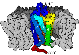
Cell surface receptors are receptors that are embedded in the plasma membrane of cells. They act in cell signaling by receiving extracellular molecules. They are specialized integral membrane proteins that allow communication between the cell and the extracellular space. The extracellular molecules may be hormones, neurotransmitters, cytokines, growth factors, cell adhesion molecules, or nutrients; they react with the receptor to induce changes in the metabolism and activity of a cell. In the process of signal transduction, ligand binding affects a cascading chemical change through the cell membrane.

The Arc system is a two-component system found in some bacteria that regulates gene expression in faculatative anaerobes such as Escheria coli. Two-component system means that it has a sensor molecule and a response regulator. Arc is an abbreviation for Anoxic Redox Control system. Arc systems are instrumental in maintaining energy metabolism during transcription of bacteria. The ArcA response regulator looks at growth conditions and expresses genes to best suit the bacteria. The Arc B sensor kinase, which is a tripartite protein, is membrane bound and can autophosphorylate.

The CHASE domain is an extracellular protein domain, which is found in transmembrane receptor from bacteria, lower eukaryotes and plants. It has been named CHASE because of its presence in diverse receptor-like proteins with histidine kinase and nucleotide cyclase domains. The CHASE domain is 200-230 amino acids long and always occurs N-terminally in extracellular or periplasmic locations, followed by an intracellular tail housing diverse enzymatic signalling domains such as histidine kinase, adenyl cyclase, GGDEF-type nucleotide cyclase and EAL-type phosphodiesterase domains, as well as non-enzymatic domains such PAS, GAF, phosphohistidine and response regulatory domains. The CHASE domain is predicted to bind diverse low molecular weight ligands, such as the cytokinin-like adenine derivatives or peptides, and mediate signal transduction through the respective receptors.

<span class="mw-page-title-main">Sda protein domain</span>

In molecular biology, the protein domain Sda is short for suppressor of dnaA or otherwise known as sporulation inhibitor A. It is found only in bacteria. This protein domain is highly important to cell survival. When starved of nutrients, the cell is under extreme stress so undergoes a series of reactions to increase the chances of survival. One method is to form endospores which can withstand a large amount of environmental pressure. Sda protein domain is a checkpoint which prevents the formation of spores. The Sda domain affects cell signalling. It prevents the cell communicating the stress that it is under, which is crucial if the cell is to survive.

<span class="mw-page-title-main">LuxR-type DNA-binding HTH domain</span>

In molecular biology, the LuxR-type DNA-binding HTH domain is a DNA-binding, helix-turn-helix (HTH) domain of about 65 amino acids. It is present in transcription regulators of the LuxR/FixJ family of response regulators. The domain is named after Vibrio fischeri luxR, a transcriptional activator for quorum-sensing control of luminescence. LuxR-type HTH domain proteins occur in a variety of organisms. The DNA-binding HTH domain is usually located in the C-terminal region of the protein; the N-terminal region often containing an autoinducer-binding domain or a response regulatory domain. Most luxR-type regulators act as transcription activators, but some can be repressors or have a dual role for different sites. LuxR-type HTH regulators control a wide variety of activities in various biological processes.

EnvZ/OmpR is a two-component regulatory system widely distributed in bacteria and particularly well characterized in Escherichia coli. Its function is in osmoregulation, responding to changes in environmental osmolality by regulating the expression of the outer membrane porins OmpF and OmpC. EnvZ is a histidine kinase which also possesses a cytoplasmic osmosensory domain, and OmpR is its corresponding response regulator protein.

<span class="mw-page-title-main">Histidine phosphotransfer domain</span>

Histidine phosphotransfer domains and histidine phosphotransferases are protein domains involved in the "phosphorelay" form of two-component regulatory systems. These proteins possess a phosphorylatable histidine residue and are responsible for transferring a phosphoryl group from an aspartate residue on an intermediate "receiver" domain, typically part of a hybrid histidine kinase, to an aspartate on a final response regulator.

A cytokinin signaling and response regulator protein is a plant protein that is involved in a two step cytokinin signaling and response regulation pathway.

<span class="mw-page-title-main">RopB transcriptional regulator</span>

RopB transcriptional regulator, also known as RopB/Rgg transcriptional regulator is a transcriptional regulator protein that regulates expression of the extracellularly secreted cysteine protease streptococcal pyrogenic exotoxin B (speB) [See Also: erythrogenic toxins] which is an important virulence factor of Streptococcus pyogenes and is responsible for the dissemination of a host of infectious diseases including strep throat, impetigo, streptococcal toxic shock syndrome, necrotizing fasciitis, and scarlet fever. Functional studies suggest that the ropB multigene regulon is responsible for not only global regulation of virulence but also a wide range of functions from stress response, metabolic function, and two-component signaling. Structural studies implicate ropB's regulatory action being reliant on a complex interaction involving quorum sensing with the leaderless peptide signal speB-inducing peptide (SIP) acting in conjunction with a pH sensitive histidine switch.

<span class="mw-page-title-main">Ethylene signaling pathway</span> Signaling pathway

Ethylene signaling pathway is a signal transduction in plant cells to regulate important growth and developmental processes. Acting as a plant hormone, the gas ethylene is responsible for promoting the germination of seeds, ripening of fruits, the opening of flowers, the abscission of leaves and stress responses. It is the simplest alkene gas and the first gaseous molecule discovered to function as a hormone.

References

  1. 1 2 Stock AM, Robinson VL, Goudreau PN (2000). "Two-component signal transduction". Annual Review of Biochemistry. 69: 183–215. doi:10.1146/annurev.biochem.69.1.183. PMID   10966457.
  2. 1 2 3 West AH, Stock AM (June 2001). "Histidine kinases and response regulator proteins in two-component signaling systems". Trends in Biochemical Sciences. 26 (6): 369–76. doi:10.1016/s0968-0004(01)01852-7. PMID   11406410.
  3. 1 2 Galperin MY (June 2005). "A census of membrane-bound and intracellular signal transduction proteins in bacteria: bacterial IQ, extroverts and introverts". BMC Microbiology. 5: 35. doi: 10.1186/1471-2180-5-35 . PMC   1183210 . PMID   15955239.
  4. 1 2 3 4 5 6 Capra EJ, Laub MT (2012). "Evolution of two-component signal transduction systems". Annual Review of Microbiology. 66: 325–47. doi:10.1146/annurev-micro-092611-150039. PMC   4097194 . PMID   22746333.
  5. Zhao X, Copeland DM, Soares AS, West AH (January 2008). "Crystal structure of a complex between the phosphorelay protein YPD1 and the response regulator domain of SLN1 bound to a phosphoryl analog". Journal of Molecular Biology. 375 (4): 1141–51. doi:10.1016/j.jmb.2007.11.045. PMC   2254212 . PMID   18076904.
  6. Rowland MA, Deeds EJ (April 2014). "Crosstalk and the evolution of specificity in two-component signaling". Proceedings of the National Academy of Sciences of the United States of America. 111 (15): 5550–5. Bibcode:2014PNAS..111.5550R. doi: 10.1073/pnas.1317178111 . PMC   3992699 . PMID   24706803.
  7. 1 2 Galperin MY (June 2006). "Structural classification of bacterial response regulators: diversity of output domains and domain combinations". Journal of Bacteriology. 188 (12): 4169–82. doi:10.1128/jb.01887-05. PMC   1482966 . PMID   16740923.
  8. Barbieri CM, Wu T, Stock AM (May 2013). "Comprehensive analysis of OmpR phosphorylation, dimerization, and DNA binding supports a canonical model for activation". Journal of Molecular Biology. 425 (10): 1612–26. doi:10.1016/j.jmb.2013.02.003. PMC   3646996 . PMID   23399542.
  9. Kenney, Linda J (2002-04-01). "Structure/function relationships in OmpR and other winged-helix transcription factors". Current Opinion in Microbiology. 5 (2): 135–141. doi:10.1016/S1369-5274(02)00310-7. PMID   11934608.
  10. Rajeev L, Luning EG, Dehal PS, Price MN, Arkin AP, Mukhopadhyay A (October 2011). "Systematic mapping of two component response regulators to gene targets in a model sulfate reducing bacterium". Genome Biology. 12 (10): R99. doi: 10.1186/gb-2011-12-10-r99 . PMC   3333781 . PMID   21992415.
  11. Baikalov I, Schröder I, Kaczor-Grzeskowiak M, Grzeskowiak K, Gunsalus RP, Dickerson RE (August 1996). "Structure of the Escherichia coli response regulator NarL". Biochemistry. 35 (34): 11053–61. CiteSeerX   10.1.1.580.6078 . doi:10.1021/bi960919o. PMID   8780507.
  12. 1 2 Galperin MY (April 2010). "Diversity of structure and function of response regulator output domains". Current Opinion in Microbiology. 13 (2): 150–9. doi:10.1016/j.mib.2010.01.005. PMC   3086695 . PMID   20226724.
  13. 1 2 Sarkar MK, Paul K, Blair D (May 2010). "Chemotaxis signaling protein CheY binds to the rotor protein FliN to control the direction of flagellar rotation in Escherichia coli". Proceedings of the National Academy of Sciences of the United States of America. 107 (20): 9370–5. Bibcode:2010PNAS..107.9370S. doi: 10.1073/pnas.1000935107 . PMC   2889077 . PMID   20439729.
  14. "Census of prokaryotic response regulators". www.ncbi.nlm.nih.gov. Retrieved 2017-10-08.
  15. 1 2 Alm E, Huang K, Arkin A (November 2006). "The evolution of two-component systems in bacteria reveals different strategies for niche adaptation". PLOS Computational Biology. 2 (11): e143. Bibcode:2006PLSCB...2..143A. doi: 10.1371/journal.pcbi.0020143 . PMC   1630713 . PMID   17083272.