SLC25A5

Last updated
SLC25A5
Identifiers
Aliases SLC25A5 , 2F1, AAC2, ANT2, T2, T3, solute carrier family 25 member 5
External IDs MGI: 1353496 HomoloGene: 55557 GeneCards: SLC25A5
Gene location (Human)
Ideogram human chromosome X.svg
Chr. X chromosome (human) [1]
Human chromosome X ideogram.svg
HSR 1996 II 3.5e.svg
Red rectangle 2x18.png
Band Xq24Start119,468,400 bp [1]
End119,471,319 bp [1]
Orthologs
SpeciesHumanMouse
Entrez
Ensembl
UniProt
RefSeq (mRNA)

NM_001152

NM_007451

RefSeq (protein)

NP_001143
NP_001143.2

NP_031477

Location (UCSC) Chr X: 119.47 – 119.47 Mb Chr X: 36.8 – 36.8 Mb
PubMed search [3] [4]
Wikidata
View/Edit Human View/Edit Mouse

Solute carrier family 25 (mitochondrial carrier; adenine nucleotide translocator), member 5 is a protein that in humans is encoded by the SLC25A5 gene on the X chromosome. [5]

Protein biological molecule consisting of chains of amino acid residues

Proteins are large biomolecules, or macromolecules, consisting of one or more long chains of amino acid residues. Proteins perform a vast array of functions within organisms, including catalysing metabolic reactions, DNA replication, responding to stimuli, providing structure to cells and organisms, and transporting molecules from one location to another. Proteins differ from one another primarily in their sequence of amino acids, which is dictated by the nucleotide sequence of their genes, and which usually results in protein folding into a specific three-dimensional structure that determines its activity.

Gene basic physical and functional unit of heredity

In biology, a gene is a sequence of nucleotides in DNA or RNA that codes for a molecule that has a function. During gene expression, the DNA is first copied into RNA. The RNA can be directly functional or be the intermediate template for a protein that performs a function. The transmission of genes to an organism's offspring is the basis of the inheritance of phenotypic trait. These genes make up different DNA sequences called genotypes. Genotypes along with environmental and developmental factors determine what the phenotypes will be. Most biological traits are under the influence of polygenes as well as gene–environment interactions. Some genetic traits are instantly visible, such as eye color or number of limbs, and some are not, such as blood type, risk for specific diseases, or the thousands of basic biochemical processes that constitute life.

Contents

This protein functions as an antiporter for ADP/ATP exchange between the mitochondrial matrix and cytoplasm. [5] [6] [7] As a result, it plays a key role in maintaining mitochondrial membrane potential and inhibiting apoptosis and has been targeted for treating cancer. [5] [6]

Antiporter

An antiporter (also called exchanger or counter-transporter) is a cotransporter and integral membrane protein involved in secondary active transport of two or more different molecules or ions across a phospholipid membrane such as the plasma membrane in opposite directions. Na+/H+ antiporters have been reviewed.

Adenosine triphosphate chemical compound

Adenosine triphosphate (ATP) is a complex organic chemical that provides energy to drive many processes in living cells, e.g. muscle contraction, nerve impulse propagation, and chemical synthesis. Found in all forms of life, ATP is often referred to as the "molecular unit of currency" of intracellular energy transfer. When consumed in metabolic processes, it converts either to adenosine diphosphate (ADP) or to adenosine monophosphate (AMP). Other processes regenerate ATP so that the human body recycles its own body weight equivalent in ATP each day. It is also a precursor to DNA and RNA, and is used as a coenzyme.

Mitochondrial matrix The gel-like material, with considerable fine structure, that lies in the matrix space, or lumen, of a mitochondrion. It contains the enzymes of the tricarboxylic acid cycle and, in some organisms, the enzymes concerned with fatty acid oxidation.

In the mitochondrion, the matrix is the space within the inner membrane. The word "matrix" stems from the fact that this space is viscous, compared to the relatively aqueous cytoplasm. The mitochondrial matrix contains the mitochondria's DNA, ribosomes, soluble enzymes, small organic molecules, nucleotide cofactors, and inorganic ions.[1] The enzymes in the matrix facilitate reactions responsible for the production of ATP, such as the citric acid cycle, oxidative phosphorylation, oxidation of pyruvate and the beta oxidation of fatty acids.

Structure

The SLC25A5 gene belongs to the ANT gene family, which itself belongs to the superfamily that includes genes encoding brown fat mitochondrial uncoupling proteins and mitochondrial phosphate carrier proteins. Compared to the other gene isoforms, SLC25A5 possesses different motifs, including a CCACT sequence rather than the canonical CCAAT sequence upstream of the TATA box, as well as five SP1 binding sites. [8] This gene consists of 4 exons, while its encoded protein forms a homodimer embedded in the inner mitochondrial membrane. [5] [6] The entire protein is composed of 300-320 amino acid residues folded into six transmembrane helices. [6] [9] The human genome contains four differentially expressed isoforms, as well as several non-transcribed pseudogenes, of this gene. [5] [10] [11]

An exon is any part of a gene that will encode a part of the final mature RNA produced by that gene after introns have been removed by RNA splicing. The term exon refers to both the DNA sequence within a gene and to the corresponding sequence in RNA transcripts. In RNA splicing, introns are removed and exons are covalently joined to one another as part of generating the mature messenger RNA. Just as the entire set of genes for a species constitutes the genome, the entire set of exons constitutes the exome.

Inner mitochondrial membrane

The inner mitochondrial membrane (IMM) is the mitochondrial membrane which separates the mitochondrial matrix from the intermembrane space.

Amino acid Organic compounds containing amine and carboxyl groups

Amino acids are organic compounds containing amine (-NH2) and carboxyl (-COOH) functional groups, along with a side chain (R group) specific to each amino acid. The key elements of an amino acid are carbon (C), hydrogen (H), oxygen (O), and nitrogen (N), although other elements are found in the side chains of certain amino acids. About 500 naturally occurring amino acids are known (though only 20 appear in the genetic code) and can be classified in many ways. They can be classified according to the core structural functional groups' locations as alpha- (α-), beta- (β-), gamma- (γ-) or delta- (δ-) amino acids; other categories relate to polarity, pH level, and side chain group type (aliphatic, acyclic, aromatic, containing hydroxyl or sulfur, etc.). In the form of proteins, amino acid residues form the second-largest component (water is the largest) of human muscles and other tissues. Beyond their role as residues in proteins, amino acids participate in a number of processes such as neurotransmitter transport and biosynthesis.

Function

This gene is a member of the mitochondrial carrier subfamily of solute carrier protein genes. The product of this gene, adenine nucleotide translocator 2 (ANT2), functions as a major constituent of the mitochondrial permeability-transition pore complex that catalyzes the exchange of mitochondrial ATP with cytosolic ADP. [5] [9] [12] As a result of its antiporter function, ANT2 maintains mitochondrial membrane potential by regulating ADP/ATP ratios in oxidative phosphorylation. ANT2 facilitates uncoupling of the mitochondrial membrane when acylated by SIRT4. [6] [7] Though uncoupling the membrane potential typically leads to apoptosis, ANT2 was found to be antiapoptotic. As a result, it is postulated to mediate the TFIIH-dependent response to DNA damage as a component of the MMS19-XPD. [12] Alternatively, suppressing the expression of this gene has been shown to induce apoptosis and inhibit tumor growth. [5] [6]

Membrane potential physical quantity

Membrane potential is the difference in electric potential between the interior and the exterior of a biological cell. With respect to the exterior of the cell, typical values of membrane potential, normally given in millivolts, range from –40 mV to –80 mV.

MMS19 protein-coding gene in the species Homo sapiens

MMS19 nucleotide excision repair protein homolog is a protein that in humans is encoded by the MMS19 gene.

<i>XPD</i> novel by Len Deighton

XPD is a spy novel by Len Deighton, published in 1981, and set in 1979, roughly contemporaneous with the time it was written. It concerns a plan by a group of former SS officers to seize power in West Germany, in which they intend to publish some wartime documents about a (fictional) secret meeting between Winston Churchill and Adolf Hitler in June 1940, and the efforts of a British agent, Boyd Stuart, to prevent the documents becoming public.

Though ANT2 is highly conserved and ubiquitously expressed, its expression levels and, accordingly, biological function, may vary depending on tissue type. [7] [9] [11] It is specifically expressed in undifferentiated cells and renewable tissues while maintaining low expression levels in differentiated cells. Due to its expression profile, it has been used as a growth marker and targeted for studies in tumor cell growth. [6] [11]

Clinical Significance

The SLC25A5 enzyme is an important constituent in apoptotic signaling and oxidative stress, most notably as part of the mitochondrial death pathway and cardiac myocyte apoptosis signaling. [13] Programmed cell death is a distinct genetic and biochemical pathway essential to metazoans. An intact death pathway is required for successful embryonic development and the maintenance of normal tissue homeostasis. Apoptosis has proven to be tightly interwoven with other essential cell pathways. The identification of critical control points in the cell death pathway has yielded fundamental insights for basic biology, as well as provided rational targets for new therapeutics a normal embryologic processes, or during cell injury (such as ischemia-reperfusion injury during heart attacks and strokes) or during developments and processes in cancer, an apoptotic cell undergoes structural changes including cell shrinkage, plasma membrane blebbing, nuclear condensation, and fragmentation of the DNA and nucleus. This is followed by fragmentation into apoptotic bodies that are quickly removed by phagocytes, thereby preventing an inflammatory response. [14] It is a mode of cell death defined by characteristic morphological, biochemical and molecular changes. It was first described as a "shrinkage necrosis", and then this term was replaced by apoptosis to emphasize its role opposite mitosis in tissue kinetics. In later stages of apoptosis the entire cell becomes fragmented, forming a number of plasma membrane-bounded apoptotic bodies which contain nuclear and or cytoplasmic elements. The ultrastructural appearance of necrosis is quite different, the main features being mitochondrial swelling, plasma membrane breakdown and cellular disintegration. Apoptosis occurs in many physiological and pathological processes. It plays an important role during embryonal development as programmed cell death and accompanies a variety of normal involutional processes in which it serves as a mechanism to remove "unwanted" cells.

Oxidative stress

Oxidative stress reflects an imbalance between the systemic manifestation of reactive oxygen species and a biological system's ability to readily detoxify the reactive intermediates or to repair the resulting damage. Disturbances in the normal redox state of cells can cause toxic effects through the production of peroxides and free radicals that damage all components of the cell, including proteins, lipids, and DNA. Oxidative stress from oxidative metabolism causes base damage, as well as strand breaks in DNA. Base damage is mostly indirect and caused by reactive oxygen species (ROS) generated, e.g. O2 (superoxide radical), OH (hydroxyl radical) and H2O2 (hydrogen peroxide). Further, some reactive oxidative species act as cellular messengers in redox signaling. Thus, oxidative stress can cause disruptions in normal mechanisms of cellular signaling.

Cancer disease of uncontrolled, unregulated and abnormal cell growth

Cancer is a group of diseases involving abnormal cell growth with the potential to invade or spread to other parts of the body. These contrast with benign tumors, which do not spread to other parts of the body. Possible signs and symptoms include a lump, abnormal bleeding, prolonged cough, unexplained weight loss and a change in bowel movements. While these symptoms may indicate cancer, they may have other causes. Over 100 types of cancers affect humans.

DNA Molecule that encodes the genetic instructions used in the development and functioning of all known living organisms and many viruses

Deoxyribonucleic acid is a molecule composed of two chains that coil around each other to form a double helix carrying the genetic instructions used in the growth, development, functioning, and reproduction of all known living organisms and many viruses. DNA and ribonucleic acid (RNA) are nucleic acids; alongside proteins, lipids and complex carbohydrates (polysaccharides), nucleic acids are one of the four major types of macromolecules that are essential for all known forms of life.

The SLC25A5 gene is important for the coding of the most abundant mitochondrial protein Ancp which represents 10% of the proteins of the inner membrane of bovine heart mitochondria. [15] [16] Ancp is encoded by four different genes: SLC25A4 (also known as ANC1 or ANT1), SLC25A5 (ANC3 or ANT2), SLC25A6 (ANC2 or ANT3) and SLC25A31 (ANC4 or ANT4). Their expression is tissue specific and highly regulated and adapted to particular cellular energetic demand. Indeed, human ANC expression patterns depend on the tissue and cell types, the developmental stage and the status of cell proliferation. Furthermore, expression of the genes is modulated by different transcriptional elements in the promoter regions. Therefore, Ancp emerges as a logical candidate to regulate the cellular dependence on oxidative energy metabolism. [15]

Overexpression of ANT2 has been linked to tumor cell growth and attributed to its anti-apoptotic function. One study found that specific silencing of the ANT2 gene failed to induce apoptosis to tumor cells without a combining treatment with lonidamine, an anti-tumor drug, thus indicating that additional factors may be involved to mediate membrane permeability and programmed cell death. [6] [10] According to a study by Oishi et al., knockdown of ANT2 upregulated DR5, resulting in Apo2L/TRAIL-induced apoptosis. [10] Moreover, studies by Ji-Young Jang et al. confirmed the effectiveness of silencing ANT2 in breast cancer and hepatocellular carcinoma using small hairpin RNAs (shRNA). [11] [17] Thus, ANT2 inhibitors could contribute to anticancer therapies. [10] [11]

In the brain, ANT2 participates as part of the post-synaptic density (PSD) and, thus, has been associated with X-linked intellectual disability (XLID). [9]

Interactions

SLC25A5 has been shown to interact with:

Related Research Articles

Hsp70

The 70 kilodalton heat shock proteins are a family of conserved ubiquitously expressed heat shock proteins. Proteins with similar structure exist in virtually all living organisms. The Hsp70s are an important part of the cell's machinery for protein folding, and help to protect cells from stress.

Thermogenin protein-coding gene in the species Homo sapiens

Thermogenin is an uncoupling protein found in the mitochondria of brown adipose tissue (BAT). It is used to generate heat by non-shivering thermogenesis, and makes a quantitatively important contribution to countering heat loss in babies which would otherwise occur due to their high surface area-volume ratio.

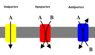
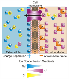
A membrane transport protein is a membrane protein involved in the movement of ions, small molecules, or macromolecules, such as another protein, across a biological membrane. Transport proteins are integral transmembrane proteins; that is they exist permanently within and span the membrane across which they transport substances. The proteins may assist in the movement of substances by facilitated diffusion or active transport. The two main types of proteins involved in such transport are broadly categorized as either channels or carriers. The solute carriers and atypical SLCs are secondary active or facilitative transporters in humans.

Substrate-level phosphorylation is a metabolic reaction that results in the formation of ATP or GTP by the direct transfer of a phosphoryl (PO3) group to ADP or GDP from another phosphorylated compound.

Bcl-2-associated X protein protein-coding gene in the species Homo sapiens

Apoptosis regulator BAX, also known as bcl-2-like protein 4, is a protein that in humans is encoded by the BAX gene. BAX is a member of the Bcl-2 gene family. BCL2 family members form hetero- or homodimers and act as anti- or pro-apoptotic regulators that are involved in a wide variety of cellular activities. This protein forms a heterodimer with BCL2, and functions as an apoptotic activator. This protein is reported to interact with, and increase the opening of, the mitochondrial voltage-dependent anion channel (VDAC), which leads to the loss in membrane potential and the release of cytochrome c. The expression of this gene is regulated by the tumor suppressor P53 and has been shown to be involved in P53-mediated apoptosis.

Adenine nucleotide translocator (ANT), also known as the ADP/ATP translocase or mitochondrial ADP/ATP carrier, exchanges free ATP with free ADP across the inner mitochondrial membrane. ANT is the most abundant protein in the inner mitochondrial membrane and belongs to mitochondrial carrier family.

Mitochondrial carrier

Mitochondrial carriers are proteins from a solute carrier family which transfer molecules across the membranes of the mitochondria. Mitochondrial carriers are also classified in the Transporter Classification Database. The Mitochondrial Carrier (MC) Superfamily has been expanded to include both the original Mitochondrial Carrier (MC) family and the Mitochondrial Inner/Outer Membrane Fusion (MMF) family.

SLC25A3 protein-coding gene in the species Homo sapiens

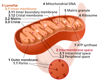
Phosphate carrier protein, mitochondrial is a protein that in humans is encoded by the SLC25A3 gene. The encoded protein is a transmembrane protein located in the mitochondrial inner membrane and catalyzes the transport of phosphate ions across it for the purpose of oxidative phosphorylation. There are two significant isoforms of this gene expressed in human cells, which differ slightly in structure and function. Mutations in this gene can cause mitochondrial phosphate carrier deficiency (MPCD), a fatal disorder of oxidative phosphorylation symptomized by lactic acidosis, neonatal hypotonia, hypertrophic cardiomyopathy, and death within the first year of life.

SLC25A14 protein-coding gene in the species Homo sapiens

Brain mitochondrial carrier protein 1 is a protein that in humans is encoded by the SLC25A14 gene.

SLC25A27 protein-coding gene in the species Homo sapiens

Mitochondrial uncoupling protein 4 is a protein that in humans is encoded by the SLC25A27 gene.

PPIF protein-coding gene in the species Homo sapiens

Peptidyl-prolyl cis-trans isomerase, mitochondrial (PPIF) is an enzyme that in humans is encoded by the PPIF gene. It has also been referred to as, but should not be confused with, cyclophilin D (CypD), which is encoded by the PPID gene. As a member of the peptidyl-prolyl cis-trans isomerase (PPIase) family, this protein catalyzes the cis-trans isomerization of proline imidic peptide bonds, which allows it to facilitate folding or repair of proteins. PPIF is a major component of the mitochondrial permeability transition pore (MPTP) and, thus, highly involved in mitochondrial metabolism and apoptosis, as well as in mitochondrial diseases and related conditions, including cardiac diseases, neurodegenerative diseases, and muscular dystrophy. In addition, PPIF participates in inflammation, as well as in ischemic reperfusion injury, AIDS, and cancer.

BCL2L13 protein-coding gene in the species Homo sapiens

BCL2-like 13 , also known as BCL2L13 or Bcl-rambo, is a protein which in humans is encoded by the BCL2L13 gene on chromosome 22. This gene encodes a mitochondrially-localized protein which is classified under the Bcl-2 protein family. Overexpression of the encoded protein results in apoptosis. As a result, it has been implicated in cancers such as childhood acute lymphoblastic leukemia (ALL) and glioblastoma multiforme (GBM). Alternatively spliced transcript variants have been observed for this gene, such as Bcl-rambo beta.

AIFM2 protein-coding gene in the species Homo sapiens

Apoptosis-inducing factor 2 (AIFM2), also known as apoptosis-inducing factor-homologous mitochondrion-associated inducer of death (AMID), is a protein that in humans is encoded by the AIFM2 gene, also known as p53-responsive gene 3 (PRG3), on chromosome 10.

SLC25A4 protein-coding gene in the species Homo sapiens

ADP/ATP translocase 1 is an enzyme that in humans is encoded by the SLC25A4 gene or adenine nucleotide translocator, ANT.

TXN2 protein-coding gene in the species Homo sapiens

Thioredoxin, mitochondrial also known as thioredoxin-2 is a protein that in humans is encoded by the TXN2 gene on chromosome 22. This nuclear gene encodes a mitochondrial member of the thioredoxin family, a group of small multifunctional redox-active proteins. The encoded protein may play important roles in the regulation of the mitochondrial membrane potential and in protection against oxidant-induced apoptosis.

SLC25A31 protein-coding gene in the species Homo sapiens

ADP/ATP translocase 4 (ANT4) is an enzyme that in humans is encoded by the SLC25A31 gene on chromosome 4. This enzyme inhibits apoptosis by catalyzing ADP/ATP exchange across the mitochondrial membranes and regulating membrane potential. In particular, ANT4 is essential to spermatogenesis, as it imports ATP into sperm mitochondria to support their development and survival. Outside this role, the SLC25AC31 gene has not been implicated in any human disease.

SLC25A6 protein-coding gene in the species Homo sapiens

ADP/ATP translocase 3 also known as solute carrier family 25 member 6 is a protein that in humans is encoded by the SLC25A6 gene.

ADP/ATP translocase

ADP/ATP translocases, also known as adenine nucleotide translocases (ANT) and ADP/ATP carrier proteins (AAC), are transporter proteins that enable the exchange of cytosolic adenosine diphosphate (ADP) and mitochondrial adenosine triphosphate (ATP) across the inner mitochondrial membrane. Free ADP is transported from the cytoplasm to the mitochondrial matrix, while ATP produced from oxidative phosphorylation is transported from the mitochondrial matrix to the cytoplasm, thus providing the cells with its main energy currency.

Necroptosis

Necroptosis is a programmed form of necrosis, or inflammatory cell death. Conventionally, necrosis is associated with unprogrammed cell death resulting from cellular damage or infiltration by pathogens, in contrast to orderly, programmed cell death via apoptosis. The discovery of necroptosis showed that cells can execute necrosis in a programmed fashion and that apoptosis is not always the preferred form of cell death. Furthermore, the immunogenic nature of necroptosis favors its participation in certain circumstances, such as aiding defense pathogens by the immune system. Necroptosis is well defined as a viral defense mechanism, allowing the cell to undergo "cellular suicide" in a caspase-independent fashion in the presence of viral caspase inhibitors to restrict virus replication. In addition to being a response to disease, necroptosis has also been characterized as a component of inflammatory diseases such as Crohn's disease, pancreatitis, and myocardial infarction.

SLC25A24 protein-coding gene in the species Homo sapiens

Solute carrier family 25, member 24 is a protein that in humans is encoded by the SLC25A24 gene.

References

  1. 1 2 3 GRCh38: Ensembl release 89: ENSG00000005022 - Ensembl, May 2017
  2. 1 2 3 GRCm38: Ensembl release 89: ENSMUSG00000016319 - Ensembl, May 2017
  3. "Human PubMed Reference:".
  4. "Mouse PubMed Reference:".
  5. 1 2 3 4 5 6 7 "Entrez Gene: Solute carrier family 25 (mitochondrial carrier; adenine nucleotide translocator), member 5" . Retrieved 2013-09-18.
  6. 1 2 3 4 5 6 7 8 Chevrollier A, Loiseau D, Reynier P, Stepien G (Jun 2011). "Adenine nucleotide translocase 2 is a key mitochondrial protein in cancer metabolism". Biochimica et Biophysica Acta. 1807 (6): 562–7. doi:10.1016/j.bbabio.2010.10.008. PMID   20950584.
  7. 1 2 3 4 Ho L, Titus AS, Banerjee KK, George S, Lin W, Deota S, Saha AK, Nakamura K, Gut P, Verdin E, Kolthur-Seetharam U (Nov 2013). "SIRT4 regulates ATP homeostasis and mediates a retrograde signaling via AMPK". Aging. 5 (11): 835–49. doi:10.18632/aging.100616. PMC   3868726 Lock-green.svg. PMID   24296486.
  8. Ku DH, Kagan J, Chen ST, Chang CD, Baserga R, Wurzel J (Sep 1990). "The human fibroblast adenine nucleotide translocator gene. Molecular cloning and sequence". The Journal of Biological Chemistry. 265 (27): 16060–3. PMID   2168878.
  9. 1 2 3 4 Vandewalle J, Bauters M, Van Esch H, Belet S, Verbeeck J, Fieremans N, Holvoet M, Vento J, Spreiz A, Kotzot D, Haberlandt E, Rosenfeld J, Andrieux J, Delobel B, Dehouck MB, Devriendt K, Fryns JP, Marynen P, Goldstein A, Froyen G (Oct 2013). "The mitochondrial solute carrier SLC25A5 at Xq24 is a novel candidate gene for non-syndromic intellectual disability". Human Genetics. 132 (10): 1177–85. doi:10.1007/s00439-013-1322-3. PMID   23783460.
  10. 1 2 3 4 Oishi M, Iizumi Y, Taniguchi T, Goi W, Miki T, Sakai T (2013). "Apigenin sensitizes prostate cancer cells to Apo2L/TRAIL by targeting adenine nucleotide translocase-2". PLOS ONE. 8 (2): e55922. doi:10.1371/journal.pone.0055922. PMC   3576345 Lock-green.svg. PMID   23431365.
  11. 1 2 3 4 5 Jang JY, Choi Y, Jeon YK, Kim CW (2008). "Suppression of adenine nucleotide translocase-2 by vector-based siRNA in human breast cancer cells induces apoptosis and inhibits tumor growth in vitro and in vivo". Breast Cancer Research. 10 (1): R11. doi:10.1186/bcr1857. PMC   2374967 Lock-green.svg. PMID   18267033.
  12. 1 2 Ito S, Tan LJ, Andoh D, Narita T, Seki M, Hirano Y, Narita K, Kuraoka I, Hiraoka Y, Tanaka K (Aug 2010). "MMXD, a TFIIH-independent XPD-MMS19 protein complex involved in chromosome segregation". Molecular Cell. 39 (4): 632–40. doi:10.1016/j.molcel.2010.07.029. PMID   20797633.
  13. Danial NN, Korsmeyer SJ (2004). "Cell death: critical control points". Cell. 116 (2): 205–19. doi:10.1016/s0092-8674(04)00046-7. PMID   14744432.
  14. Kerr JF, Wyllie AH, Currie AR (Aug 1972). "Apoptosis: a basic biological phenomenon with wide-ranging implications in tissue kinetics". British Journal of Cancer. 26 (4): 239–57. doi:10.1038/bjc.1972.33. PMC   2008650 Lock-green.svg. PMID   4561027.
  15. 1 2 Clémençon B, Babot M, Trézéguet V (2013). "The mitochondrial ADP/ATP carrier (SLC25 family): pathological implications of its dysfunction". Mol. Aspects Med. 34 (2-3): 485–93. doi:10.1016/j.mam.2012.05.006. PMID   23506884.
  16. De Marcos Lousa C, Trézéguet V, Dianoux AC, Brandolin G, Lauquin GJ (2002). "The human mitochondrial ADP/ATP carriers: kinetic properties and biogenesis of wild-type and mutant proteins in the yeast S. cerevisiae". Biochemistry. 41 (48): 14412–20. doi:10.1021/bi0261490. PMID   12450408.
  17. Jang JY, Jeon YK, Lee CE, Kim CW (Feb 2013). "ANT2 suppression by shRNA may be able to exert anticancer effects in HCC further by restoring SOCS1 expression". International Journal of Oncology. 42 (2): 574–82. doi:10.3892/ijo.2012.1736. PMID   23242177.

Further reading

This article incorporates text from the United States National Library of Medicine, which is in the public domain.