Salt and pepper syndrome | |
---|---|
Other names | Amish infantile epilepsy syndrome, ST3GAL5-CDG, Salt and pepper mental retardation syndrome, Infantile-onset symptomatic epilepsy syndrome - developmental stagnation - blindness, Epilepsy syndrome, infantile-onset symptomatic [1] [2] [3] |
Specialty | Medical genetics |
Usual onset | Childhood |
Causes | Genetic mutation |
Prevention | none |
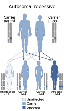
Salt and pepper developmental regression syndrome, also known as Amish infantile epileptic syndrome [4] or GM3 deficiency syndrome, [5] is a rare autosomal recessive progressive neurological disorder characterized by developmental delay, severe intellectual disability, seizures, and skin pigmentation irregularities. [6] [7] [8] [9] The clinical symptoms of this condition start manifesting soon after birth, during the newborn/neo-natal stage of life. [10]
The clinical course of this condition, as mentioned before, typically starts soon after birth.[ citation needed ]
The first features of the syndrome start off with feeding difficulties, constant irritability, frequent vomiting, and hypotonia. [11] [6] [10]
The second manifestations of this condition are frequent seizures whose type can vary between patients, but usually consist of tonic-clonic seizures. These usually start sometime during the baby's first year of life. [11] [6] [10]
As the disease progresses, intellectual abilities, developmental milestones, and the senses of hearing and vision start deteriorating, this pathology will cause a phenomenon known as developmental regression (a condition where all developmental abilities a baby has acquired through their short life-time start deteriorating), severe intellectual disabilities, and will eventually lead to severe visual and hearing impairments in some to most patients. [11] [6] [10]
Some patients with the condition start developing involuntary movements of the arms and/or legs that are similar to those shown by patients with choreoathetosis. [11] [6] [10]
Another relatively benign symptom shown by patients with this condition is localized hyperpigmentation spots, which resemble freckles and are usually spread all over a patient's body, there is no accurate way of predicting whether these spots will fade or not in a patient. As previously mentioned, these cutaneous spots are benign, meaning that they don't cause any symptoms in and on themselves. [11] [6] [10]
Rare features of the disorder include self-harming behaviors and icthyosis. [8]
The feeding difficulties and regular vomiting characteristic of this condition cause failure to thrive, [12] a condition characterized by lower-than-average weight gain in babies and toddlers which can in and on itself cause even more complications, such as malnutrition.[ medical citation needed ]
This condition is caused by a mutation known under the name of Arg288Ter (also known as R288X), said mutation is found in the ST3GAL5 gene, located on the chromosome 2. ST3GAL5 is responsible for the production of an enzyme known as GM3 synthase, this enzyme is essential for the creation of a group of molecules known as gangliosides, [13] which, although present in almost all tissues and cells of the body, are mostly concentrated on the nervous system. [14] The mutation involved in this condition results in premature termination of GM3 synthase which essentially results in a disruption affecting the production of GM3 synthase, the absence of this enzyme significantly interrupts the production of any ganglioside, [11] [15] which in turn doesn't let the brain develop and function properly. [16] The reason as to why the absence of gangliosides in children with the disorder causes abnormal brain development is still unknown. [14]
These mutations are inherited in an autosomal recessive fashion, which means that for a child to show the signs of the disorder, both of their parents must have transmitted them at least one copy of the dysfunctional ST3GAL5 gene. This transmission can occur whether or not the parents are affected with the disease themselves or not. If the parents are heterozygous (meaning they only carry one copy of the mutation) carriers of the gene for the disorder, there is a 1 in 4 (or 25%) chance of one of their offspring inheriting both copies of the defective gene which would make the child a homozygous carrier of the mutation, consequently giving the child the disorder. [17] [18]
This condition can be diagnosed through genetic testing methods such as whole exome sequencing or targeted sequencing of the ST3GAL5 gene for mutations, MRIs, electroencephalograms, IQ tests, and physical examination.[ citation needed ]
Genetic testing is a very essential part of diagnosing salt pepper syndrome, without a genetic test, the only proof one would have for a diagnosis of the syndrome would be physical and intellectual anomalies, which wouldn't be sufficient to warrant a diagnosis.[ citation needed ]
Jamal et al. (2021) found a homozygous frameshift mutation (c.1030_1031del) in the ST3GAL5 gene of an Iranian child with the disorder born to consanguineous, first-cousin parents. [19]
Luigi et al. (2014) found a homozygous missense mutation (c.994G>A, also called p.E332K) in the ST3GAL5 gene of two American siblings previously described in the medical literature through the genetic testing methods of whole exome sequencing and Sanger sequencing. [20]
Jin et al. (2016) found compound heterozygous missense mutations through whole exome sequencing and Sanger sequencing in two South Korean sisters who were originally thought to have a Rett-like syndrome. [21]
In 2013, Konstantina et al. described 2 siblings, born of consanguineous French parents. The both of them showed signs of salt pepper syndrome. Brain MRIs done on them came back with results showing cortical atrophy and lesions of the brain's white matter. [22]
Electroencephalograms done on the affected patients reported by Simpson et al. showed "multifocal epileptiform discharges" which were "superimposed on a diffuse slow background activity." [23]
An IQ score of less than 70 obtained through an IQ test is typically sufficient to warrant a diagnosis of intellectual disability in children with the condition. [24]
Physical examination done in a clinical environment is also necessary for a diagnosis of salt & pepper syndrome.[ citation needed ]
In 2006, Fahhad et al. published the results of an ophthalmoscopy study done on 4 children from 2 sibships of an Amish family; they showed optic nerve paleness on both eyes, indicative of bilateral optic atrophy, a condition which can lead to visual impairment and is usually associated with other disorders (including salt pepper syndrome). [25]
Anomaly scan is a prenatal diagnostic method which can be used to detect congenital anomalies in fetuses under ultrasound.[ citation needed ]
Jamal et al. (2021) found microcephaly on a child with the disorder prior to her birth using this diagnostic method. [19]
The parents of the patients with SPS reported by Simpson et al. used gastric tube feeding to feed their affected children, this treatment method is usually needed by most children with the disorder due to their feeding difficulties. [23]
Seizures shown by patients with this condition cannot be treated with conventional anticonvulsant medication.[ citation needed ]
A study done in 2019 by Bowser et al. on 50 Amish patients with the condition showed that prescribed antiseizure medications such as phenobarbital, benzodiazepines, and valproic acid had some effect at treating the patients' seizures, insomnia, and irritability, which helped decrease their parents' overall stress of taking care of them. [26]
2 of the children reported by Simpson et al. had their seizures managed with vagus nerve stimulators. [23]
According to OMIM, around 100-200 cases of salt and pepper syndrome have been described in the medical literature. Most of these cases were from very large Old Order Amish families in Pennsylvania and Ohio. [27] The mutations are an example of the Founder effect in action. [28]
As of 2022, only 26 cases from non-Amish patients affected with salt & pepper syndrome have been described in the medical literature. [28]
This condition was first discovered in 2004 by Simpson et al. Their patients were 9 members from 2 sibships belonging to a single large, endogamous Amish family from Geauga County, Ohio. Their symptoms started between the ages of 2 weeks old and 3 months old, with irritability, vomiting, feeding difficulties and a failure to thrive followed by seizures in the first year of their life. They had developmental regression and couldn't do almost anything without the help of someone else. They had choreoathetosic non-purposeful hand movements. Brain studies on the oldest of the 9 affected members showed brain atrophy (which younger patients didn't have). Electroencephalograms gave back abnormal results. All members of the family shared a common great-great-grandparent. The disorder shown by this family clearly followed an autosomal recessive inheritance pattern. A homozygosity mapping study done afterwards on the affected family members showed linkage to the 2p12-p11.2 locus on chromosome 2, and after sequencing genes in said chromosomal region, they found a mutation of the nonsense type in their ST3GAL5 gene. [23]
A follow-up study done on the family in 2004 by Simpson, Cross, Proukakis, et.al., revealed that one of the studied members had died (nearly 3 years old) while the younger members had made it to their teens, with the oldest of them being 18 years old. [23]
Although salt and pepper developmental regression syndrome is prevalent among the Old Order Amish, further studies in 2016, 2021, and in 2023, found it in two Korean sisters, in an Iranian patient, and in a family in Saudi Arabia. [29] [30] [31]
This condition has various names, but the three main ones are:[ citation needed ]
Propionic acidemia, also known as propionic aciduria or propionyl-CoA carboxylase deficiency, is a rare autosomal recessive metabolic disorder, classified as a branched-chain organic acidemia.
Ellis–Van Creveld syndrome is a rare genetic disorder of the skeletal dysplasia type.
Sandhoff disease is a lysosomal genetic, lipid storage disorder caused by the inherited deficiency to create functional beta-hexosaminidases A and B. These catabolic enzymes are needed to degrade the neuronal membrane components, ganglioside GM2, its derivative GA2, the glycolipid globoside in visceral tissues, and some oligosaccharides. Accumulation of these metabolites leads to a progressive destruction of the central nervous system and eventually to death. The rare autosomal recessive neurodegenerative disorder is clinically almost indistinguishable from Tay–Sachs disease, another genetic disorder that disrupts beta-hexosaminidases A and S. There are three subsets of Sandhoff disease based on when first symptoms appear: classic infantile, juvenile and adult late onset.
Biotinidase deficiency is an autosomal recessive metabolic disorder in which biotin is not released from proteins in the diet during digestion or from normal protein turnover in the cell. This situation results in biotin deficiency.
2-hydroxyglutaric aciduria is a rare neurometabolic disorder characterized by the significantly elevated levels of hydroxyglutaric acid in one's urine. It is either autosomal recessive or autosomal dominant.
The GM1 gangliosidoses, usually shortened to GM1, are gangliosidoses caused by mutation in the GLB1 gene resulting in a deficiency of beta-galactosidase. The deficiency causes abnormal storage of acidic lipid materials in cells of the central and peripheral nervous systems, but particularly in the nerve cells, resulting in progressive neurodegeneration. GM1 is a rare lysosomal storage disorder with a prevalence of 1 to every 100,000 to 200,000 live births worldwide, although rates are higher in some regions.
Rhizomelic chondrodysplasia punctata is a rare developmental brain disorder characterized by abnormally short arms and legs (rhizomelia), seizures, recurrent respiratory tract infections and congenital cataracts.
Vici syndrome, also called immunodeficiency with cleft lip/palate, cataract, hypopigmentation and absent corpus callosum, is a rare autosomal recessive congenital disorder characterized by albinism, agenesis of the corpus callosum, cataracts, cardiomyopathy, severe psychomotor retardation, seizures, immunodeficiency and recurrent severe infections. To date, about 50 cases have been reported.
Intermembrane lipid transfer protein VPS13B, also known as vacuolar protein sorting-associated 13B, and Cohen syndrome protein 1 is a protein that in humans is encoded by the VPS13B gene. It is a giant protein associated with the Golgi apparatus that is believed to be involved in post-Golgi apparatus sorting and trafficking. Mutations in the human VPS13B gene cause Cohen syndrome.
Lactosylceramide alpha-2,3-sialyltransferase is an enzyme that in humans is encoded by the ST3GAL5 gene.
De Barsy syndrome is a rare autosomal recessive genetic disorder. Symptoms include cutis laxa as well as other eye, musculoskeletal, and neurological abnormalities. It is usually progressive, manifesting side effects that can include clouded corneas, cataracts, short stature, dystonia, or progeria.
Epilepsy-intellectual disability in females also known as PCDH19 gene-related epilepsy or epileptic encephalopathy, early infantile, 9 (EIEE9), is a rare type of epilepsy that affects predominately females and is characterized by clusters of brief seizures, which start in infancy or early childhood, and is occasionally accompanied by varying degrees of cognitive impairment. The striking pattern of onset seizures at a young age, genetic testing and laboratory results, potential developmental delays or developmental regression and associated disorders, eases diagnosis.
Mitochondrial DNA depletion syndrome, or Alper's disease, is any of a group of autosomal recessive disorders that cause a significant drop in mitochondrial DNA in affected tissues. Symptoms can be any combination of myopathic, hepatopathic, or encephalomyopathic. These syndromes affect tissue in the muscle, liver, or both the muscle and brain, respectively. The condition is typically fatal in infancy and early childhood, though some have survived to their teenage years with the myopathic variant and some have survived into adulthood with the SUCLA2 encephalomyopathic variant. There is currently no curative treatment for any form of MDDS, though some preliminary treatments have shown a reduction in symptoms.
CDK13-related disorder, also known as congenital heart defects, dysmorphic facial features and intellectual developmental disorder (CHDFIDD), is a very rare autosomal dominant genetic condition characterised by congenital heart defects, intellectual disability and characteristic facial features. Those affected typically have motor and language delays, low muscle tone and gastrointestinal dysmotility. Facial features include a wide nasal bridge, widely-spaced eyes, prominent, low-set ears, a flat nose tip and a small mouth. Less common features include congenital spinal abnormalities, hearing loss or seizures.
Chudley–Mccullough syndrome is a rare genetic disorder which is characterized by bilateral congenital hearing loss associated with brain malformations. It is a type of syndromic deafness.
Severe intellectual disability-progressive spastic diplegia syndrome is a rare novel genetic disorder characterized by severe intellectual disabilities, ataxia, craniofacial dysmorphisms, and muscle spasticity. It is a type of autosomal dominant syndromic intellectual disability.
SLC13A5 citrate transporter disorder, or SLC13A5 Epilepsy, is a rare genetic spectrum disorder that presents with neurological symptoms. Symptoms include severe seizures, ataxia, dystonia, teeth hypoplasia, poor communication skills, difficulty standing or walking, as well as developmental delay. Other names associated with SLC13A5 Epilepsy include SLC13A5 Citrate Transporter Disorder, Citrate Transporter Disorder, SLC13A5 Deficiency, Early Infantile Epilepsy Encephalopathy 25 (EIEE25), Developmental Epilepsy Encephalopathy 25 (DEE25), and Kohlschutter-Tonz Syndrome (non-ROGDI).
Spondyloenchondrodysplasia is the medical term for a rare spectrum of symptoms that are inherited following an autosomal recessive inheritance pattern. Skeletal anomalies are the usual symptoms of the disorder, although its phenotypical nature is highly variable among patients with the condition, including symptoms such as muscle spasticity or thrombocytopenia purpura. It is a type of immunoosseous dysplasia.
Isolated hyperchlorhidrosis, also known as Carbonic anhydrase XII deficiency, is a rare autosomal recessive genetic condition characterized by a lifelong tendency to lose massive amounts of sodium and chloride through sweat which leads to various symptoms.
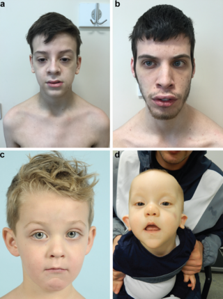
Beck–Fahrner syndrome, also known as BEFAHRS and TET3 deficiency, is a rare genetic disorder caused by mutations of the TET3 gene. The clinical presentation varies among individuals, but typically includes global developmental delay, slow progress in mental and physical activities, autism, decreased muscle tone, epilepsy and dysmorphic features.