Figure 9. Human mammary gland stroma: TEM; original magnification 9,100x. A: Lymphocyte establishing a multicontact synapse (MS) with a TC. The blue rectangle shows the synaptic ‘kiss and run’ region. The synaptic membranes appear traced in B (violet - TC, orange - lymphocyte). The distances between membranes are shown in C. Note (asterisk) a peculiar conformation of ER connecting mitochondria with the cell surface, suggestive for a possible role in synaptic Ca2+ homeostasis. Reproduced with permission from [22]
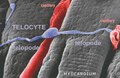
Figure 10. Scanning electron micrograph of monkey left ventricular myocardium. A typical TC is located across the cardiomyocytes, in close contacts with blood capillaries. Note, the cardiomyocytes striations and the openings of T tubules.
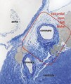
Figure 11. Digitally coloured electron micrograph of mouse ventricular endocardium (burgundy). TC (blue) make an interstitial network in the heart. Subendocardial telocytes (TC1) sends Tp between cardiomyocytes (CM) and communicate with TC2. Cap, blood capillary. Scale bar 5 μm. Reproduced with permission from [4]
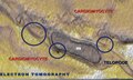
Figure 12. This electron tomography (thick section of about 300 nm) shows nanostructures connecting the TC and cardiomyocytes in adult mouse heart. The bridging structures (encircled) have 10-15 nm and suggest a molecular interaction between the Tp of one TC and the two adjacent cardiomyocytes. The dilated segment of Tp involved in the heterocellular connection (podom) - contains a mitochondrion (m).
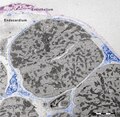
Figure 13. High resolution light microscopy on toluidine blue stained semithin section (~1 μm thick ultramicrotome)
Figure 14. Electron micrographs illustrates the relationships of TC (blue) with cardiomyocyte progenitors (CMP, brown). The Tp run parallel with the main axis of the CMP and seem to establish their direction of development.
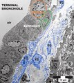
Figure 15. Mice lung. Terminal bronchiole. At least 4 TC with their extensive Tp are visible between the epithelium and an arteriole (SMC - smooth muscle cells). Note, the striking labyrinthine network formed by Tp. In the upper part a mitosis (prophase) is obvious (orange circle). In addition, a putative stem cell (SC, green oval) is in close contacts with telocytes prolongations, establishing a heterocellular junctions, visible at higher magnification only). The tandem TC-SC forms, presumably, a TC-SC niche. In the lower part, a macrophage (MF) makes a stromal synapse with Tp.
Figure 16. Rat striated skeletal muscle (diaphargm). A typical TC (blue) with two convoluted Tp is shown, by transmission electron microscopy. Note, two shed vesicles (sv, violet). m-mitochondria, Ly-lymphocyte. The asterisks indicate, presumably, two empty exosomes, which probably released their vesicle content. BV-small blood vessel.
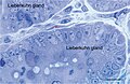
Figure 17. Rat jejunum. Toluidine blue stained Semithin Epon sections of jejunum mucosa showing the bottom of Lieberkuhn glands in transverse section and a telocyte (red star) surrounding one of the gland. Note the spindle-shape body sending off two telopodes, one of which measure at least 50 μm in the section plane.
Figure 18. Rat jejunum muscularis mucosa. The photo is a colour-enhanced digital micrograph of a black and white transmission electron microscopy image. A blue telopode of 14.2 μm in the section plane is illustrated around a nerve ending (green) between smooth muscle cells (brown).
Figure 19. Rat jejunum mucosa. A. This electron microscope image disclose a telopode (blue) in the profound region of lamina propria, close to the muscularis mucosa (brown) and in the proximity of a nerve ending (green). Note the alternating podom and podomere. B. Inset disclosing the organelle details of the podomere – intermediate filaments and free ribosomes, and of the podom – mitochondrion and endoplasmic reticulum cisternae. C. High resolution image illustrating in detail multiple mitochondria, endoplasmic reticulum cisterne and caveolae (arrow).
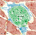
Figure 20. Rat jejunum. A. Photomicrograph of an interstitial cell of Cajal (violet) in muscularis externa. Note the large cell body which extend slender and relatively short connection towards the nerve endings (green). B. Digitally coloured TEM image showing a fibroblast (garnet) and a telocyte (blue) in the lamina propria. C. Coloured transmission electron micrograph (TEM) of a tangential section through a fibroblast cell. The internal structure can be seen, including the dilated rough endoplasmic reticulum (blue). responsible for synthesising collagen. In blue a telopode underlying the intestinal epithelium.
Figure 21. Rat jejunum mucosa. A telocyte (blue) telopode is engaged in different types of synapses with a plasma cell: two plain synapses (PS) and one multicontact synapse (MS) are seen.
Figure 22. Rat jejunum. A-E. 3-D image reconstruction from 5 serial sections of telocytes (blue) in lamina propria: telopodes branching in a 3-D pattern. Telocyte's nucleus is colored in violet. F-J. Computer-aided volume rendering and different-angle stereoscopic views of a telocyte (blue) surrounding a nerve fiber (green) in muscularis mucosa (dark red).
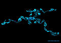
Figure 23. 3D reconstruction of a telocyte with its long telopodes.
Figure 24. A podom is a dilated portion of a telopode. Note the endoplasmic reticulum in yellow and mitochondria in red.
Figure 25. A color representation of convoluted telopodes (blue) and a shedding vesicle (magenta).
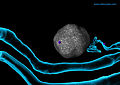
Figure 26. Shedding vesicles (magenta) emerged from the telopodes (blue) and are heading towards a stem cell (gray).


![Figure 5. Human term placenta. The TC (blue) has few organelles in the perinuclear area and 3 emerging Tp (red arrows); black arrowheads mark the dichotomic branching points. Note the podoms and podomeres. Black arrow points the junction between a Tp and a smooth muscle cell (SMC, colored in brown). Reproduced with permission from [2]. Telocytes-Fig 5.tif](http://upload.wikimedia.org/wikipedia/commons/thumb/0/04/Telocytes-Fig_5.tif/lossy-page1-250px-Telocytes-Fig_5.tif.jpg)
![Figure 6. Non-pregnant myometrium. Digitally colored TC (blue) with 3 Tp that encircle bundles of cross-cut smooth muscle cells (SMC, Sienna brown); N - nuclei. Reproduced with permission from [1]. Telocytes - Fig 6.tif](http://upload.wikimedia.org/wikipedia/commons/thumb/0/02/Telocytes_-_Fig_6.tif/lossy-page1-250px-Telocytes_-_Fig_6.tif.jpg)

![Figure 8. Rat stomach, multicontact stromal synapses between two TC, a plasma cell and an eosinophil, respectively. 3-D image computer-aided reconstruction from 9 serial ultrathin sections; original magnification 1,500x. The upper inset shows contact points where the distance between both cell membranes (Tp membrane and plasma-cell membrane) is 15 nm or less (in violet), seen from the plasma cell cytoplasm. In the lower inset Tp were rendered transparent in order to depict the same synapse. Reproduced with permission from [22]. Telocytes-Fig 8.tif](http://upload.wikimedia.org/wikipedia/commons/thumb/c/cb/Telocytes-Fig_8.tif/lossy-page1-250px-Telocytes-Fig_8.tif.jpg)