Trichodysplasia spinulosa

Last updated
Trichodysplasia spinulosa
Other namesViral-associated trichodysplasia spinulosa
Van der Meijden plospathogens 2010 TSPyV nose.png
The nose of a patient diagnosed with trichodysplasia spinulosa. Left panel shows characteristic findings of facial papules, protrusive keratotic spicules, and thickened skin; right panel shows appearance following treatment with topical cidofovir. [1]
Specialty Infectious disease, dermatology

Trichodysplasia spinulosa (also known by many other names, including viral-associated trichodysplasia spinulosa, viral-associated trichodysplasia, pilomatrix dysplasia and ciclosporin-induced folliculodystrophy, although the last is a misnomer) is a rare cutaneous condition that has been described almost exclusively in immunocompromised patients, usually organ transplant recipients, on regimens of immunosuppressive drugs. [2] [3] As of early 2016, a total of 32 cases had been reported in the medical literature. [2] Despite its rarity, TS is believed to be underdiagnosed, and the growing population of patients on immunosuppressive drug regimens suggests its incidence may rise. [2] [3] TS has been described as an emerging infectious disease. [4]

Contents

Symptoms and signs

The disease is characterized by flesh-colored to erythematous (reddened) papules occurring in the central region of the face and sometimes elsewhere on the body, often accompanied by protrusive "spicules" or spines made of keratin and by alopecia of affected skin, typically the eyebrows and sometimes the eyelashes or scalp hairs. Pruritus (itching) has been described in about a third of reported cases. [2] Facial papules are generally 1- to 3-mm in size. [5] :415 The condition is considered to be benign, but can be disfiguring; the spines are often prominent, and in later stages the affected facial skin thickens noticeably. [2] [3]

Causes

TS has been reported almost exclusively in immunocompromised patients, primarily organ transplant recipients on regimens of immunosuppressive drugs, and also in patients with hematolymphoid malignancies. [2] [3] As of 2016 there were no case reports in the literature describing cases of TS in patients with HIV-AIDS. [2]

There is compelling evidence that trichodysplasia spinulosa is caused by a polyomavirus called trichodysplasia spinulosa polyomavirus (TSPyV) or Human polyomavirus 8. [2] [3] [1] [6] There is evidence that exposure to the virus is common among healthy adults; estimates of seroprevalence (that is, prevalence of detectable antibodies against viral proteins) in immunocompetent adults range from 70 to 80% in different sample populations. [3] [7] [8] TSPyV infects the skin, but viral DNA is rarely detectable there in asymptomatic individuals even if they possess antibodies to the virus indicating exposure. [3] It is not known whether TS represents new primary infection or opportunistic reactivation of a latent infection. [3]

Mechanism

The hyperproliferation of keratinocyte inner root sheath cells in which large aggregates of viral particles can be found suggests that TSPyV actively replicates in these cells. This is thought to underlie the clinical manifestations of TS. However, the precise mechanism is not well characterized. [3] There is limited evidence implicating the large tumor antigen as responsible for inducing cellular proliferation through pathways involving phosphorylated retinoblastoma protein (pRB). [9]

Diagnosis

Overview of histological findings in trichodysplasia spinulosa. Top row (A1-A3) shows healthy control; bottom row (B1-B3) shows TS skin. Low-magnification-power overview (A1, B1); high-power representative examples of epidermis showing thickening of the skin (acanthosis) in TS (A2, B2); high-power representative examples of hair follicles showing enlarged and dysmorphic appearance in TS (A3, B3). Inset in B3 shows characteristic eosinophilic protein granules, probably trichohyalin (arrowheads). H&E stain. Scale bars are 100mm. Kazem plosone 2014 TSPyV histology.png
Overview of histological findings in trichodysplasia spinulosa. Top row (A1-A3) shows healthy control; bottom row (B1-B3) shows TS skin. Low-magnification-power overview (A1, B1); high-power representative examples of epidermis showing thickening of the skin (acanthosis) in TS (A2, B2); high-power representative examples of hair follicles showing enlarged and dysmorphic appearance in TS (A3, B3). Inset in B3 shows characteristic eosinophilic protein granules, probably trichohyalin (arrowheads). H&E stain. Scale bars are 100μm.

TS can be diagnosed based on clinical observations, but is usually confirmed by histopathology of a lesional biopsy or a plucked spicule. Characteristic histological findings include enlarged and abnormally organized hair follicles and hyperproliferation of inner root sheath cells containing large eosinophilic trichohyalin granules. Antibodies against major capsid protein VP1, the major component of the viral capsid, can be used to confirm the presence of viral particles in cell nuclei. Electron microscopy can also be used to detect viral particles. Quantification of viral load can be performed using quantitative PCR, as affected skin demonstrates much higher viral loads compared to unaffected skin or to asymptomatic individuals who test positive for viral DNA. [2] [3]

Differential diagnosis includes other visually similar conditions affecting the hair follicles, many of which appear as drug side effects. [2] A proposed classification system lists TS as one of a group of cutaneous conditions with similar manifestations and distinct etiologies, collectively called the digitate keratoses. [10] Although confirmed TS is rare, the condition is thought to be underdiagnosed. [2]

Treatment

There have been too few cases of TS reported for a standard treatment to be established. In some cases, improvement in immune function has been noted to produce spontaneous improvement in TS symptoms. This pattern is consistent with the behavior of other viral diseases found in immunocompromised patients, most relevantly with the nephropathy associated in kidney transplant recipients with the polyomavirus BK virus. Antiviral drugs such as valganciclovir and cidofovir have shown benefit in treating this disorder in case reports. [2] [3] [11] [12]

Outcomes

TS is considered to be a benign dysplasia, although it can be disfiguring and is sometimes itchy. It is not known whether TS lesions have the potential to develop into cancer; while this outcome has never been reported, some polyomaviruses are oncogenic. The natural history of untreated TS is not known and no long-term studies of its progress have been performed. Improvement in immune function has been reported to resolve symptoms in some individual cases. Treatment with antiviral drugs has also been reported to improve symptoms, but only as long as treatment continues. [2] [3]

History

TS was first described in a 1995 case report as "ciclosporin-induced folliculodystrophy", thought at the time to be an adverse effect of ciclosporin treatment. [3] [13] A subsequent report in 1999, which introduced the term "trichodysplasia spinulosa", used electron microscopy to identify the presence of virus particles in affected cells consistent with what were at the time known as papovaviruses. [14] (The group has since been divided into the papillomavirus and polyomavirus families.) In 2010, researchers used rolling circle amplification to recover viral DNA from TS lesions and thus discovered a novel polyomavirus, trichodysplasia spinulosa polyomavirus (TSPyV). There is compelling evidence that TSPyV is the direct causative agent of TS. [2] [3]

Related Research Articles

Progressive multifocal leukoencephalopathy Viral disease affecting human brains

Progressive multifocal leukoencephalopathy (PML) is a rare and often fatal viral disease characterized by progressive damage (-pathy) or inflammation of the white matter (leuko-) of the brain (-encephalo-) at multiple locations (multifocal). It is caused by the JC virus, which is normally present and kept under control by the immune system. The JC virus is harmless except in cases of weakened immune systems. In general, PML has a mortality rate of 30–50% in the first few months, and those who survive can be left with varying degrees of neurological disabilities.

Psoriasis Skin disease

Psoriasis is a long-lasting, noncontagious autoimmune disease characterized by raised areas of abnormal skin. These areas are red, or purple on some people with darker skin, dry, itchy, and scaly. Psoriasis varies in severity from small, localized patches to complete body coverage. Injury to the skin can trigger psoriatic skin changes at that spot, which is known as the Koebner phenomenon.

<i>Polyomaviridae</i> Family of viruses

Polyomaviridae is a family of viruses whose natural hosts are primarily mammals and birds. As of 2020, there are six recognized genera and 117 species, five of which are unassigned to a genus. 14 species are known to infect humans, while others, such as Simian Virus 40, have been identified in humans to a lesser extent. Most of these viruses are very common and typically asymptomatic in most human populations studied. BK virus is associated with nephropathy in renal transplant and non-renal solid organ transplant patients, JC virus with progressive multifocal leukoencephalopathy, and Merkel cell virus with Merkel cell cancer.

Merkel-cell carcinoma Rare and highly aggressive skin cancer

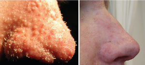
Merkel cell carcinoma (MCC) is a rare and aggressive skin cancer occurring in about 3 people per 1,000,000 members of the population. It is also known as cutaneous APUDoma, primary neuroendocrine carcinoma of the skin, primary small cell carcinoma of the skin, and trabecular carcinoma of the skin. Factors involved in the development of MCC include the Merkel cell polyomavirus, a weakened immune system, and exposure to ultraviolet radiation. Merkel-cell carcinoma usually arises on the head, neck, and extremities, as well as in the perianal region and on the eyelid. It is more common in people over 60 years old, Caucasian people, and males. MCC is less common in children.

Pyoderma gangrenosum Medical condition

Pyoderma gangrenosum is a rare, inflammatory skin disease where painful pustules or nodules become ulcers that progressively grow. Pyoderma gangrenosum is not infectious.

BK virus Member of the polyomavirus family

The BK virus is a member of the polyomavirus family. Past infection with the BK virus is widespread, but significant consequences of infection are uncommon, with the exception of the immunocompromised and the immunosuppressed. BK virus is an abbreviation of the name of the first patient from whom the virus was isolated in 1971.

Solar urticaria Medical condition

Solar urticaria (SU) is a rare condition in which exposure to ultraviolet or UV radiation, or sometimes even visible light, induces a case of urticaria or hives that can appear in both covered and uncovered areas of the skin. It is classified as a type of physical urticaria. The classification of disease types is somewhat controversial. One classification system distinguished various types of SU based on the wavelength of the radiation that causes the breakout; another classification system is based on the type of allergen that initiates a breakout.

Merkel cell polyomavirus was first described in January 2008 in Pittsburgh, Pennsylvania. It was the first example of a human viral pathogen discovered using unbiased metagenomic next-generation sequencing with a technique called digital transcriptome subtraction. MCV is one of seven currently known human oncoviruses. It is suspected to cause the majority of cases of Merkel cell carcinoma, a rare but aggressive form of skin cancer. Approximately 80% of Merkel cell carcinoma (MCC) tumors have been found to be infected with MCV. MCV appears to be a common—if not universal—infection of older children and adults. It is found in respiratory secretions suggesting that it may be transmitted by a respiratory route. But it also can be found shedding from healthy skin, and in gastrointestinal tract tissues and elsewhere, and so its precise mode of transmission remains unknown. In addition, recent studies suggest that this virus may latently infect the human sera and PBMCs.

Trichodysplasia spinulosa polyomavirus is a member virus of Human polyomavirus 8 that infects human hosts. First discovered in 2010, TSPyV is associated with Trichodysplasia spinulosa, a rare skin disease only seen in immunocompromised patients. The virus causes hyperproliferation and enlargement of hair follicles by modulating PP2A protein phosphatase signaling pathways. TSPyV was the eighth human polyomavirus to be discovered, and one of four associated with human disease, out of 13 human polyomaviruses known as of the 2015 update to polyomavirus taxonomy released by the International Committee on Taxonomy of Viruses.

Topical steroid withdrawal Medical condition

Topical steroid withdrawal, also known as red burning skin and steroid dermatitis, has been reported in users of topical steroids after they stop the use, following usually any more than 2 weeks in duration. Symptoms include redness of the skin, a burning sensation, and itchiness. This may then be followed by skin peeling.

Hamster polyomavirus is an unenveloped double-stranded DNA virus of the polyomavirus family whose natural host is the hamster. It was originally described in 1967 by Arnold Graffi as a cause of epithelioma in Syrian hamsters.

WU polyomavirus is a virus of the family Polyomaviridae. It was discovered in 2007 in samples of human respiratory secretions, originally from a child patient in Australia who presented with clinical signs of pneumonia and in whom other common respiratory viruses were not detected. Follow-up studies identified the presence of WU virus in respiratory secretion samples from patients in Australia and the United States, suggesting that, like other human polyomaviruses, WU virus is widely distributed.

KI polyomavirus is a virus of the family Polyomaviridae. It was discovered in 2007 in stored samples of human respiratory secretions collected by the Karolinska Institute, after which the virus is named.

Human polyomavirus 7 (HPyV7) is a virus of the polyomavirus family that infects human hosts. It was discovered in 2010 and is a common component of the skin flora in healthy adults. There is limited evidence from case reports linking the virus to a skin rash occurring in immunocompromised organ transplant recipients.

Human polyomavirus 6 (HPyV6) is a virus of the polyomavirus family that infects human hosts. It was discovered in 2010 and is a component of the skin flora in healthy adults.

Human polyomavirus 9 (HPyV9) is a virus of the polyomavirus family that infects human hosts. It was discovered in 2011 and is a component of the skin flora in healthy adults.

Sorex araneus polyomavirus 1, formerly known as Human polyomavirus 12 (HPyV12), is a virus of the polyomavirus family that was first identified in human hosts and also infects shrews. It was discovered and reported in 2013 after isolation from the organs of the gastrointestinal tract, particularly the liver. The virus was renamed to Sorex araneus polyomavirus 1 in 2018, after discovery of the same virus in shrews. Infecting multiple hosts is rare among mammalian polyomaviruses.

New Jersey polyomavirus is a virus of the polyomavirus family that infects human hosts. It was first identified in 2014 in a pancreatic transplant patient in New Jersey. It is the 13th and most recent human polyomavirus to be described.

The middle tumor antigen is a protein encoded in the genomes of some polyomaviruses, which are small double-stranded DNA viruses. MTag is expressed early in the infectious cycle along with two other related proteins, the small tumor antigen and large tumor antigen. MTag occurs only in a few known polyomaviruses, while STag and LTag are universal - it was first identified in mouse polyomavirus (MPyV), the first polyomavirus discovered, and also occurs in hamster polyomavirus. In MPyV, MTag is an efficient oncoprotein that can be sufficient to induce neoplastic transformation in some cells.

Cutaneous manifestations of COVID-19 are characteristic signs or symptoms of the Coronavirus disease 2019 that occur in the skin. The American Academy of Dermatology reports that skin lesions such as morbilliform, pernio, urticaria, macular erythema, vesicular purpura, papulosquamous purpura and retiform purpura are seen in people with COVID-19. Pernio-like lesions were more common in mild disease while retiform purpura was seen only in critically ill patients. The major dermatologic patterns identified in individuals with COVID-19 are urticarial rash, confluent erythematous/morbilliform rash, papulovesicular exanthem, chilbain-like acral pattern, livedo reticularis and purpuric “vasculitic” pattern. Chilblains and Multisystem inflammatory syndrome in children are also cutaneous manifestations of COVID-19.

References

  1. 1 2 van der Meijden E, Janssens RW, Lauber C, Bouwes Bavinck JN, Gorbalenya AE, Feltkamp MC (July 2010). "Discovery of a new human polyomavirus associated with trichodysplasia spinulosa in an immunocompromized patient". PLOS Pathog. 6 (7): e1001024. doi:10.1371/journal.ppat.1001024. PMC   2912394 . PMID   20686659.
  2. 1 2 3 4 5 6 7 8 9 10 11 12 13 14 Wu, J.H.; Nguyen, H.P.; Rady, P.L.; Tyring, S.K. (March 2016). "Molecular insight into the viral biology and clinical features of trichodysplasia spinulosa". British Journal of Dermatology. 174 (3): 490–498. doi:10.1111/bjd.14239. PMID   26479880. S2CID   26367357.
  3. 1 2 3 4 5 6 7 8 9 10 11 12 13 14 Kazem, Siamaque; van der Meijden, Els; Feltkamp, Mariet C. W. (August 2013). "The trichodysplasia spinulosa-associated polyomavirus: virological background and clinical implications". APMIS. 121 (8): 770–782. doi:10.1111/apm.12092. PMID   23593936. S2CID   13734654.
  4. Nawas, Zeena Y.; Tong, Yun; Kollipara, Ramya; Peranteau, Andrew J.; Woc-Colburn, Laila; Yan, Albert C.; Lupi, Omar; Tyring, Stephen K. (July 2016). "Emerging infectious diseases with cutaneous manifestations". Journal of the American Academy of Dermatology. 75 (1): 1–16. doi:10.1016/j.jaad.2016.04.033. PMID   27317512.
  5. James, William D.; Berger, Timothy G.; et al. (2006). Andrews' Diseases of the Skin: clinical Dermatology. Saunders Elsevier. ISBN   0-7216-2921-0.
  6. Kazem, Siamaque; van der Meijden, Els; Kooijman, Sander; Rosenberg, Arlene S.; Hughey, Lauren C.; Browning, John C.; Sadler, Genevieve; Busam, Klaus; Pope, Elena; Benoit, Taylor; Fleckman, Philip; de Vries, Esther; Eekhof, Just A.; Feltkamp, Mariet C.W. (March 2012). "Trichodysplasia spinulosa is characterized by active polyomavirus infection". Journal of Clinical Virology. 53 (3): 225–230. doi:10.1016/j.jcv.2011.11.007. PMID   22196870.
  7. van der Meijden, E; Kazem, S; Burgers, MM; Janssens, R; Bouwes Bavinck, JN; de Melker, H; Feltkamp, MC (August 2011). "Seroprevalence of trichodysplasia spinulosa-associated polyomavirus". Emerging Infectious Diseases. 17 (8): 1355–63. doi:10.3201/eid1708.110114. PMC   3381547 . PMID   21801610.
  8. Gossai, A; Waterboer, T; Nelson, HH; Michel, A; Willhauck-Fleckenstein, M; Farzan, SF; Hoen, AG; Christensen, BC; Kelsey, KT; Marsit, CJ; Pawlita, M; Karagas, MR (1 January 2016). "Seroepidemiology of Human Polyomaviruses in a US Population". American Journal of Epidemiology. 183 (1): 61–9. doi:10.1093/aje/kwv155. PMC   5006224 . PMID   26667254.
  9. 1 2 Kazem, Siamaque; van der Meijden, Els; Wang, Richard C.; Rosenberg, Arlene S.; Pope, Elena; Benoit, Taylor; Fleckman, Philip; Feltkamp, Mariet C. W.; Deb, Sumitra (7 October 2014). "Polyomavirus-Associated Trichodysplasia Spinulosa Involves Hyperproliferation, pRB Phosphorylation and Upregulation of p16 and p21". PLOS ONE. 9 (10): e108947. Bibcode:2014PLoSO...9j8947K. doi: 10.1371/journal.pone.0108947 . PMC   4188587 . PMID   25291363.
  10. Caccetta, Tony Philip; Dessauvagie, Ben; McCallum, Dugald; Kumarasinghe, Sujith Prasad (July 2012). "Multiple minute digitate hyperkeratosis: A proposed algorithm for the digitate keratoses". Journal of the American Academy of Dermatology. 67 (1): e49–e55. doi:10.1016/j.jaad.2010.07.023. PMID   21050621.
  11. Holzer, Aton M.; Hughey, Lauren C. (January 2009). "Trichodysplasia of immunosuppression treated with oral valganciclovir". Journal of the American Academy of Dermatology. 60 (1): 169–172. doi:10.1016/j.jaad.2008.07.051. PMC   2708076 . PMID   19103376.
  12. Wanat, Karolyn A. (1 February 2012). "Viral-Associated Trichodysplasia". Archives of Dermatology. 148 (2): 219–23. doi:10.1001/archdermatol.2011.1413. PMC   3346264 . PMID   22351821.
  13. Izakovic, J; Büchner, SA; Düggelin, M; Guggenheim, R; Itin, PH (December 1995). "[Hair-like hyperkeratoses in patients with kidney transplants. A new cyclosporin side-effect]". Der Hautarzt; Zeitschrift für Dermatologie, Venerologie, und verwandte Gebiete. 46 (12): 841–6. doi:10.1007/s001050050350. PMID   8567267. S2CID   6750161.
  14. Haycox, CL; Kim, S; Fleckman, P; Smith, LT; Piepkorn, M; Sundberg, JP; Howell, DN; Miller, SE (December 1999). "Trichodysplasia spinulosa--a newly described folliculocentric viral infection in an immunocompromised host". The Journal of Investigative Dermatology. Symposium Proceedings. 4 (3): 268–71. doi: 10.1038/sj.jidsp.5640227 . PMID   10674379.
Classification
D
External resources