Large tumor antigen

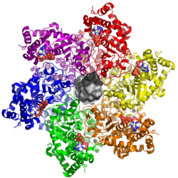
Last updated
The zinc-binding and ATPase/helicase domains of the large tumor antigen in hexameric form, shown with bound ADP (white), zinc (black spheres), and double-stranded DNA (center, light and dark gray). 5tct LTag DNA.png
The zinc-binding and ATPase/helicase domains of the large tumor antigen in hexameric form, shown with bound ADP (white), zinc (black spheres), and double-stranded DNA (center, light and dark gray).

The large tumor antigen (also called the large T-antigen and abbreviated LTag or LT) is a protein encoded in the genomes of polyomaviruses, which are small double-stranded DNA viruses. LTag is expressed early in the infectious cycle and is essential for viral proliferation. Containing four well-conserved protein domains as well as several intrinsically disordered regions, LTag is a fairly large multifunctional protein; in most polyomaviruses, it ranges from around 600-800 amino acids in length. LTag has two primary functions, both related to replication of the viral genome: it unwinds the virus's DNA to prepare it for replication, and it interacts with proteins in the host cell to dysregulate the cell cycle so that the host's DNA replication machinery can be used to replicate the virus's genome. Some polyomavirus LTag proteins - most notably the well-studied SV40 large tumor antigen from the SV40 virus - are oncoproteins that can induce neoplastic transformation in the host cell. [2] [3]

Contents

Domain structure

Polyomavirus LTag proteins contain four well-conserved, globular protein domains: from N- to C-terminus, these are the J domain, the origin-binding domain (OBD), the zinc-binding domain, and the AAA+ ATPase domain. The domains are linked by intrinsically disordered regions, which are themselves often functionally important and whose length varies among polyomaviruses; both the folded globular domains and the disordered regions form protein-protein interactions with a number of host cell proteins. Some LTag homologs also have a disordered C-terminal tail called the host range domain, which can be phosphorylated and in some strains is essential, although the molecular mechanism of its essentiality is unclear. [3]

In some polyomaviruses, truncated variants of the LTag protein are produced through alternative splicing that do not include the helicase (zinc-binding and ATPase) components. These truncated LTags retain their ability to interact with some cell cycle regulatory proteins and are involved in cell transformation but not in viral genome replication. [2] [4]

J domain

The J domain is a DnaJ molecular chaperone that is required for viral genome replication in vivo (but is dispensable in cell-free laboratory experiments). The J domain interacts with Hsc70 heat-shock proteins. In many polyomavirus LTags, N-terminal to the J domain is a sequence motif that mediates binding of LTag to the host cell retinoblastoma protein, a key determinant of cell cycle progression. This unstructured linker region also contains a nuclear localization sequence, which triggers the host cell to transport the protein from the cytoplasm where is it translated to the nucleus where it performs its replication-related functions. [3]

Origin-binding domain

The OBD binds the viral genome's origin of replication by recognizing specific sequences that occur in the portion of the viral genome known as the non-coding control region. It also forms interactions with host cell proteins, such as replication protein A and Nbs1. The OBD is required for viral replication. [3]

Zinc-binding domain

The zinc-binding and ATPase domains together comprise the helicase portion of the LTag protein. The primary function of the zinc-binding domain is oligomerization of LTag. Formation of dodecamer structures (two hexameric rings) is required for helicase activity, which begins at the origin of replication through coordination between the OBD, zinc-binding, and ATPase domains. [2] [3]

ATPase domain

The zinc-binding and helicase domains of LTag shown bound to p53. 2h1l LTag p53.png
The zinc-binding and helicase domains of LTag shown bound to p53.

The ATPase domain is a member of the AAA+ ATPase family and contains conserved motifs such as the ATP-binding Walker A box. Energy from ATP hydrolysis is required for helicase activity. The ATPase domain also contains regions responsible for protein-protein interactions with host cell proteins, most notably topoisomerase 1 and the cell cycle regulator p53. LTag is unique among known AAA+ ATPases in that it is capable of initiating the melting of DNA around the origin; in most such cases a distinct initiator protein is responsible for this step, after which the helicase continues unwinding. [2] [3]

Function

The major functions of LTag in the viral life cycle involve dysregulation of the host cell's cell cycle and replication of the virus's circular DNA genome. Because polyomavirus genome replication relies on the DNA replication machinery of the host cell, the cell must be in S phase (the part of the cell cycle in which the host cell's genome is normally replicated) in order to provide the necessary molecular machinery for viral DNA replication. The SV40 LTag can induce S phase and activate the host cell's DNA damage response. [3] Coordinated actions of the OBD and helicase regions result in physical manipulation of the viral genome, melting the DNA double helix at the origin of replication and unwinding the circular DNA chromosome in a bidirectional fashion. [2] [3] The structure and function of LTag resembles that of the human papillomavirus oncoproteins. [2]

Expression

Genome structure of WU virus, a typical human polyomavirus. The early genes are at left, comprising LTag (purple) and STag (blue); the late genes are at right, and the origin of replication is shown at the top of the figure. Gaynor plospathogens 2007 WUvirusgenome.png
Genome structure of WU virus, a typical human polyomavirus. The early genes are at left, comprising LTag (purple) and STag (blue); the late genes are at right, and the origin of replication is shown at the top of the figure.

LTag is encoded in the "early region" of the polyomavirus genome, so named because this region of the genome is expressed early in the infectious process. (The "late region" contains genes encoding the viral capsid proteins.) The early region typically contains at least two genes and is transcribed as a single messenger RNA processed by alternative splicing. The LTag gene is usually encoded in two exons, of which the first overlaps with the gene for the small tumor antigen (STag); as a result, the two proteins share an N-terminal sequence of around 80 residues, while the remaining ~90 residues of STag are unshared. [4] [7] In a few polyomaviruses - most notably murine polyomavirus, the first member of the family discovered and an efficient oncovirus - an additional protein called middle tumor antigen is expressed from the early region and is highly efficient at cellular transformation. [8]

Cellular transformation

Some, but not all, polyomaviruses are oncoviruses capable of inducing neoplastic transformation in some cells. In oncogenic polyomaviruses, the tumor antigens are responsible for the transformation activity, although the exact molecular mechanisms vary from one virus to another. [3] [2] [9]

SV40

The SV40 large T antigen from the SV40 virus is the most well-studied member of the LTag family. SV40, also known as Macaca mulatta polyomavirus 1, natively infects monkeys and does not cause disease; however, it is oncogenic in some rodents and can immortalize some human cells in primary cell culture. SV40 has three early proteins, the large tumor antigen, the small tumor antigen, and a small protein called 17kT that shares most of its sequence with the N-terminus of LTag. Of these, LTag is primarily responsible for cellular transformation. STag alone cannot transform cells, but improves the efficiency with which LTag performs this function. LTag's transforming effect can largely be attributed to its ability to bind the retinoblastoma protein (Rb) and the tumor suppressor protein p53; abrogating either binding site renders LTag unable to transform primary cultured cells. [10] In fact, p53 - now established as a key driver in carcinogenesis - was originally discovered by its ability to bind LTag. [10] [11] [12]

Murine polyomavirus

Murine polyomavirus (MPyV), described in the 1950s, was the first polyomavirus discovered and can cause tumors in rodents. MPyV has three early proteins; in addition to LTag and STag it also expresses middle tumor antigen, which is primarily responsible for the virus's transforming activity. [10]

Merkel cell polyomavirus

Merkel cell polyomavirus (MCPyV), also known as Human polyomavirus 5, naturally infects humans and is associated with Merkel cell carcinoma (MCC), a rare form of skin cancer originating from Merkel cells. Although MCPyV infection is common and usually asymptomatic, a large majority of MCC tumors possess a genomically integrated copy of the polyomavirus genome. [9] [13] MCPyV possesses four early proteins, including a 57kT alternative splicing isoform and an alternate protein called ALTO. In Merkel cell polyomavirus, unlike in SV40, LTag alone does not support efficient viral replication and STag is required. Comparison of the sequences of MCPyV and SV40 LTag predicts that they have similar capacities for protein-protein interactions, including preservation of the Rb and p53 binding sites. [9] Mutations in MCPyV LTag associated with tumors consist of large C-terminal truncations that eliminate the DNA replication functions of the protein by removing the zinc-binding and ATPase/helicase domains, without affecting these protein-protein interaction sites. [14] [15]

Role in taxonomy

LTag is a large protein whose domains can be detected and annotated bioinformatically. As a result, it is often used in comparing and determining relationships among polyomaviruses. The International Committee on Taxonomy of Viruses currently classifies polyomaviruses primarily according to the sequence identity of their LTag genes. [16] This system has been questioned by phylogenetic studies suggesting that the evolutionary histories of LTag and major capsid protein VP1 are divergent and that some modern polyomavirus represent chimeric lineages. [17]

Related Research Articles

<i>Polyomaviridae</i> Family of viruses

Polyomaviridae is a family of viruses whose natural hosts are primarily mammals and birds. As of 2020, there are six recognized genera and 117 species, five of which are unassigned to a genus. 14 species are known to infect humans, while others, such as Simian Virus 40, have been identified in humans to a lesser extent. Most of these viruses are very common and typically asymptomatic in most human populations studied. BK virus is associated with nephropathy in renal transplant and non-renal solid organ transplant patients, JC virus with progressive multifocal leukoencephalopathy, and Merkel cell virus with Merkel cell cancer.

SV40 is an abbreviation for simian vacuolating virus 40 or simian virus 40, a polyomavirus that is found in both monkeys and humans. Like other polyomaviruses, SV40 is a DNA virus that has the potential to cause tumors in animals, but most often persists as a latent infection. SV40 has been widely studied as a model eukaryotic virus, leading to many early discoveries in eukaryotic DNA replication and transcription.

Oncovirus Viruses that can cause cancer

An oncovirus or oncogenic virus is a virus that can cause cancer. This term originated from studies of acutely transforming retroviruses in the 1950–60s, when the term "oncornaviruses" was used to denote their RNA virus origin. With the letters "RNA" removed, it now refers to any virus with a DNA or RNA genome causing cancer and is synonymous with "tumor virus" or "cancer virus". The vast majority of human and animal viruses do not cause cancer, probably because of longstanding co-evolution between the virus and its host. Oncoviruses have been important not only in epidemiology, but also in investigations of cell cycle control mechanisms such as the retinoblastoma protein.

SV40 large T antigen

SV40 large T antigen is a hexamer protein that is a dominant-acting oncoprotein derived from the polyomavirus SV40. TAg is capable of inducing malignant transformation of a variety of cell types. The transforming activity of TAg is due in large part to its perturbation of the retinoblastoma (pRb) and p53 tumor suppressor proteins. In addition, TAg binds to several other cellular factors, including the transcriptional co-activators p300 and CBP, which may contribute to its transformation function.

Merkel cell polyomavirus was first described in January 2008 in Pittsburgh, Pennsylvania. It was the first example of a human viral pathogen discovered using unbiased metagenomic next-generation sequencing with a technique called digital transcriptome subtraction. MCV is one of seven currently known human oncoviruses. It is suspected to cause the majority of cases of Merkel cell carcinoma, a rare but aggressive form of skin cancer. Approximately 80% of Merkel cell carcinoma (MCC) tumors have been found to be infected with MCV. MCV appears to be a common—if not universal—infection of older children and adults. It is found in respiratory secretions suggesting that it may be transmitted by a respiratory route. But it also can be found shedding from healthy skin, and in gastrointestinal tract tissues and elsewhere, and so its precise mode of transmission remains unknown. In addition, recent studies suggest that this virus may latently infect the human sera and PBMCs.

Trichodysplasia spinulosa polyomavirus is a member virus of Human polyomavirus 8 that infects human hosts. First discovered in 2010, TSPyV is associated with Trichodysplasia spinulosa, a rare skin disease only seen in immunocompromised patients. The virus causes hyperproliferation and enlargement of hair follicles by modulating PP2A protein phosphatase signaling pathways. TSPyV was the eighth human polyomavirus to be discovered, and one of four associated with human disease, out of 13 human polyomaviruses known as of the 2015 update to polyomavirus taxonomy released by the International Committee on Taxonomy of Viruses.

Murine polyomavirus Species of virus

Murine polyomavirus is an unenveloped double-stranded DNA virus of the polyomavirus family. The first member of the family discovered, it was originally identified by accident in the 1950s. A component of mouse leukemia extract capable of causing tumors, particularly in the parotid gland, in newborn mice was reported by Ludwik Gross in 1953 and identified as a virus by Sarah Stewart and Bernice Eddy at the National Cancer Institute, after whom it was once called "SE polyoma". Stewart and Eddy would go on to study related polyomaviruses such as SV40 that infect primates, including humans. These discoveries were widely reported at the time and formed the early stages of understanding of oncoviruses.

Major capsid protein VP1

Major capsid protein VP1 is a viral protein that is the main component of the polyomavirus capsid. VP1 monomers are generally around 350 amino acids long and are capable of self-assembly into an icosahedral structure consisting of 360 VP1 molecules organized into 72 pentamers. VP1 molecules possess a surface binding site that interacts with sialic acids attached to glycans, including some gangliosides, on the surfaces of cells to initiate the process of viral infection. The VP1 protein, along with capsid components VP2 and VP3, is expressed from the "late region" of the circular viral genome.

Hamster polyomavirus is an unenveloped double-stranded DNA virus of the polyomavirus family whose natural host is the hamster. It was originally described in 1967 by Arnold Graffi as a cause of epithelioma in Syrian hamsters.

WU polyomavirus is a virus of the family Polyomaviridae. It was discovered in 2007 in samples of human respiratory secretions, originally from a child patient in Australia who presented with clinical signs of pneumonia and in whom other common respiratory viruses were not detected. Follow-up studies identified the presence of WU virus in respiratory secretion samples from patients in Australia and the United States, suggesting that, like other human polyomaviruses, WU virus is widely distributed.

KI polyomavirus is a virus of the family Polyomaviridae. It was discovered in 2007 in stored samples of human respiratory secretions collected by the Karolinska Institute, after which the virus is named.

Agnoprotein InterPro Family

Agnoprotein is a protein expressed by some members of the polyomavirus family from a gene called the agnogene. Polyomaviruses in which it occurs include two human polyomaviruses associated with disease, BK virus and JC virus, as well as the simian polyomavirus SV40.

Human polyomavirus 7 (HPyV7) is a virus of the polyomavirus family that infects human hosts. It was discovered in 2010 and is a common component of the skin flora in healthy adults. There is limited evidence from case reports linking the virus to a skin rash occurring in immunocompromised organ transplant recipients.

Human polyomavirus 6 (HPyV6) is a virus of the polyomavirus family that infects human hosts. It was discovered in 2010 and is a component of the skin flora in healthy adults.

MW polyomavirus is a virus of the polyomavirus family that infects human hosts. It was discovered in 2012 and reported independently by several research groups. It has been identified mostly in stool samples from children and has been detected in a variety of geographic locations.

STL polyomavirus is a virus of the polyomavirus family that infects human hosts. It was first reported in 2013 and is most closely related to MW polyomavirus. It has been identified mostly in stool samples from children and has been detected in a variety of geographic locations.

Small tumor antigen

The small tumor antigen is a protein encoded in the genomes of polyomaviruses, which are small double-stranded DNA viruses. STag is expressed early in the infectious cycle and is usually not essential for viral proliferation, though in most polyomaviruses it does improve replication efficiency. The STag protein is expressed from a gene that overlaps the large tumor antigen (LTag) such that the two proteins share an N-terminal DnaJ-like domain but have distinct C-terminal regions. STag is known to interact with host cell proteins, most notably protein phosphatase 2A (PP2A), and may activate the expression of cellular proteins associated with the cell cycle transition to S phase. In some polyomaviruses - such as the well-studied SV40, which natively infects monkeys - STag is unable to induce neoplastic transformation in the host cell on its own, but its presence may increase the transforming efficiency of LTag. In other polyomaviruses, such as Merkel cell polyomavirus, which causes Merkel cell carcinoma in humans, STag appears to be important for replication and to be an oncoprotein in its own right.

The middle tumor antigen is a protein encoded in the genomes of some polyomaviruses, which are small double-stranded DNA viruses. MTag is expressed early in the infectious cycle along with two other related proteins, the small tumor antigen and large tumor antigen. MTag occurs only in a few known polyomaviruses, while STag and LTag are universal - it was first identified in mouse polyomavirus (MPyV), the first polyomavirus discovered, and also occurs in hamster polyomavirus. In MPyV, MTag is an efficient oncoprotein that can be sufficient to induce neoplastic transformation in some cells.

Minor capsid protein VP2 and minor capsid protein VP3 are viral proteins that are components of the polyomavirus capsid. Polyomavirus capsids are composed of three proteins; the major component is major capsid protein VP1, which self-assembles into pentamers that in turn self-assemble into enclosed icosahedral structures. The minor components are VP2 and VP3, which bind in the interior of the capsid.

<i>Monodnaviria</i> Realm of viruses

Monodnaviria is a realm of viruses that includes all single-stranded DNA viruses that encode an endonuclease of the HUH superfamily that initiates rolling circle replication of the circular viral genome. Viruses descended from such viruses are also included in the realm, including certain linear single-stranded DNA (ssDNA) viruses and circular double-stranded DNA (dsDNA) viruses. These atypical members typically replicate through means other than rolling circle replication.

References

  1. Gai, Dahai; Wang, Damian; Li, Shu-Xing; Chen, Xiaojiang S. (2016-12-06). "The structure of SV40 large T hexameric helicase in complex with AT-rich origin DNA". eLife. 5. doi:10.7554/eLife.18129. ISSN   2050-084X. PMC   5140265 . PMID   27921994.
  2. 1 2 3 4 5 6 7 Topalis, D.; Andrei, G.; Snoeck, R. (February 2013). "The large tumor antigen: A "Swiss Army knife" protein possessing the functions required for the polyomavirus life cycle". Antiviral Research. 97 (2): 122–136. doi:10.1016/j.antiviral.2012.11.007. PMID   23201316.
  3. 1 2 3 4 5 6 7 8 9 An, Ping; Sáenz Robles, Maria Teresa; Pipas, James M. (13 October 2012). "Large T Antigens of Polyomaviruses: Amazing Molecular Machines". Annual Review of Microbiology. 66 (1): 213–236. doi:10.1146/annurev-micro-092611-150154. PMID   22994493.
  4. 1 2 Moens, U.; Van Ghelue, M.; Johannessen, M. (5 May 2007). "Oncogenic potentials of the human polyomavirus regulatory proteins". Cellular and Molecular Life Sciences. 64 (13): 1656–1678. doi:10.1007/s00018-007-7020-3. PMID   17483871. S2CID   31314244.
  5. Lilyestrom, Wayne; Klein, Michael G.; Zhang, Rongguang; Joachimiak, Andrzej; Chen, Xiaojiang S. (2006-09-01). "Crystal structure of SV40 large T-antigen bound to p53: interplay between a viral oncoprotein and a cellular tumor suppressor". Genes & Development. 20 (17): 2373–2382. doi:10.1101/gad.1456306. ISSN   0890-9369. PMC   1560412 . PMID   16951253.
  6. Gaynor, Anne M.; Nissen, Michael D.; Whiley, David M.; Mackay, Ian M.; Lambert, Stephen B.; Wu, Guang; Brennan, Daniel C.; Storch, Gregory A.; Sloots, Theo P. (2007-05-04). "Identification of a novel polyomavirus from patients with acute respiratory tract infections". PLOS Pathogens. 3 (5): e64. doi:10.1371/journal.ppat.0030064. ISSN   1553-7374. PMC   1864993 . PMID   17480120.
  7. Van Ghelue, Marijke; Khan, Mahmud Tareq Hassan; Ehlers, Bernhard; Moens, Ugo (November 2012). "Genome analysis of the new human polyomaviruses". Reviews in Medical Virology. 22 (6): 354–377. doi: 10.1002/rmv.1711 . PMID   22461085.
  8. Fluck, M. M.; Schaffhausen, B. S. (31 August 2009). "Lessons in Signaling and Tumorigenesis from Polyomavirus Middle T Antigen". Microbiology and Molecular Biology Reviews. 73 (3): 542–563. doi:10.1128/MMBR.00009-09. PMC   2738132 . PMID   19721090.
  9. 1 2 3 Stakaitytė, Gabrielė; Wood, Jennifer J.; Knight, Laura M.; Abdul-Sada, Hussein; Adzahar, Noor Suhana; Nwogu, Nnenna; Macdonald, Andrew; Whitehouse, Adrian (2014-06-27). "Merkel Cell Polyomavirus: Molecular Insights into the Most Recently Discovered Human Tumour Virus". Cancers. 6 (3): 1267–1297. doi: 10.3390/cancers6031267 . PMC   4190541 . PMID   24978434.
  10. 1 2 3 Cheng, Jingwei; DeCaprio, James A.; Fluck, Michele M.; Schaffhausen, Brian S. (2009). "Cellular transformation by Simian Virus 40 and Murine Polyoma Virus T antigens". Seminars in Cancer Biology. 19 (4): 218–228. doi:10.1016/j.semcancer.2009.03.002. PMC   2694755 . PMID   19505649.
  11. Lane, D. P.; Crawford, L. V. (1979-03-15). "T antigen is bound to a host protein in SY40-transformed cells". Nature. 278 (5701): 261–263. Bibcode:1979Natur.278..261L. doi:10.1038/278261a0. PMID   218111. S2CID   4357967.
  12. Linzer, Daniel I.H.; Levine, Arnold J. (1979). "Characterization of a 54K Dalton cellular SV40 tumor antigen present in SV40-transformed cells and uninfected embryonal carcinoma cells". Cell. 17 (1): 43–52. doi:10.1016/0092-8674(79)90293-9. PMID   222475. S2CID   38710686.
  13. Feng, Huichen; Shuda, Masahiro; Chang, Yuan; Moore, Patrick S. (2008-02-22). "Clonal Integration of a Polyomavirus in Human Merkel Cell Carcinoma". Science. 319 (5866): 1096–1100. Bibcode:2008Sci...319.1096F. doi:10.1126/science.1152586. ISSN   0036-8075. PMC   2740911 . PMID   18202256.
  14. Shuda, Masahiro; Feng, Huichen; Kwun, Hyun Jin; Rosen, Steven T.; Gjoerup, Ole; Moore, Patrick S.; Chang, Yuan (2008-10-21). "T antigen mutations are a human tumor-specific signature for Merkel cell polyomavirus". Proceedings of the National Academy of Sciences. 105 (42): 16272–16277. Bibcode:2008PNAS..10516272S. doi: 10.1073/pnas.0806526105 . ISSN   0027-8424. PMC   2551627 . PMID   18812503.
  15. Wendzicki, Justin A.; Moore, Patrick S.; Chang, Yuan (2015-04-01). "Large T and small T antigens of Merkel cell polyomavirus". Current Opinion in Virology. 11: 38–43. doi:10.1016/j.coviro.2015.01.009. ISSN   1879-6265. PMC   4456251 . PMID   25681708.
  16. Calvignac-Spencer, Sébastien; Feltkamp, Mariet C. W.; Daugherty, Matthew D.; Moens, Ugo; Ramqvist, Torbjörn; Johne, Reimar; Ehlers, Bernhard (29 February 2016). "A taxonomy update for the family Polyomaviridae". Archives of Virology. 161 (6): 1739–1750. doi: 10.1007/s00705-016-2794-y . PMID   26923930.
  17. Buck, Christopher B.; Doorslaer, Koenraad Van; Peretti, Alberto; Geoghegan, Eileen M.; Tisza, Michael J.; An, Ping; Katz, Joshua P.; Pipas, James M.; McBride, Alison A. (2016-04-19). "The Ancient Evolutionary History of Polyomaviruses". PLOS Pathogens. 12 (4): e1005574. doi:10.1371/journal.ppat.1005574. ISSN   1553-7374. PMC   4836724 . PMID   27093155.