Related Research Articles

In mammals and other animals, the vagina is the elastic, muscular reproductive organ of the female genital tract. In humans, it extends from the vulval vestibule to the cervix. The vaginal introitus is normally partly covered by a thin layer of mucosal tissue called the hymen. The vagina allows for copulation and birth. It also channels menstrual flow, which occurs in humans and closely related primates as part of the menstrual cycle.
The "fruit machine" was a battery of psychological tests developed in Canada by Dr. Frank Robert Wake, a psychology professor with Carleton University in the 1960s. It was hoped that Dr. Wake's research program would be able to help the Government of Canada identify gay men working in the Public Service or to prevent gay people from obtaining government jobs. The subjects were made to view erotic imagery; "homosexual words," as well as an early form of lie detector to measure perspiration and pulse. The so-called machine was supposed to measure the subject's pupil dilation, in response to the erotic images and words. The crude apparatus was constructed by the RCMP's Identification Branch.

The G-spot, also called the Gräfenberg spot, is characterized as an erogenous area of the vagina that, when stimulated, may lead to strong sexual arousal, powerful orgasms and potential female ejaculation. It is typically reported to be located 5–8 cm (2–3 in) up the front (anterior) vaginal wall between the vaginal opening and the urethra and is a sensitive area that may be part of the female prostate.

Doggy style is a sex position in which one participant bends over, crouches on all fours, or lies on their abdomen, for sexual intercourse, other forms of sexual penetration or other sexual activity. Doggy style is a form of rear-entry position, others being the spoons sex position in which the receiving partner lies on their side or the reverse cowgirl sex position. Non-penetrative sex in this position may also be regarded as doggy style.
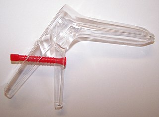
A speculum is a medical tool for investigating body orifices, with a form dependent on the orifice for which it is designed. In old texts, the speculum may also be referred to as a diopter or dioptra. Like an endoscope, a speculum allows a view inside the body; endoscopes, however, tend to have optics while a speculum is intended for direct vision.

Sexual stimulation is anything that leads to sexual arousal or orgasm. This thing can be physical or of other senses, and is known as a stimulus.
Penile plethysmography (PPG) or phallometry is a measurement of blood flow to the penis, typically used as a proxy for measurement of sexual arousal. The most commonly reported methods of conducting penile plethysmography involves the measurement of the circumference of the penis with a mercury-in-rubber or electromechanical strain gauge, or the volume of the penis with an airtight cylinder and inflatable cuff at the base of the penis. Corpora cavernosa nerve penile plethysmographs measure changes in response to inter-operative electric stimulation during surgery. The volumetric procedure was invented by Kurt Freund and is considered to be particularly sensitive at low arousal levels. The easier to use circumferential measures are more widely used, however, and more common in studies using erotic film stimuli. A corresponding device in women is the vaginal photoplethysmograph.
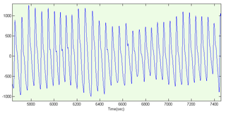
A photoplethysmogram (PPG) is an optically obtained plethysmogram that can be used to detect blood volume changes in the microvascular bed of tissue. A PPG is often obtained by using a pulse oximeter which illuminates the skin and measures changes in light absorption. A conventional pulse oximeter monitors the perfusion of blood to the dermis and subcutaneous tissue of the skin.
The human sexual response cycle is a four-stage model of physiological responses to sexual stimulation, which, in order of their occurrence, are the excitement, plateau, orgasmic, and resolution phases. This physiological response model was first formulated by William H. Masters and Virginia E. Johnson, in their 1966 book Human Sexual Response. Since that time, other models regarding human sexual response have been formulated by several scholars who have criticized certain inaccuracies in the human sexual response cycle model.
Sexual arousal disorder is characterized by a lack or absence of sexual fantasies and desire for sexual activity in a situation that would normally produce sexual arousal, or the inability to attain or maintain typical responses to sexual arousal. The disorder is found in the DSM-IV. The condition should not be confused with a sexual desire disorder.

Vaginal discharge is a mixture of liquid, cells, and bacteria that lubricate and protect the vagina. This mixture is constantly produced by the cells of the vagina and cervix, and it exits the body through the vaginal opening. The composition, amount, and quality of discharge varies between individuals and can vary throughout the menstrual cycle and throughout the stages of sexual and reproductive development. Normal vaginal discharge may have a thin, watery consistency or a thick, sticky consistency, and it may be clear or white in color. Normal vaginal discharge may be large in volume but typically does not have a strong odor, nor is it typically associated with itching or pain. While most discharge is considered physiologic or represents normal functioning of the body, some changes in discharge can reflect infection or other pathological processes. Infections that may cause changes in vaginal discharge include vaginal yeast infections, bacterial vaginosis, and sexually transmitted infections. The characteristics of abnormal vaginal discharge vary depending on the cause, but common features include a change in color, a foul odor, and associated symptoms such as itching, burning, pelvic pain, or pain during sexual intercourse.

A G-spot vibrator is a sex toy with female and male varieties. The female version of the device is built to massage the G-spot, described as a bean-shaped area of the vagina. Some women report that it is an erogenous zone which, when stimulated, can lead to strong sexual arousal, powerful orgasms and female ejaculation. The male version of the G-spot vibrator is used for massaging the prostate for both sexual and health-related reasons.

Clitoral erection is a physiological phenomenon where the clitoris becomes enlarged and firm.

Sexual arousal describes the physiological and psychological responses in preparation for sexual intercourse or when exposed to sexual stimuli. A number of physiological responses occur in the body and mind as preparation for sexual intercourse, and continue during intercourse. Male arousal will lead to an erection, and in female arousal, the body's response is engorged sexual tissues such as nipples, clitoris, vaginal walls, and vaginal lubrication.
Clitoral photoplethysmography uses light to measure clitoral blood volume (CBV).
The labial thermistor clip is a device used measure the skin temperature of the labia minora and is associated blood engorgement. This device consists of a thermistor affixed to a small metal clip that can be attached to the labia minora. The labial thermistor clip is the second most commonly used physiological measure of female genital response, next to the vaginal photoplethysmograph (VPG). Both devices can be used simultaneously. The labial thermistor clip has some advantages over VPG, including better test-retest reliability, greater correlation between genital and self-reported sexual arousal, and an absolute unit of change (temperature). Like VPG, the labial thermistor clip has discriminant validity; that is, it detects differences between sexual and nonsexual stimuli. It is also sensitive to different levels of sexual arousal. The labial thermistor clip has some disadvantages because participants have difficulty with placing the device correctly and some report discomfort with using the device.

Laser Doppler imaging (LDI) is an imaging method that uses a laser beam to image live tissue. When the laser light reaches the tissue, the moving blood cells generate Doppler components in the reflected (backscattered) light. The light that comes back is detected using a photodiode that converts it into an electrical signal. Then the signal is processed to calculate a signal that is proportional to the tissue perfusion in the imaged area. When the process is completed, the signal is processed to generate an image that shows the perfusion on a screen.

The dimensions and shape of human vaginas are of great importance in medicine and surgery, in addition to their relevance to sexual pleasure and childbirth; there appears to be no one way, however, to characterize the vagina's size and shape. In addition to variations from individual to individual, the size and shape of a vagina in the baseline state can vary substantially during sexual arousal and intercourse.
Sexual concordance refers to the degree of correlation between subjective sexual arousal and physiological genital response. This phenomenon is often studied within the fields of sexology and psychology to understand the complex relationship between the mind and body during sexual activity.

Meredith L. Chivers is a Canadian sexologist and clinical psychologist noted for her research on female sexuality, sexual orientation, paraphilias, sex differences, gender identity, and the physiology of sexual arousal. She is an associate professor in the Department of Psychology at Queen's University in Kingston, Ontario, Canada.
References
- 1 2 3 Chivers ML, Seto MC, Lalumière ML, Laan E, Grimbos T (2010). "Agreement of Self-Reported and Genital Measures of Sexual Arousal in Men and Women: A Meta-Analysis". Archives of Sexual Behavior. 39 (1): 5–56. doi:10.1007/s10508-009-9556-9. PMC 2811244 . PMID 20049519.
- 1 2 3 Jannsen, Erick; Prause, Nicole; Geer, James H. (2007). "Chapter 11: The Sexual Response". In Cacioppo, John T.; Tassinary, Louis G.; Berntson, Gary (eds.). Handbook of Psychophysiology. Cambridge University Press. p. 254. ISBN 9781139461931.
- 1 2 Hatch, J. P. [“Vaginal photoplethysmography: Methodological considerations”],”Archives of Sexual Behavior, 8, 357–374”, 1979
- ↑ Laan, E., Everaerd, W., & Evers, A. “Assessment of female sexual arousal: Response specificity and construct validity”, “Psychophysiology, 32, 476–485”, 1995
- 1 2 Molenkamp, Bert (2016). "SexLab - Equipment & Instruments - Vaginal Photoplethysmography". Indiana University. Retrieved 10 February 2018.
- ↑ Palti, Y. & Bercovici, B. [“Photoplethysmographic study of the vaginal blood pulse”], “American Journal of Obstetrics & Gynecology, 97, 143–53”, 1967
- ↑ Sintchak, G; Geer, JH (January 1975). "A vaginal plethysmograph system". Psychophysiology. 12 (1): 113–5. doi:10.1111/j.1469-8986.1975.tb03074.x. PMID 1114202.