Birdshot chorioretinopathy

Last updated
Birdshot chorioretinopathy (HLA-A29 uveitis)
Other namesMultiple small, cream-colored lesions, symmetrically scattered mainly around the optic disk, Vitiliginous choroiditis
Introductieslide.png
Characteristic hypo-pigmentation in birdshot chorioretinopathy
Specialty Ophthalmology   OOjs UI icon edit-ltr-progressive.svg

Birdshot chorioretinopathy, now commonly named birdshot uveitis or HLA-A29 uveitis, [1] is a rare form of bilateral posterior uveitis affecting both eyes. It causes severe, progressive inflammation of both the choroid and retina. [2] [3] [4]

Contents

Affected individuals are almost exclusively Caucasian and usually diagnosed in the fourth to sixth decade of their lives. [5]

Symptoms and signs

Symptoms of this disorder include floaters, blurred vision, photopsia (flashing lights in eyes), loss of color vision and nyctalopia. In an eye examination, light-colored spots on the retina are seen. Complete loss of visual acuity may happen[ citation needed ] The name of the condition comes from the small light-colored fundus spots on the retina, scattered in a pattern like birdshot from a shotgun, but these spots might not be present in early stages.[ citation needed ]

Pathophysiology

Birdshot chorioretinopathy is a rare form of posterior uveitis and accounts for 1–3% of uveitis cases in general. Birdshot chorioretinopathy is thought to be an autoimmune disease. The disease has strong association with the human leukocyte antigen haplotype (HLA)-A29, which is the strongest association between a disease and HLA class I documented (>99% of patients are HLA-A29 positive by molecular testing and HLA-A29-negative cases are controversial [1] ). This indicates a role for T-lymphocytes in the pathogenesis. Birdshot chorioretinopathy is associated with IL-17, a hallmark cytokine of TH17 cells that play an important role in autoimmunity. [6] [7] The disease affects typically middle-aged or elderly caucasians. HLA-A29 is less prevalent in Asia and no birdshot chorioretinopathy cases have been reported in Asia. When birdshot chorioretinopathy is suspected, a person is usually tested to determine if they are HLA-A29 positive. Although previously HLA-A29 testing was not considered necessary for definitive diagnosis, because HLA-A29 is also common in the general healthy population (7%). An increasing number of specialists consider the presence of HLA-A29 critical for diagnosis. [1] Additional (genetic or environmental) or unknown factors may be associated with HLA-A29 in the pathogenesis.

In 2014, Kuiper et al. conducted a genome-wide association study in birdshot chorioretinopathy and studied the entire genome of Dutch, Spanish and English patients. This large genetic study ascertained HLA-A29:02 as the primary risk factor and identified the endoplasmic reticulum aminopeptidase (ERAP) 2 gene strongly associated with birdshot chorioretinopathy. [8] Genetic variants near ERAP2 on chromosome 5 resulted in high mRNA and protein expression of this aminopeptidase in BSCR patients. ERAP2 is an aminopeptidase that, together with the closely related ERAP1, trims peptides in the endoplasmic reticulum and loads these peptides on HLA molecules for presentation to T cells of the immune system. ERAP-HLA associations have also been reported in Ankylosing spondylitis and Behcet's disease, suggesting shared pathogenic pathways among these diseases. The combined Birdshot risk polymorphisms at 5q15 linked to ERAP1 and ERAP2 genes (T-rs10044354-C-rs2287987) are only observed in HLA-A29-positive individuals in populations in which BU is "endemic", explaining why BU is reported merely in populations of Western European Ancestry (Spain, France, The Netherlands, Great Britain, but also non-european countries such as Puerto Rico). [9] Functional analysis of patient-derived immune cells revealed that ERAP2 increases the abundance of peptides that specifically bind to HLA-A29 and further supports that the antigen presentation pathway drives the etiology of BSCR. [10]

Diagnosis

Treatment

Birdshot chorioretinopathy may show resistance to treatment. Immunosuppressant therapy along with oral corticosteroid has been somewhat effective in slowing down the progressive inflammation associated with the disorder, preserving visual integrity as much as possible. Long-term use of such medications must be closely monitored, however, due to the discomforting and potentially debilitating and life-threatening side-effects. [11] [12]

Immunosuppressive drugs such as the therapeutic monoclonal antibody daclizumab, ciclosporin, methotrexate and Adalimumab, sold under the trade name Humira, have proven to be effective treatment options for birdshot chorioretinopathy. Substantial reduction and even stabilization of both vitreous inflammation and retinal vasculitis have been evident via electroretinography, during daclizumab (IL-2 receptor blocker) therapy. This is also supported by the observation of elevated levels of IL-2 in the eyes of patients. [6] Loss of visual acuity unrelated to the inflammation caused by the disorder, however, often remains unchanged despite usage of the drug. This is reflected by the lack of difference in visual acuity and the vision-related quality of life among various treatment categories in birdshot patients. [13] Contraindications and adverse side-effects are always a factor, as well. [14]

A corticosteroid implant surgically placed within the eye can be used for patients who are not able to achieve remission on or tolerate immunomodulatory therapy, or in cases where this option presents less of a burden on life than long-term medical therapy.

Related Research Articles

<span class="mw-page-title-main">Chorioretinitis</span> Medical condition

Chorioretinitis is an inflammation of the choroid and retina of the eye. It is a form of posterior uveitis. Inflammation of these layers can lead to vision-threatening complications. If only the choroid is inflamed, not the retina, the condition is termed choroiditis. The ophthalmologist's goal in treating these potentially blinding conditions is to eliminate the inflammation and minimize the potential risk of therapy to the patient.

Daclizumab is a therapeutic humanized monoclonal antibody which was used for the treatment of adults with relapsing forms of multiple sclerosis (MS). Daclizumab works by binding to CD25, the alpha subunit of the IL-2 receptor of T-cells.
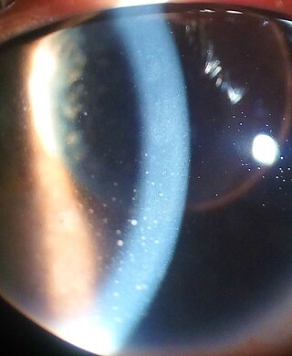
<span class="mw-page-title-main">Uveitis</span> Inflammation of the uvea of the eye

Uveitis is inflammation of the uvea, the pigmented layer of the eye between the inner retina and the outer fibrous layer composed of the sclera and cornea. The uvea consists of the middle layer of pigmented vascular structures of the eye and includes the iris, ciliary body, and choroid. Uveitis is described anatomically, by the part of the eye affected, as anterior, intermediate or posterior, or panuveitic if all parts are involved. Anterior uveitis (iridocyclitis) is the most common, with the incidence of uveitis overall affecting approximately 1:4500, most commonly those between the ages of 20-60. Symptoms include eye pain, eye redness, floaters and blurred vision, and ophthalmic examination may show dilated ciliary blood vessels and the presence of cells in the anterior chamber. Uveitis may arise spontaneously, have a genetic component, or be associated with an autoimmune disease or infection. While the eye is a relatively protected environment, its immune mechanisms may be overcome resulting in inflammation and tissue destruction associated with T-cell activation.

<span class="mw-page-title-main">Central serous chorioretinopathy</span> Eye disease characterized by leakage of fluid under the retina

Central serous chorioretinopathy, also known as central serous retinopathy (CSR), is an eye disease that causes visual impairment, often temporary, usually in one eye. When the disorder is active it is characterized by leakage of fluid under the retina that has a propensity to accumulate under the central macula. This results in blurred or distorted vision (metamorphopsia). A blurred or gray spot in the central visual field is common when the retina is detached. Reduced visual acuity may persist after the fluid has disappeared.

<span class="mw-page-title-main">Choroideremia</span> Medical condition

Choroideremia is a rare, X-linked recessive form of hereditary retinal degeneration that affects roughly 1 in 50,000 males. The disease causes a gradual loss of vision, starting with childhood night blindness, followed by peripheral vision loss and progressing to loss of central vision later in life. Progression continues throughout the individual's life, but both the rate of change and the degree of visual loss are variable among those affected, even within the same family.
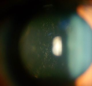
<span class="mw-page-title-main">Intermediate uveitis</span> Medical condition

Intermediate uveitis is a form of uveitis localized to the vitreous and peripheral retina. Primary sites of inflammation include the vitreous of which other such entities as pars planitis, posterior cyclitis, and hyalitis are encompassed. Intermediate uveitis may either be an isolated eye disease or associated with the development of a systemic disease such as multiple sclerosis or sarcoidosis. As such, intermediate uveitis may be the first expression of a systemic condition. Infectious causes of intermediate uveitis include Epstein–Barr virus infection, Lyme disease, HTLV-1 virus infection, cat scratch disease, and hepatitis C.

<span class="mw-page-title-main">HLA-DR1</span>

HLA-DR1 (DR1) is a HLA-DR serotype that recognizes the DRB1*01 gene products. It has been observed to be common among centenarians.

<span class="mw-page-title-main">Acute posterior multifocal placoid pigment epitheliopathy</span> Eye disease causing lesions in retina

Acute posterior multifocal placoid pigment epitheliopathy (APMPPE) is an acquired inflammatory uveitis that belongs to the heterogenous group of white dot syndromes in which light-coloured (yellowish-white) lesions begin to form in the macular area of the retina. Early in the course of the disease, the lesions cause acute and marked vision loss that ranges from mild to severe but is usually transient in nature. APMPPE is classified as an inflammatory disorder that is usually bilateral and acute in onset but self-limiting. The lesions leave behind some pigmentation, but visual acuity eventually improves even without any treatment.
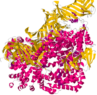
<span class="mw-page-title-main">ERAP1</span> Protein-coding gene in the species Homo sapiens

Type 1 tumor necrosis factor receptor shedding aminopeptidase regulator, also known as endoplasmic reticulum aminopeptidase 1 (ARTS-1), is a protein which in humans is encoded by the ARTS-1 gene.

Acute retinal necrosis (ARN) is a medical inflammatory condition of the eye. The condition presents itself as a necrotizing retinitis. The inflammation onset is due to certain herpes viruses, varicella zoster virus (VZV), herpes simplex virus (HSV-1 and HSV-2) and Epstein–Barr virus (EBV).
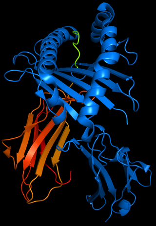
<span class="mw-page-title-main">HLA-B51</span> Serotype

HLA-B51 (B51) is an HLA-B serotype. The serotype identifies the more common HLA-B*51 gene products.

White dot syndromes are inflammatory diseases characterized by the presence of white dots on the fundus, the interior surface of the eye. The majority of individuals affected with white dot syndromes are younger than fifty years of age. Some symptoms include blurred vision and visual field loss. There are many theories for the etiology of white dot syndromes including infectious, viral, genetics and autoimmune.

Douglas Jabs is an American ophthalmologist and an expert in clinical research in the fields of ophthalmology and uveitis.

<span class="mw-page-title-main">Retinal vasculitis</span> Medical condition

Retinal vasculitis is inflammation of the vascular branches of the retinal artery, caused either by primary ocular disease processes, or as a specific presentation of any systemic form of vasculitis such as Behçet's disease, sarcoidosis, multiple sclerosis, or any form of systemic necrotizing vasculitis such as temporal arteritis, polyarteritis nodosa, and granulomatosis with polyangiitis, or due to lupus erythematosus, or rheumatoid arthritis. Eales disease, pars planitis, birdshot retinochoroidopathy, and Fuchs heterochromic iridocyclitis (FHI) can also cause retinal vasculitis. Infectious pathogens such as Mycobacterium tuberculosis, visceral larva migrans can also cause retinal vasculitis. Drug-induced vasculitis may involve retina as well, as seen in methamphetamine induced vasculitis.

<span class="mw-page-title-main">Behçet's disease</span> Inflammatory disorder

Behçet's disease (BD) is a type of inflammatory disorder which affects multiple parts of the body. The most common symptoms include painful sores on the mucous membranes of the mouth and other parts of the body, inflammation of parts of the eye, and arthritis. The sores can last from a few days, up to a week or more. Less commonly there may be inflammation of the brain or spinal cord, blood clots, aneurysms, or blindness. Often, the symptoms come and go.

<span class="mw-page-title-main">Toxoplasmic chorioretinitis</span> Type of eye infection

Toxoplasma chorioretinitis, more simply known as ocular toxoplasmosis, is possibly the most common cause of infections in the back of the eye worldwide. The causitive agent is Toxoplasma gondii, and in the United States, most cases are acquired congenitally. The most common symptom is decreased visual acuity in one eye. The diagnosis is made by examination of the eye, using ophthalmoscopy. Sometimes serologic testing is used to rule out the disease, but due to high rates of false positives, serologies are not diagnostic of toxoplasmic retinitis.

Multiple evanescent white dot syndrome (MEWDS) is an uncommon inflammatory condition of the retina that typically affects otherwise healthy young females in the second to fourth decades of life.
<span class="mw-page-title-main">Vogt–Koyanagi–Harada disease</span> Medical condition

Vogt–Koyanagi–Harada disease (VKH) is a multisystem disease of presumed autoimmune cause that affects melanin-pigmented tissues. The most significant manifestation is bilateral, diffuse uveitis, which affects the eyes. VKH may variably also involve the inner ear, with effects on hearing, the skin, and the meninges of the central nervous system.

Occult macular dystrophy (OMD) is a rare inherited degradation of the retina, characterized by progressive loss of function in the most sensitive part of the central retina (macula), the location of the highest concentration of light-sensitive cells (photoreceptors) but presenting no visible abnormality. "Occult" refers to the degradation in the fundus being difficult to discern. The disorder is called "dystrophy" instead of "degradation" to distinguish its genetic origin from other causes, such as age. OMD was first reported by Y. Miyake et al. in 1989.

<span class="mw-page-title-main">Uveitic glaucoma</span> Glaucoma caused by uveitis or its treatments

Uveitic glaucoma is most commonly a progression stage of noninfectious anterior uveitis or iritis.

References

  1. 1 2 3 Herbort CP et al. (2017). "Why birdshot retinochoroiditis should rather be called 'HLA-A29 uveitis'?". Br J Ophthalmol. 101 (7): 2016–309764. doi:10.1136/bjophthalmol-2016-309764. PMC   5530806 . PMID   28314830.
  2. RESERVED, INSERM US14 -- ALL RIGHTS. "Orphanet: Birdshot chorioretinopathy". www.orpha.net. Retrieved 2017-07-02.{{cite web}}: CS1 maint: numeric names: authors list (link)
  3. "Birdshot chorioretinopathy | Genetic and Rare Diseases Information Center (GARD) – an NCATS Program". rarediseases.info.nih.gov. Archived from the original on 2018-03-04. Retrieved 2017-07-02.
  4. "Birdshot Retinopathy: Background, Pathophysiology, Epidemiology". 2017-05-02.{{cite journal}}: Cite journal requires |journal= (help)
  5. Cassoux N, Lehoan GP (2000). "Birdshot retinochoroidopathy". Ann Med Interne. 151 (Suppl. 1): 1s45–1s47. PMID   10896989.
  6. 1 2 Kuiper JJW, Mutis T, de Jager W, de Groot-Mijnes JD, Rothova A (2011). "Intraocular interleukin-17 and proinflammatory cytokines in HLA-A29-associated birdshot chorioretinopathy". Am J Ophthalmol. 152 (2): 177–182. doi:10.1016/j.ajo.2011.01.031. PMID   21570674.
  7. Kuiper JJW, Emmelot ME, Rothova A, Mutis T (2013). "Interleukin-17 production and T helper 17 cells in peripheral blood mononuclear cells in response to ocular lysate in patients with birdshot chorioretinopathy". Mol Vis. 19: 2606–14. PMC   3874049 . PMID   24379648.
  8. Kuiper JJW, van Setten J, Ripke S, Van't Slot R, Mulder F, Missotten T, Baarsma GS, Francioli LC, Pulit SL, de Kovel CG, Ten Dam-van Loon N, den Hollander AI, Huis In Het Veld P, Hoyng CB, Cordero-Coma M, Martín J, Llorenç V, Arya B, Thomas D, Bakker SC, Ophoff RA, Rothova A, de Bakker PI, Mutis T, Koeleman BP (2014). "A genome-wide association study identifies a functional ERAP2 haplotype associated with birdshot chorioretinopathy". Hum Mol Genet. 23 (22): 6081–6087. doi:10.1093/hmg/ddu307. PMC   4204766 . PMID   24957906.
  9. Kuiper JJW, Venema WJ (2020). "HLA-A29 and Birdshot Uveitis: Further Down the Rabbit Hole". Front. Immunol. 11: 599558. doi: 10.3389/fimmu.2020.599558 . PMC   7687429 . PMID   33262772.
  10. Venema WJ, Hiddingh S, De Boer JH, Claas FH, Mulder A, Den Hollander AI, Stratikos E, Sarkizova S, Van Der Veken LT, Janssen GM, Van Veelen PA, Kuiper JJ (2021). "ERAP2 increases the abundance of a distinct peptide submotif highly selective for the Birdshot Uveitis-associated HLA-A29". Front. Immunol. 12: 634441. doi: 10.3389/fimmu.2021.634441 . PMC   7950316 . PMID   33717175.
  11. Kiss S, Ahmed M, Letko E, Foster CS (2005). "Long-term follow-up of patients with birdshot retinochoroidopathy treated with corticosteroid-sparing systemic immunomodulatory therapy". Ophthalmology. 112 (6): 1066–1071. doi:10.1016/j.ophtha.2004.12.036. PMID   15936442.
  12. Becker MD, Wertheim MS, Smith JR, Rosenbaum JT (2005). "Long-term follow-up of patients with birdshot retinochoroidopathy treated with systemic immunosuppressants". Ocul Immuno Inflamm. 13 (4): 289–293. doi:10.1080/09273940490912407. PMID   16159719. S2CID   1554209.
  13. Kuiper JJW, Missotten T, Baarsma SG, Rothova A (2013). "Vision-related quality of life in patients with birdshot chorioretinopathy". Acta Ophthalmol. 91 (1): 177–182. doi:10.1111/aos.12054. PMID   23289541. S2CID   6475153.
  14. Sobrin L, Huang JJ, Christen W, Kafkala C, Choopong P, Foster CS (2008). "Daclizumab for treatment of birdshot chorioretinopathy". Arch Ophthalmol. 126 (2): 186–191. doi:10.1001/archophthalmol.2007.49. PMID   18268208.