Orbital cellulitis

Last updated
Orbital cellulitis
Orbital cellulitis.jpg
Orbital Cellulitis
Specialty Ophthalmology   OOjs UI icon edit-ltr-progressive.svg

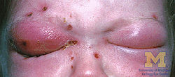
Orbital cellulitis is inflammation of eye tissues behind the orbital septum. It is most commonly caused by an acute spread of infection into the eye socket from either the adjacent sinuses or through the blood. It may also occur after trauma. When it affects the rear of the eye, it is known as retro-orbital cellulitis.

Contents

It should not be confused with periorbital cellulitis, which refers to cellulitis anterior to the septum.

Without proper treatment, orbital cellulitis may lead to serious consequences, including permanent loss of vision or even death.

Signs and symptoms

Orbital cellulitis commonly presents with painful eye movement, sudden vision loss, chemosis, bulging of the infected eye, and limited eye movement. Along with these symptoms, patients typically have redness and swelling of the eyelid, pain, discharge, inability to open the eye, occasional fever and lethargy.

Complications

Complications include hearing loss, blood infection, meningitis, cavernous sinus thrombosis, cerebral abscess, and blindness. [1] [2] It is possible that children experience more severe complications due to their immature immune system and because they have thinner orbital bones, which makes the infection easier to spread. [3] [4]

Causes

Gram-positive stain, possibly showing staphylococcus aureus, which is one of the primary causes of orbital cellulitis. Gram-positive stain.jpg
Gram-positive stain, possibly showing staphylococcus aureus, which is one of the primary causes of orbital cellulitis.

Orbital cellulitis occurs commonly from bacterial infection spread via the paranasal sinuses, usually from a previous sinus infection. Other ways in which orbital cellulitis may occur are from blood stream infections or from eyelid skin infections. Upper respiratory infection, sinus infection, trauma to the eye, ocular or periocular infection, and systemic infection all increase one's risk of orbital cellulitis.

Staphylococcus aureus , Haemophilus influenzae B, Moraxella catarrhalis, Streptococcus pneumoniae, and beta-hemolytic streptococci are bacteria that can be responsible for orbital cellulitis. [5] [4]

Risk Factors

Risk factors for the development of orbital cellulitis include, but are not limited to: [6] [7]

Diagnosis

Early diagnosis of orbital cellulitis is urgent, and it involves a complete and thorough physical examination. Common presenting signs include: a protruding eye (proptosis), eyelid edema (swelling), eye pain, vision loss, inability to move the eye completely (ophthalmoplegia), and fever. It is important to correlate physical findings with patient history and reported symptoms. [8]

CT scan and MRI of the orbits are two imaging modalities that are commonly used to aid in the diagnosis and monitoring of orbital cellulitis, as they can provide detailed images that can show the extent of inflammation along with possible abscess location, size, and involvement of surrounding structures. [3] Ultrasound has also been used as an imaging modality in the past, but it cannot provide the same level of detail as CT or MRI. [3]

Blood cultures, electrolytes, and a complete blood count (CBC) with differential showing elevated white blood cell count is a useful laboratory test that may aid in diagnosis. [8] [5]

Differential Diagnosis

A variety of pathologies and diseases can present similarly to orbital cellulitis, including: [9]

Treatment

Immediate treatment is very important, and it typically involves intravenous (IV) antibiotics in the hospital and frequent observation (every 4–6 hours). [2] [10] Several lab tests should be ordered, including a complete blood count, differential, and blood culture.

Prognosis

Although orbital cellulitis is considered an ophthalmic emergency, the prognosis is good if prompt medical treatment is received.

Death and blindness rates without treatment

Bacterial infections of the orbit have long been associated with a risk of devastating outcomes and intracranial spread.

The natural course of the disease, as documented by Gamble (1933), in the pre-antibiotic era, resulted in death in 17% of patients and permanent blindness in 20%. [12]

Epidemiology

Orbital cellulitis is an uncommon medical condition, with the reported rates being much higher among the pediatric population compared to the adult population. [3] One study reported that children are approximately 16 times more likely to suffer from orbital cellulitis compared to adults. [13] It is twice as common among male children compared to female children. [1] Some studies reported that orbital cellulitis follows a seasonal pattern, with the highest rates occurring during the fall and winter, which coincides with the higher rates of sinus infection during the colder months. [14]

Related Research Articles

<span class="mw-page-title-main">Abscess</span> Localized collection of pus that has built up within the tissue of the body

An abscess is a collection of pus that has built up within the tissue of the body. Signs and symptoms of abscesses include redness, pain, warmth, and swelling. The swelling may feel fluid-filled when pressed. The area of redness often extends beyond the swelling. Carbuncles and boils are types of abscess that often involve hair follicles, with carbuncles being larger. A cyst is related to an abscess, but it contains a material other than pus, and a cyst has a clearly defined wall.

<span class="mw-page-title-main">Brain abscess</span> Medical condition

Brain abscess is an abscess within the brain tissue caused by inflammation and collection of infected material coming from local or remote infectious sources. The infection may also be introduced through a skull fracture following a head trauma or surgical procedures. Brain abscess is usually associated with congenital heart disease in young children. It may occur at any age but is most frequent in the third decade of life.

<span class="mw-page-title-main">Sinusitis</span> Inflammation of the mucous membrane that lines the sinuses resulting in symptoms

Sinusitis, also known as rhinosinusitis, is inflammation of the mucous membranes that line the sinuses resulting in symptoms that may include thick nasal mucus, a plugged nose, and facial pain. Other signs and symptoms may include fever, headaches, a poor sense of smell, sore throat, a feeling that phlegm is oozing out from the back of the nose to the throat along with a necessity to clear the throat frequently and frequent attacks of cough.

<span class="mw-page-title-main">Lemierre's syndrome</span> Medical condition

Lemierre's syndrome is infectious thrombophlebitis of the internal jugular vein. It most often develops as a complication of a bacterial sore throat infection in young, otherwise healthy adults. The thrombophlebitis is a serious condition and may lead to further systemic complications such as bacteria in the blood or septic emboli.

Bloodstream infections (BSIs), septicemia which include bacteremias when the infections are bacterial and fungemias when the infections are fungal, are infections present in the blood. Blood is normally a sterile environment, so the detection of microbes in the blood is always abnormal. A bloodstream infection is different from sepsis, which is the host response to bacteria.

<span class="mw-page-title-main">Cellulitis</span> Bacterial infection of the inner layers of the skin called the dermis

Cellulitis is usually a bacterial infection involving the inner layers of the skin. It specifically affects the dermis and subcutaneous fat. Signs and symptoms include an area of redness which increases in size over a few days. The borders of the area of redness are generally not sharp and the skin may be swollen. While the redness often turns white when pressure is applied, this is not always the case. The area of infection is usually painful. Lymphatic vessels may occasionally be involved, and the person may have a fever and feel tired.

<span class="mw-page-title-main">Boil</span> Medical condition (infection)

A boil, also called a furuncle, is a deep folliculitis, which is an infection of the hair follicle. It is most commonly caused by infection by the bacterium Staphylococcus aureus, resulting in a painful swollen area on the skin caused by an accumulation of pus and dead tissue. Boils are therefore basically pus-filled nodules. Individual boils clustered together are called carbuncles. Most human infections are caused by coagulase-positive S. aureus strains, notable for the bacteria's ability to produce coagulase, an enzyme that can clot blood. Almost any organ system can be infected by S. aureus.

<span class="mw-page-title-main">Epiglottitis</span> Inflammation of the epiglottis

Epiglottitis is the inflammation of the epiglottis—the flap at the base of the tongue that prevents food entering the trachea (windpipe). Symptoms are usually rapid in onset and include trouble swallowing which can result in drooling, changes to the voice, fever, and an increased breathing rate. As the epiglottis is in the upper airway, swelling can interfere with breathing. People may lean forward in an effort to open the airway. As the condition worsens, stridor and bluish skin may occur.

<span class="mw-page-title-main">Mastoiditis</span> Middle ear disease

Mastoiditis is the result of an infection that extends to the air cells of the skull behind the ear. Specifically, it is an inflammation of the mucosal lining of the mastoid antrum and mastoid air cell system inside the mastoid process. The mastoid process is the portion of the temporal bone of the skull that is behind the ear. The mastoid process contains open, air-containing spaces. Mastoiditis is usually caused by untreated acute otitis media and used to be a leading cause of child mortality. With the development of antibiotics, however, mastoiditis has become quite rare in developed countries where surgical treatment is now much less frequent and more conservative, unlike former times.

<span class="mw-page-title-main">Sultamicillin</span> Chemical compound

Sultamicillin, sold under the brand name Unasyn among others, is an oral form of the penicillin antibiotic combination ampicillin/sulbactam. It is used for the treatment of bacterial infections of the upper and lower respiratory tract, the kidneys and urinary tract, skin and soft tissues, among other organs. It contains esterified ampicillin and sulbactam.

<span class="mw-page-title-main">Lymphangitis</span> Medical condition

Lymphangitis is an inflammation or an infection of the lymphatic channels that occurs as a result of infection at a site distal to the channel. The most common cause of lymphangitis in humans is Streptococcus pyogenes, hemolytic streptococci, and in some cases, mononucleosis, cytomegalovirus, tuberculosis, syphilis, and the fungus Sporothrix schenckii. Lymphangitis is sometimes mistakenly called "blood poisoning". In reality, "blood poisoning" is synonymous with sepsis.

<span class="mw-page-title-main">Cavernous sinus thrombosis</span> Medical condition

Cavernous sinus thrombosis (CST) is the formation of a blood clot within the cavernous sinus, a cavity at the base of the brain which drains deoxygenated blood from the brain back to the heart. This is a rare disorder and can be of two types–septic cavernous thrombosis and aseptic cavernous thrombosis. The most common form is septic cavernous sinus thrombosis. The cause is usually from a spreading infection in the nose, sinuses, ears, or teeth. Staphylococcus aureus and Streptococcus are often the associated bacteria.

<span class="mw-page-title-main">Periorbital cellulitis</span> Medical condition

Periorbital cellulitis, or preseptal cellulitis, is an inflammation and infection of the eyelid and portions of skin around the eye anterior to the orbital septum. It may be caused by breaks in the skin around the eye, and subsequent spread to the eyelid; infection of the sinuses around the nose (sinusitis); or from spread of an infection elsewhere through the blood.

<span class="mw-page-title-main">Dacryocystitis</span> Medical condition

Dacryocystitis is an infection of the lacrimal sac, secondary to obstruction of the nasolacrimal duct at the junction of lacrimal sac. The term derives from the Greek dákryon (tear), cysta (sac), and -itis (inflammation). It causes pain, redness, and swelling over the inner aspect of the lower eyelid and epiphora. When nasolacrimal duct obstruction is secondary to a congenital barrier it is referred to as dacryocystocele. It is most commonly caused by Staphylococcus aureus and Streptococcus pneumoniae. The most common complication is corneal ulceration, frequently in association with S. pneumoniae. The mainstays of treatment are oral antibiotics, warm compresses, and relief of nasolacrimal duct obstruction by dacryocystorhinostomy.

<span class="mw-page-title-main">Staphylococcal infection</span> Medical condition

A staphylococcal infection or staph infection is an infection caused by members of the Staphylococcus genus of bacteria.

<span class="mw-page-title-main">Dacryocystocele</span> Medical condition

Dacryocystocele (Dacryocystitis) or timo cyst is a benign, bluish-gray mass in the inferomedial canthus that develops within a few days or weeks after birth. The uncommon condition forms as a result as a consequence of narrowing or obstruction of the nasolacrimal duct, usually during prenatal development. Nasolacrimal duct obstruction disrupts the lacrimal drainage system, eventually creating a swelling cyst in the lacrimal sac area by the nasal cavity. The location of the cyst can cause respiratory dysfunction, compromising the airway. The obstruction ultimately leads to epiphora, an abundance of tear production.

Pneumococcal infection is an infection caused by the bacterium Streptococcus pneumoniae.

Acute infectious thyroiditis (AIT) also known as suppurative thyroiditis, microbial inflammatory thyroiditis, pyrogenic thyroiditis and bacterial thyroiditis.

Perianal cellulitis, also known as perianitis or perianal streptococcal dermatitis, is a bacterial infection affecting the lower layers of the skin (cellulitis) around the anus. It presents as bright redness in the skin and can be accompanied by pain, difficulty defecating, itching, and bleeding. This disease is considered a complicated skin and soft tissue infection (cSSTI) because of the involvement of the deeper soft tissues.

Necrotizing pneumonia (NP), also known as cavitary pneumonia or cavitatory necrosis, is a rare but severe complication of lung parenchymal infection. In necrotizing pneumonia, there is a substantial liquefaction following death of the lung tissue, which may lead to gangrene formation in the lung. In most cases patients with NP have fever, cough and bad breath, and those with more indolent infections have weight loss. Often patients clinically present with acute respiratory failure. The most common pathogens responsible for NP are Streptococcus pneumoniae, Staphylococcus aureus, Klebsiella pneumoniae. Diagnosis is usually done by chest imaging, e.g. chest X-ray, CT scan. Among these CT scan is the most sensitive test which shows loss of lung architecture and multiple small thin walled cavities. Often cultures from bronchoalveolar lavage and blood may be done for identification of the causative organism(s). It is primarily managed by supportive care along with appropriate antibiotics. However, if patient develops severe complications like sepsis or fails to medical therapy, surgical resection is a reasonable option for saving life.

References

  1. 1 2 Nageswaran, Savithri; Woods, Charles R.; Benjamin, Daniel K.; Givner, Laurence B.; Shetty, Avinash K. (2006). "Orbital Cellulitis in Children". The Pediatric Infectious Disease Journal. 25 (8): 695–699. doi:10.1097/01.inf.0000227820.36036.f1. ISSN   0891-3668. PMID   16874168. S2CID   23453070.
  2. 1 2 Garcia, G (2000-08-01). "Criteria for nonsurgical management of subperiosteal abscess of the orbit Analysis of outcomes 1988–1998". Ophthalmology. 107 (8): 1454–1456. doi:10.1016/s0161-6420(00)00242-6. ISSN   0161-6420. PMID   10919887.
  3. 1 2 3 4 Tsirouki, Theodora; Dastiridou, Anna I.; Ibánez flores, Nuria; Cerpa, Johnny Castellar; Moschos, Marilita M.; Brazitikos, Periklis; Androudi, Sofia (2018). "Orbital cellulitis". Survey of Ophthalmology. 63 (4): 534–553. doi:10.1016/j.survophthal.2017.12.001. ISSN   0039-6257. PMID   29248536. S2CID   3534108.
  4. 1 2 Ruiz Carrillo, José Daniel; Vázquez Guerrero, Edwin; Mercado Uribe, Mónica Cecilia (2017-03-01). "Orbital cellulitis complicated by subperiosteal abscess due to Streptococcus pyogenes infection". Boletín Médico del Hospital Infantil de México (English Edition). 74 (2): 134–140. doi: 10.1016/j.bmhime.2017.11.020 . ISSN   2444-3409.
  5. 1 2 Howe, L.; Jones, N. S. (2004-12-01). "Guidelines for the management of periorbital cellulitis/abscess". Clinical Otolaryngology and Allied Sciences. 29 (6): 725–728. doi:10.1111/j.1365-2273.2004.00889.x. ISSN   1365-2273. PMID   15533168.
  6. Branson, Sara V.; McClintic, Elysa; Yeatts, R. Patrick (2018). "Septic Cavernous Sinus Thrombosis Associated With Orbital Cellulitis". Ophthalmic Plastic and Reconstructive Surgery. 35 (3): 272–280. doi:10.1097/iop.0000000000001231. ISSN   0740-9303. PMID   30320718. S2CID   52980567.
  7. O., Chaudhry, Imtiaz A. Al-Rashed, Waleed Arat, Yonca (2012). "The Hot Orbit: Orbital Cellulitis". Middle East African Journal of Ophthalmology. Medknow Publications & Media Pvt Ltd. 19 (1): 34–42. doi: 10.4103/0974-9233.92114 . OCLC   806537339. PMC   3277022 . PMID   22346113.{{cite journal}}: CS1 maint: multiple names: authors list (link)
  8. 1 2 Mejia, Ernesto; Braiman, Melvyn (2018), "Ocular Cellulitis", StatPearls, StatPearls Publishing, PMID   30020691 , retrieved 2018-11-25
  9. Hood, C T (2009-07-24). "The Wills eye manual: office and emergency room diagnosis and treatment of eye disease". British Journal of Ophthalmology. 93 (8): 1127–1128. doi:10.1136/bjo.2008.152355. ISSN   0007-1161. S2CID   72653095.
  10. Mbbs, M P Ferguson; Fraco, A A McNab (1999). "Current treatment and outcome in orbital cellulitis". Australian and New Zealand Journal of Ophthalmology. 27 (6): 375–379. doi:10.1046/j.1440-1606.1999.00242.x. ISSN   0814-9763. PMID   10641894.
  11. Kornelsen, Emily; Mahant, Sanjay; Parkin, Patricia; Ren, Lily Yuxi; Reginald, Yohann A; Shah, Samir S; Gill, Peter J (2021-04-28). Cochrane Eyes and Vision Group (ed.). "Corticosteroids for periorbital and orbital cellulitis". Cochrane Database of Systematic Reviews. 2021 (4): CD013535. doi:10.1002/14651858.CD013535.pub2. PMC   8092453 . PMID   33908631.
  12. GAMBLE, R. C. (1933-10-01). "Acute Inflammations of the Orbit in Children". Archives of Ophthalmology. 10 (4): 483–497. doi:10.1001/archopht.1933.00830050059008. ISSN   0003-9950.
  13. Murphy, C; Livingstone, I; Foot, B; Murgatroyd, H; MacEwen, C J (2014-06-17). "Orbital cellulitis in Scotland: current incidence, aetiology, management and outcomes: Table 1". British Journal of Ophthalmology. 98 (11): 1575–1578. doi:10.1136/bjophthalmol-2014-305222. ISSN   0007-1161. PMID   24939424. S2CID   206873221.
  14. Ivanišević, Milan; Ivanišević, Petar; Lešin, Mladen (2018-10-29). "Epidemiological characteristics of orbital cellulitis among adult population in the Split region, Croatia". Wiener Klinische Wochenschrift. 131 (9–10): 205–208. doi:10.1007/s00508-018-1402-4. ISSN   0043-5325. PMID   30374774. S2CID   53102990.