Posterior ischemic optic neuropathy

Last updated
Posterior ischemic optic neuropath
Specialty Ophthalmology   OOjs UI icon edit-ltr-progressive.svg

Posterior ischemic optic neuropathy (PION) is a medical condition characterized by damage to the retrobulbar portion of the optic nerve due to inadequate blood flow (ischemia) to the optic nerve. Despite the term posterior, this form of damage to the eye's optic nerve due to poor blood flow also includes cases where the cause of inadequate blood flow to the nerve is anterior, as the condition describes a particular mechanism of visual loss as much as the location of damage in the optic nerve. In contrast, anterior ischemic optic neuropathy (AION) is distinguished from PION by the fact that AION occurs spontaneously and on one side in affected individuals with predisposing anatomic or cardiovascular risk factors.[ citation needed ]

Contents

Signs and symptoms

PION is characterized by moderate to severe painless vision loss of abrupt onset. One or both eyes may be affected and color vision is typically impaired. [1] [2] [3]

Ophthalmoscopic exam

Looking inside the person's eyes at the time of onset, ophthalmoscope exam reveals no visible changes to the optic nerve head. Weeks after ischemic insult, nerve atrophy originating from the damaged posterior optic nerve progresses to involve the anterior optic nerve head. Four to eight weeks after onset, atrophy of the optic nerve head is observable upon ophthalmoscope exam. [4]

Pupils

If both eyes are affected by PION, the pupils may look symmetrical. However, if the eyes are asymmetrically affected, i.e. one eye's optic nerve is more damaged than the other, it will produce an important sign called an afferent pupillary defect.[ citation needed ]

Defective light perception in one eye causes an asymmetrical pupillary constriction reflex called the afferent pupillary defect (APD).[ citation needed ]

Arteritic PION

A-PION most commonly affects Caucasian women, with an average age of 73. [2] [5] At onset vision loss is unilateral, but without treatment it rapidly progresses to involve both eyes. Vision loss is usually severe, ranging from counting fingers to no light perception. Associated symptoms are jaw pain exacerbated by chewing, scalp tenderness, shoulder and hip pain, headache and fatigue. [3] [4]

Perioperative PION

Vision loss is usually apparent upon waking from general anesthesia. Signs observable to a bystander include long surgery duration and facial swelling. Vision loss is usually bilateral and severe, ranging from counting fingers to no light perception. [1] [2] [3] [4] [6] [7] [ improper synthesis? ]

Cause

PION is a watershed infarction of the optic nerve that may cause either unilateral or, more often, bilateral blindness. PION typically occurs in two categories of people:[ citation needed ]

The combination of anemia and low blood pressure means that the blood is carrying less oxygen to the tissues. The optic nerve can be at very high risk for damage from insufficient blood supply due to swelling (from lack of oxygen) in a confined bony space resulting in a compartment syndrome. Restricted blood flow can lead to permanent damage to the optic nerve and result in blindness (often in both eyes). For technical reasons this occurs more frequently with spinal surgeries. [8]

Cardiovascular risk factors

Perioperative PION patients have a higher prevalence of cardiovascular risk factors than in the general population. Documented cardiovascular risks in people affected by perioperative PION include high blood pressure, diabetes mellitus, high levels of cholesterol in the blood, tobacco use, abnormal heart rhythms, stroke, and obesity. Men are also noted to be at higher risk, which is in accordance with the trend, as men are at higher risk of cardiovascular disease. [9] [1] [3] [6] [7] [10] [11] [12] [13] These cardiovascular risks all interfere with adequate blood flow, and also may suggest a contributory role of defective vascular autoregulation. [1] [4] [6] [7] [ improper synthesis? ]

Perioperative PION

As illustrated by the risk factors above, perioperative hypoxia is a multifactorial problem. Amidst these risk factors it may be difficult to pinpoint the optic nerve's threshold for cell death, and the exact contribution of each factor. [14]

Low blood pressure and anemia are cited as perioperative complications in nearly all reports of PION, which suggests a causal relationship. However, while low blood pressure and anemia are relatively common in the perioperative setting, PION is exceedingly rare. Spine and cardiac bypass surgeries have the highest estimated incidences of PION, 0.028% and 0.018% respectively, and this is still extremely low. [8] [15] [16] This evidence suggests that optic nerve injury in PION patients is caused by more than just anemia and low blood pressure. [14]

Evidence suggests that the multifactorial origin of perioperative PION involves the risks discussed above and perhaps other unknown factors. Current review articles of PION propose that vascular autoregulatory dysfunction and anatomic variation are under-investigated subjects that may contribute to patient-specific susceptibility. [4] [6]

Pathogenesis

PION

In both types of PION, decreased blood flow leads to the death of optic nerve cells. Ischemic injury to the optic nerve causes inflammation and swelling. Because the posterior optic nerve passes through the optic canal, a bony tunnel leading to the brain, swelling in this rigid space causes compression of the optic nerve. This compression worsens ischemia and perpetuates the cycle of injury, and swelling, and compression. [1]

A-PION

A-PION is caused by an inflammatory disease called giant cell arteritis (GCA). GCA is an inflammatory disease of blood vessels. It is believed to be an autoimmune disease caused by inappropriate T-cell activity. [4] [17] When T-cells damage arteries supplying the optic nerve, a blood clot forms and stops blood flow. When blood flow stops, oxygen delivery stops and optic nerve fibers die.[ citation needed ]

Perioperative PION

The exact cause of perioperative PION is unknown. Many risk factors have been identified, all of which contribute to inadequate delivery of oxygen to optic nerve cells. Alone, none of these risk factors is enough to cause PION. However, in susceptible individuals, a combination of these risk factors produces devastating blindness. This evidence suggests that PION is a disease of multifactorial origin.[ citation needed ]

Risks of perioperative PION can be divided into two categories, intraoperative ischemic pressures, and cardiovascular risk factors.[ citation needed ]

Intraoperative ischemic pressures

Many causes of decreased blood flow during surgery are systemic, i.e. they decrease blood flow throughout the body. Studies have shown that nearly all perioperative PION patients had prolonged periods of low blood pressure during the operation and postoperative anemia. The average perioperative PION patient loses 4 liters of blood during surgery, and the majority receive blood transfusions. Massive blood loss is just one cause of low blood pressure. Medications used for general anesthesia can also lower blood pressure. The average surgery duration in PION cases is 7 to 9 hours, which increases the risk of prolonged low blood pressure. [8] [1] [4] [6]

Other intraoperative ischemic pressures are local, i.e. they decrease blood flow to the affected area, the optic nerve. Facial swelling, periorbital swelling, direct orbital compression, facedown position during surgery, and a tilted operating table in feet-above-head position, have all been reported to be associated with perioperative PION. All of these factors are believed to increase tissue pressure and venous pressure around the optic nerve, thereby decreasing local blood flow and oxygen delivery. [9] [1] [4] [6] [10] [11] [12] [14]

Surgeries with the highest estimated incidence of PION are surgeries with a higher risk of the aforementioned conditions. In spine surgery, patients are susceptible to significant blood loss, and they are positioned face down for long periods of time, which increases venous pressure, decreases arterial perfusion pressure, and often causes facial swelling (increased tissue pressure). Spine surgery is estimated to have the highest incidence of PION, 0.028%. [8] Long duration of feet-above-head position in prostate surgery has also been suggested to increase risk of PION. [18]

Diagnosis

The diagnosis of PION is often difficult since the optic nerves initially appear normal. The injury occurs posterior to that portion of the nerve visible during ophthalmoscopic examination. There may be an abnormal relative pupillary response (APD) if the injury is confined to one optic nerve, but often it is bilateral and the symmetry of pupillary responses is maintained. Furthermore, MRI scanning may not be helpful. It is not uncommon for the erroneous diagnoses of malingering or cortical blindness to be made. If possible, an urgent neuro-ophthalmology consult is most likely to lead to the correct diagnosis. [9]

There is no confirmatory test for PION. PION is a diagnosis of exclusion. To prevent impending blindness, it is urgent to rule out giant cell arteritis when a patient over 50 presents with sudden vision loss.[ citation needed ]

Differential diagnosis

In the postoperative setting, without gross eye injury, visual loss requires an assessment of the whole visual system for ischemic damage. The optic nerve is not the only tissue of the visual pathway susceptible to decreased blood flow. Decreased oxygenation of the retina or brain could also impair vision. [9]

Anterior ischemic optic neuropathy

PION is less common than Anterior Ischemic Optic Neuropathy (AION). [4] Blood supply and surrounding anatomy make the anterior and posterior portions of the optic nerve susceptible to different ischemic pressures.[ citation needed ]

The posterior optic nerve receives blood primarily from the pial branches of the ophthalmic artery. The optic canal, a boney tunnel leading to the brain, surrounds the most posterior part of this optic nerve segment.[ citation needed ]

The anterior optic nerve receives blood primarily from the posterior ciliary arteries. The anterior optic nerve, a.k.a. the optic nerve head, is surrounded by the scleral canal, and is vulnerable to crowding of nerve fibers. The portion of the optic nerve head that is visible by looking into the eye with an ophthalmoscope is called the optic disc.[ citation needed ]

PION versus AION

At the onset of symptoms, ophthalmoscope examination can differentiate AION from PION. If optic nerve head involvement is observed, it is AION. PION does not produce optic atrophy that is observable via ophthalmoscope until four to eight weeks after onset. In addition, AION often shows a characteristic altitudinal defect on a Humphrey Visual Field test.[ citation needed ]

GCA

The American College of Rheumatology has defined a combination of physical symptoms and inflammatory changes to diagnose giant cell arteritis. [19]

Prevention

Individuals with a history of high blood pressure, diabetes, and smoking are most susceptible to PION as they have a compromised system of blood vessel autoregulation. Hence, extra efforts may need to be taken for them in the form of careful or staged surgery or the controlling the anemia from blood loss (by administration of blood transfusions), and the careful maintenance of their blood pressure. [1]

Treatment

Once visual loss has occurred, it becomes more problematic, but there are reports of recovered vision if blood transfusions and agents that raise blood pressure are administered within hours. [20]

A-PION

If a diagnosis of GCA is suspected, treatment with steroids should begin immediately. A sample (biopsy) of the temporal artery should be obtained to confirm the diagnosis and guide future management, but should not delay initiation of treatment. Treatment does not recover lost vision, but prevents further progression and second eye involvement. High dose corticosteroids may be tapered down to low doses over approximately one year. [2] [3] [6] [12] [21] [22] [23] [24] [ improper synthesis? ]

Perioperative

Rapid blood transfusions, to correct anemia and raise blood pressure, may improve PION outcomes. In one report of a related disease, hypotension-induced AION, 3 out of 3 patients who received rapid transfusions reported partial recovery of vision. [20] While rapid transfusions offer some hope, the prognosis for perioperative PION remains poor. Prevention remains the best way to reduce PION.

One retrospective report proposes that incidence of PION could be reduced in high-risk cases by altering surgical management. For example, for patients undergoing spine surgery, measures could be taken to minimize intraoperative hypotension, to accelerate the process of blood replacement, and to aggressively treat facial swelling. [1]

Epidemiology

PION most commonly affects the elderly. The mean patient age was 62 years in one series (range 18 to 90 years).The mean age varies by etiology category; patients with giant cell arteritis (GCA) are older (mean 78 years, range 50 to 82 years), while those with PION in the setting of spine surgery are younger on average. [25]

There is a higher than expected prevalence of atherosclerotic risk factors and comorbid vascular disease, especially in patients with nonarteritic (idiopathic) PION, with 87 percent of patients having at least one risk factor for, or one other manifestation of, atherosclerotic vascular disease.

While anterior ischemic optic neuropathy (AION) appears to be more common than PION after cardiac surgery, PION is relatively more common in cases of spine surgery.

Related Research Articles

<span class="mw-page-title-main">Optic neuritis</span> Medical condition

Optic neuritis describes any condition that causes inflammation of the optic nerve; it may be associated with demyelinating diseases, or infectious or inflammatory processes.

<span class="mw-page-title-main">Glaucoma</span> Group of eye diseases

Glaucoma is a group of eye diseases that lead to damage of the optic nerve, which is important for transmitting visual information from the eye to the brain. This damage is often caused by increased pressure within the eye, known as intraocular pressure (IOP) and may cause vision loss if left untreated. The word glaucoma originated from the Greek word ΓλαύV̇ξ (glaukos), which means "to glow". Glaucoma has been called the "silent thief of sight" because the loss of vision usually occurs slowly over a long period of time. It is associated with old age, a family history of glaucoma, and certain medical conditions or medications.

<span class="mw-page-title-main">Optic nerve</span> Second cranial nerve, which connects the eyes to the brain

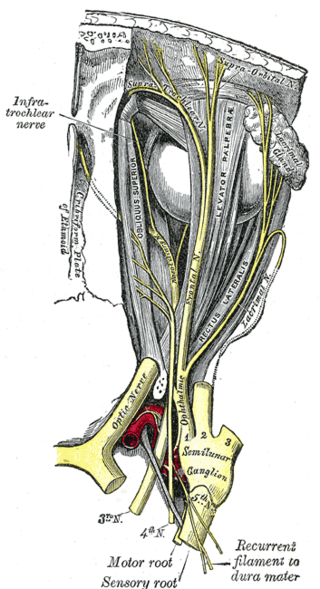
In neuroanatomy, the optic nerve, also known as the second cranial nerve, cranial nerve II, or simply CN II, is a paired cranial nerve that transmits visual information from the retina to the brain. In humans, the optic nerve is derived from optic stalks during the seventh week of development and is composed of retinal ganglion cell axons and glial cells; it extends from the optic disc to the optic chiasma and continues as the optic tract to the lateral geniculate nucleus, pretectal nuclei, and superior colliculus.

<span class="mw-page-title-main">Papilledema</span> Eye disorder

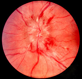
Papilledema or papilloedema is optic disc swelling that is caused by increased intracranial pressure due to any cause. The swelling is usually bilateral and can occur over a period of hours to weeks. Unilateral presentation is extremely rare.

The National Eye Institute (NEI) is part of the U.S. National Institutes of Health (NIH), an agency of the U.S. Department of Health and Human Services. The mission of NEI is "to eliminate vision loss and improve quality of life through vision research." NEI consists of two major branches for research: an extramural branch that funds studies outside NIH and an intramural branch that funds research on the NIH campus in Bethesda, Maryland. Most of the NEI budget funds extramural research.

<span class="mw-page-title-main">Amaurosis fugax</span> Medical condition

Amaurosis fugax is a painless temporary loss of vision in one or both eyes.

<span class="mw-page-title-main">Optic disc</span> Optic nerve head, the point of exit for ganglion cell axons leaving the eye

The optic disc or optic nerve head is the point of exit for ganglion cell axons leaving the eye. Because there are no rods or cones overlying the optic disc, it corresponds to a small blind spot in each eye.

Anterior ischemic optic neuropathy (AION) is a medical condition involving loss of vision caused by damage to the optic nerve as a result of insufficient blood supply (ischemia). This form of ischemic optic neuropathy is generally categorized as two types: arteritic AION, in which the loss of vision is the result of an inflammatory disease of arteries in the head called temporal arteritis, and non-arteritic AION, which is due to non-inflammatory disease of small blood vessels.

<span class="mw-page-title-main">Ischemic optic neuropathy</span> Medical condition

Ischemic optic neuropathy (ION) is the loss of structure and function of a portion of the optic nerve due to obstruction of blood flow to the nerve. Ischemic forms of optic neuropathy are typically classified as either anterior ischemic optic neuropathy or posterior ischemic optic neuropathy according to the part of the optic nerve that is affected. People affected will often complain of a loss of visual acuity and a visual field, the latter of which is usually in the superior or inferior field.

Arteritic anterior ischemic optic neuropathy is the cause of vision loss that occurs in temporal arteritis. Temporal arteritis is an inflammatory disease of medium-sized blood vessels that happens especially with advancing age. AAION occurs in about 15-20 percent of patients with temporal arteritis. Damage to the blood vessels supplying the optic nerves leads to insufficient blood supply (ischemia) to the nerve and subsequent optic nerve fiber death. Most cases of AAION result in nearly complete vision loss first to one eye. If the temporal arteritis is left untreated, the fellow eye will likely suffer vision loss as well within 1–2 weeks. Arteritic AION falls under the general category of anterior ischemic optic neuropathy, which also includes non-arteritic AION. AION is considered an eye emergency, immediate treatment is essential to rescue remaining vision.

Toxic and nutritional optic neuropathy is a group of medical disorders defined by visual impairment due to optic nerve damage secondary to a toxic substance and/or nutritional deficiency. The causes of these disorders are various, but they are linked by shared signs and symptoms, which this article will describe. In several of these disorders, both toxic and nutritional factors play a role, acting synergistically.

Optic neuropathy is damage to the optic nerve from any cause. The optic nerve is a bundle of millions of fibers in the retina that sends visual signals to the brain. [1].

Ocular ischemic syndrome is the constellation of ocular signs and symptoms secondary to severe, chronic arterial hypoperfusion to the eye. Amaurosis fugax is a form of acute vision loss caused by reduced blood flow to the eye; it may be a warning sign of an impending stroke, as both stroke and retinal artery occlusion can be caused by thromboembolism due to atherosclerosis elsewhere in the body. Consequently, those with transient blurring of vision are advised to urgently seek medical attention for a thorough evaluation of the carotid artery. Anterior segment ischemic syndrome is a similar ischemic condition of anterior segment usually seen in post-surgical cases. Retinal artery occlusion leads to rapid death of retinal cells, thereby resulting in severe loss of vision.

<span class="mw-page-title-main">Optic disc drusen</span> Medical condition

Optic disc drusen (ODD) are globules of mucoproteins and mucopolysaccharides that progressively calcify in the optic disc. They are thought to be the remnants of the axonal transport system of degenerated retinal ganglion cells. ODD have also been referred to as congenitally elevated or anomalous discs, pseudopapilledema, pseudoneuritis, buried disc drusen, and disc hyaline bodies.

Central retinal artery occlusion (CRAO) is a disease of the eye where the flow of blood through the central retinal artery is blocked (occluded). There are several different causes of this occlusion; the most common is carotid artery atherosclerosis.

<span class="mw-page-title-main">Blurred vision</span> Medical condition

Blurred vision is an ocular symptom where vision becomes less precise and there is added difficulty to resolve fine details.

Autoimmune optic neuropathy (AON), sometimes called autoimmune optic neuritis, may be a forme fruste of systemic lupus erythematosus (SLE) associated optic neuropathy. AON is more than the presence of any optic neuritis in a patient with an autoimmune process, as it describes a relatively specific clinical syndrome. AON is characterized by chronically progressive or recurrent vision loss associated with serological evidence of autoimmunity. Specifically, this term has been suggested for cases of optic neuritis with serological evidence of vasculitis by positive ANA, despite the lack of meeting criteria for SLE. The clinical manifestations include progressive vision loss that tends to be steroid-responsive and steroid dependent.

Mitohondrial optic neuropathies are a heterogenous group of disorders that present with visual disturbances resultant from mitochondrial dysfunction within the anatomy of the Retinal Ganglion Cells (RGC), optic nerve, optic chiasm, and optic tract. These disturbances are multifactorial, their aetiology consisting of metabolic and/or structural damage as a consequence of genetic mutations, environmental stressors, or both. The three most common neuro-ophthalmic abnormalities seen in mitochondrial disorders are bilateral optic neuropathy, ophthalmoplegia with ptosis, and pigmentary retinopathy.

Sickle cell retinopathy can be defined as retinal changes due to blood vessel damage in the eye of a person with a background of sickle cell disease. It can likely progress to loss of vision in late stages due to vitreous hemorrhage or retinal detachment. Sickle cell disease is a structural red blood cell disorder leading to consequences in multiple systems. It is characterized by chronic red blood cell destruction, vascular injury, and tissue ischemia causing damage to the brain, eyes, heart, lungs, kidneys, spleen, and musculoskeletal system.

Diabetic papillopathy is an ocular complication of diabetes mellitus characterized by optic disc swelling and edema of optic nerve head. The condition may affect both type 1 and type 2 diabetic patients.

References

  1. 1 2 3 4 5 6 7 8 9 Dunker S, Hsu HY, Sebag J, Sadun AA (June 2002). "Perioperative risk factors for posterior ischemic optic neuropathy". Journal of the American College of Surgeons. 194 (6): 705–10. doi:10.1016/S1072-7515(02)01210-3. PMID   12081060.
  2. 1 2 3 4 Hayreh SS (November 2004). "Posterior ischaemic optic neuropathy: clinical features, pathogenesis, and management". Eye. 18 (11): 1188–206. doi: 10.1038/sj.eye.6701562 . PMID   15534605.
  3. 1 2 3 4 5 Sadda SR, Nee M, Miller NR, Biousse V, Newman NJ, Kouzis A (November 2001). "Clinical spectrum of posterior ischemic optic neuropathy". American Journal of Ophthalmology. 132 (5): 743–50. doi:10.1016/S0002-9394(01)01199-0. PMID   11704036.
  4. 1 2 3 4 5 6 7 8 9 Hayreh SS (January 2009). "Ischemic optic neuropathy". Progress in Retinal and Eye Research. 28 (1): 34–62. doi:10.1016/j.preteyeres.2008.11.002. PMID   19063989. S2CID   41796396.
  5. Hayreh SS, Podhajsky PA, Zimmerman B (April 1998). "Ocular manifestations of giant cell arteritis". American Journal of Ophthalmology. 125 (4): 509–20. doi:10.1016/s0002-9394(99)80192-5. PMID   9559737.
  6. 1 2 3 4 5 6 7 Buono LM, Foroozan R (2005). "Perioperative posterior ischemic optic neuropathy: review of the literature". Survey of Ophthalmology. 50 (1): 15–26. doi:10.1016/j.survophthal.2004.10.005. PMID   15621075.
  7. 1 2 3 Lee LA, Roth S, Posner KL, et al. (October 2006). "The American Society of Anesthesiologists Postoperative Visual Loss Registry: analysis of 93 spine surgery cases with postoperative visual loss". Anesthesiology. 105 (4): 652–9, quiz 867–8. doi: 10.1097/00000542-200610000-00007 . PMID   17006060. S2CID   13138366.
  8. 1 2 3 4 Chang SH, Miller NR (June 2005). "The incidence of vision loss due to perioperative ischemic optic neuropathy associated with spine surgery: the Johns Hopkins Hospital Experience". Spine. 30 (11): 1299–302. doi:10.1097/01.brs.0000163884.11476.25. PMID   15928556. S2CID   25631710.
  9. 1 2 3 4 Newman NJ (April 2008). "Perioperative visual loss after nonocular surgeries". American Journal of Ophthalmology. 145 (4): 604–610. doi:10.1016/j.ajo.2007.09.016. PMC   2989384 . PMID   18358851.
  10. 1 2 Ho VT, Newman NJ, Song S, Ksiazek S, Roth S (January 2005). "Ischemic optic neuropathy following spine surgery". Journal of Neurosurgical Anesthesiology. 17 (1): 38–44. PMC   2699455 . PMID   15632541.
  11. 1 2 Alexandrakis G, Lam BL (March 1999). "Bilateral posterior ischemic optic neuropathy after spinal surgery". American Journal of Ophthalmology. 127 (3): 354–5. doi:10.1016/S0002-9394(98)00343-2. PMID   10088754.
  12. 1 2 3 Gill B, Heavner JE (April 2006). "Postoperative visual loss associated with spine surgery". European Spine Journal. 15 (4): 479–84. doi:10.1007/s00586-005-0914-6. PMC   3489312 . PMID   15926057.
  13. Pazos GA, Leonard DW, Blice J, Thompson DH (1999). "Blindness after bilateral neck dissection: case report and review". American Journal of Otolaryngology. 20 (5): 340–5. doi:10.1016/S0196-0709(99)90039-X. PMID   10512147.
  14. 1 2 3 Myers MA, Hamilton SR, Bogosian AJ, Smith CH, Wagner TA (June 1997). "Visual loss as a complication of spine surgery. A review of 37 cases". Spine. 22 (12): 1325–9. doi:10.1097/00007632-199706150-00009. PMID   9201835. S2CID   7611986.
  15. Stevens WR, Glazer PA, Kelley SD, Lietman TM, Bradford DS (June 1997). "Ophthalmic complications after spinal surgery". Spine. 22 (12): 1319–24. doi:10.1097/00007632-199706150-00008. PMID   9201834. S2CID   23877951.
  16. Sweeney PJ, Breuer AC, Selhorst JB, et al. (May 1982). "Ischemic optic neuropathy: a complication of cardiopulmonary bypass surgery". Neurology. 32 (5): 560–2. doi:10.1212/wnl.32.5.560. PMID   7200214. S2CID   28381520.
  17. Weyand CM, Goronzy JJ (July 2003). "Medium- and large-vessel vasculitis". The New England Journal of Medicine. 349 (2): 160–9. doi:10.1056/NEJMra022694. PMID   12853590.
  18. Weber ED, Colyer MH, Lesser RL, Subramanian PS (December 2007). "Posterior ischemic optic neuropathy after minimally invasive prostatectomy". Journal of Neuro-Ophthalmology. 27 (4): 285–7. doi: 10.1097/WNO.0b013e31815b9f67 . PMID   18090562.
  19. Hunder GG, Bloch DA, Michel BA, et al. (August 1990). "The American College of Rheumatology 1990 criteria for the classification of giant cell arteritis". Arthritis and Rheumatism. 33 (8): 1122–8. doi:10.1002/art.1780330810. PMID   2202311.
  20. 1 2 Connolly SE, Gordon KB, Horton JC (February 1994). "Salvage of vision after hypotension-induced ischemic optic neuropathy". American Journal of Ophthalmology. 117 (2): 235–42. doi:10.1016/s0002-9394(14)73082-x. PMID   8116753.
  21. Salvarani C, Macchioni PL, Tartoni PL, et al. (1987). "Polymyalgia rheumatica and giant cell arteritis: a 5-year epidemiologic and clinical study in Reggio Emilia, Italy". Clinical and Experimental Rheumatology. 5 (3): 205–15. PMID   3501353.
  22. Delecoeuillerie G, Joly P, Cohen de Lara A, Paolaggi JB (September 1988). "Polymyalgia rheumatica and temporal arteritis: a retrospective analysis of prognostic features and different corticosteroid regimens (11 year survey of 210 patients)". Annals of the Rheumatic Diseases. 47 (9): 733–9. doi:10.1136/ard.47.9.733. PMC   1003589 . PMID   3178314.
  23. Lundberg I, Hedfors E (October 1990). "Restricted dose and duration of corticosteroid treatment in patients with polymyalgia rheumatica and temporal arteritis". The Journal of Rheumatology. 17 (10): 1340–5. PMID   2254893.
  24. Foroozan R, Deramo VA, Buono LM, et al. (March 2003). "Recovery of visual function in patients with biopsy-proven giant cell arteritis". Ophthalmology. 110 (3): 539–42. doi:10.1016/S0161-6420(02)01775-X. PMID   12623817.
  25. Hayreh SS. Posterior ischaemic optic neuropathy: clinical features, pathogenesis, and management. Eye (Lond) 2004; 18:1188.

Further reading