Cervical intraepithelial neoplasia

Last updated
Cervical intraepithelial neoplasia
Other namesCervical dysplasia
VIAPosCIN1.gif
Positive visual inspection with acetic acid of the cervix for CIN-1
Specialty Gynecology

Cervical intraepithelial neoplasia (CIN), also known as cervical dysplasia, is the abnormal growth of cells on the surface of the cervix that could potentially lead to cervical cancer. [1] More specifically, CIN refers to the potentially precancerous transformation of cells of the cervix.

Contents

CIN most commonly occurs at the squamocolumnar junction of the cervix, a transitional area between the squamous epithelium of the vagina and the columnar epithelium of the endocervix. [2] It can also occur in vaginal walls and vulvar epithelium. CIN is graded on a 1–3 scale, with 3 being the most abnormal (see classification section below).

Human papillomavirus (HPV) infection is necessary for the development of CIN, but not all with this infection develop cervical cancer. [3] Many women with HPV infection never develop CIN or cervical cancer. Typically, HPV resolves on its own. [4] However, those with an HPV infection that lasts more than one or two years have a higher risk of developing a higher grade of CIN. [5]

Like other intraepithelial neoplasias, CIN is not cancer and is usually curable. [3] Most cases of CIN either remain stable or are eliminated by the person's immune system without need for intervention. However, a small percentage of cases progress to cervical cancer, typically cervical squamous cell carcinoma (SCC), if left untreated. [6]

Signs and symptoms

There are no specific symptoms of CIN alone.

Generally, signs and symptoms of cervical cancer include: [7]

HPV infection of the vulva and vagina can cause genital warts or be asymptomatic.

Causes

The cause of CIN is chronic infection of the cervix with HPV, especially infection with high-risk HPV types 16 or 18. It is thought that the high-risk HPV infections have the ability to inactivate tumor suppressor genes such as the p53 gene and the RB gene, thus allowing the infected cells to grow unchecked and accumulate successive mutations, eventually leading to cancer. [1]

Some groups of women have been found to be at a higher risk of developing CIN: [1] [8]

Additionally, a number of risk factors have been shown to increase an individual's likelihood of developing CIN 3/carcinoma in situ (see below): [9]

Pathophysiology

Transformation zone Diagram showing the transformation zone on the cervix CRUK 375.svg
Transformation zone

The earliest microscopic change corresponding to CIN is epithelial dysplasia, or surface lining, of the cervix, which is essentially undetectable by the woman. The majority of these changes occur at the squamocolumnar junction, or transformation zone, an area of unstable cervical epithelium that is prone to abnormal changes. [2] Cellular changes associated with HPV infection, such as koilocytes, are also commonly seen in CIN. While infection with HPV is needed for development of CIN, most women with HPV infection do not develop high-grade intraepithelial lesions or cancer. HPV is not alone enough causative. [10]

Of the over 100 different types of HPV, approximately 40 are known to affect the epithelial tissue of the anogenital area and have different probabilities of causing malignant changes. [11]

Diagnosis

A test for HPV called the Digene HPV test is highly accurate and serves as both a direct diagnosis and adjuvant to the Pap smear, which is a screening device that allows for an examination of cells but not tissue structure, needed for diagnosis. A colposcopy with directed biopsy is the standard for disease detection. Endocervical brush sampling at the time of Pap smear to detect adenocarcinoma and its precursors is necessary along with doctor/patient vigilance on abdominal symptoms associated with uterine and ovarian carcinoma. The diagnosis of CIN or cervical carcinoma requires a biopsy for histological analysis.[ citation needed ]

Classification

Normal cervical epithelium Cervical intraepithelial neoplasia (1) normal squamous epithelium.jpg
Normal cervical epithelium

Historically, abnormal changes of cervical epithelial cells were described as mild, moderate, or severe epithelial dysplasia. In 1988 the National Cancer Institute developed "The Bethesda System for Reporting Cervical/Vaginal Cytologic Diagnoses". [12] This system provides a uniform way to describe abnormal epithelial cells and determine specimen quality, thus providing clear guidance for clinical management. These abnormalities were classified as squamous or glandular and then further classified by the stage of dysplasia: atypical cells, mild, moderate, severe, and carcinoma. [13]

Depending on several factors and the location of the lesion, CIN can start in any of the three stages and can either progress or regress. [1] The grade of squamous intraepithelial lesion can vary.[ citation needed ]

CIN is classified in grades: [14]

Histology GradeCorresponding CytologyDescriptionImage
CIN 1 (Grade I)Low-grade squamous intraepithelial lesion (LSIL)
  • Mild epithelial dysplasia
  • Confined to the basal 1/3 of the epithelium
  • Typically corresponds to infection with HPV
  • High rate of regression back to normal cells
  • Usually managed expectantly
LSIL (CIN 1), Cervical Biopsy (3776284166).jpg
CIN 2/3High-grade squamous intraepithelial lesion (HSIL)
  • Represents a mix of low- and high-grade lesions not easily differentiated by histology
  • HSIL+ encompasses HSIL, AGC, and cancer
CIN 2 (Grade II)
  • Moderate dysplasia confined to the basal 2/3 of the epithelium
  • Represents a mix of low- and high-grade lesions not easily differentiated by histology
  • CIN 2+ encompasses CIN 2, CIN 3, adenocarcinoma in situ (AIS), and cancer
Cervical intraepithelial neoplasia (3) CIN2.jpg
CIN 3 (Grade III)
  • Severe dysplasia with undifferentiated neoplastic cells that span more than 2/3 of the epithelium
  • May involve the full thickness
  • May also be referred to as cervical carcinoma in situ
  • CIN 3+ encompasses CIN 3, AIS, and cancer
Histopathology of CIN 3.jpg
Locations of CIN findings can be described in terms of quadrants, or corresponding to a clock face when the subject is in supine position. Cervix quadrants and directions.svg
Locations of CIN findings can be described in terms of quadrants, or corresponding to a clock face when the subject is in supine position.
Endocervical gland invasion is associated with high-grade lesions. Histopathology of CIN 3 with endocervical gland invasion.jpg
Endocervical gland invasion is associated with high-grade lesions.

Changes in terminology

The College of American Pathologists and the American Society of Colposcopy and Cervical Pathology came together in 2012 to publish changes in terminology to describe HPV-associated squamous lesions of the anogenital tract as LSIL or HSIL as follows below: [16]

CIN 1 is referred to as LSIL.

CIN 2 that is negative for p16, a marker for high-risk HPV, is referred to as LSIL. Those that are p16-positive are referred to as HSIL.

CIN 3 is referred to as HSIL.

Screening

The two screening methods available are the Pap smear and testing for HPV. CIN is usually discovered by a screening test, the Pap smear. The purpose of this test is to detect potentially precancerous changes through random sampling of the transformation zone. Pap smear results may be reported using the Bethesda system (see above). The sensitivity and specificity of this test were variable in a systematic review looking at accuracy of the test. An abnormal Pap smear result may lead to a recommendation for colposcopy of the cervix, an in office procedure during which the cervix is examined under magnification. A biopsy is taken of any abnormal appearing areas.[ citation needed ]

Colposcopy is usually very painful and so researchers have tried to find which pain relief is best for women with CIN to use. Research suggests that the injection of a local anaesthetic and vasoconstrictor (medicine that causes blood vessels to narrow) into the cervix may lower blood loss and pain during colposcopy. [17]

HPV testing can identify most of the high-risk HPV types responsible for CIN. HPV screening happens either as a co-test with the Pap smear or can be done after a Pap smear showing abnormal cells, called reflex testing. Frequency of screening changes based on guidelines from the Society of Lower Genital Tract Disorders (ASCCP). The World Health Organization also has screening and treatment guidelines for precancerous cervical lesions and prevention of cervical cancer.[ citation needed ]

Primary prevention

HPV vaccination is the approach to primary prevention of both CIN and cervical cancer.

VaccineHPV Genotypes Protected AgainstWho Gets It?Number of DosesTiming Recommendation
Gardasil - quadrivalent6, 11 (cause genital warts) 16, 18 (cause ≈70% of cervical cancers) [18] females and males age 9-263before sexual debut or shortly thereafter
Cervarix - bivalent16, 18females age 9-253
Gardasil 9 - nonavalent vaccine6, 11, 16, 18, 31, 33, 45, 52, 58 (cause ≈20% of cervical cancers) [19] females and males ages 9–263

It is important to note that these vaccines do not protect against all types of HPV known to cause cancer. Therefore, screening is still recommended in vaccinated individuals.

Secondary prevention

Appropriate management with monitoring and treatment is the approach to secondary prevention of cervical cancer in cases of persons with CIN.[ citation needed ]

Treatment

Cervical cryotherapy Cervical Cryotherapy.png
Cervical cryotherapy

Treatment for CIN 1, mild dysplasia, is not recommended if it lasts fewer than two years. [20] Usually, when a biopsy detects CIN 1, the woman has an HPV infection, which may clear on its own within 12 months. Therefore, it is instead followed for later testing rather than treated. [20] In young women closely monitoring CIN 2 lesions also appears reasonable. [6]

The typical threshold for treatment is CIN 2+, although a more restrained approach may be taken for young persons and pregnant women. Treatment for higher-grade CIN involves removal or destruction of the abnormal cervical cells by cryocautery, electrocautery, laser cautery, loop electrical excision procedure (LEEP), or cervical conization. [21] While these surgical methods effectively reduce the risk of developing cervical cancer, [22] [23] they cause an increased risk of premature birth in future pregnancies. [24] [25] Surgical techniques that remove more cervical tissue come with less risk of the cancer recurring but a higher chance of giving birth prematurely. Due to this risk, taking into account the age, childbearing plans of the woman, the size and location of the cancer cells are crucial for choosing the right procedure. [22] [23]

While retinoids are not effective in preventing the progression of CIN, they may be effective in causing regression of disease in people with CIN2. [26] Therapeutic vaccines are currently undergoing clinical trials. The lifetime recurrence rate of CIN is about 20%,[ citation needed ] but it isn't clear what proportion of these cases are new infections rather than recurrences of the original infection.

Research to investigate if prophylactic antibiotics can help prevent infection in women undergoing excision of the cervical transformation zone found a lack of quality evidence. [27]

People with HIV and CIN 2+ should be initially managed according to the recommendations for the general population according to the 2012 updated ASCCP consensus guidelines. [28]

Outcomes

It used to be thought that cases of CIN progressed through grades 1–3 toward cancer in a linear fashion. [29] [30] [31]

However most CIN spontaneously regress. Left untreated, about 70% of CIN 1 will regress within one year; 90% will regress within two years. [32] About 50% of CIN 2 cases will regress within two years without treatment.[ citation needed ]

Progression to cervical carcinoma in situ (CIS) occurs in approximately 11% of CIN 1 and 22% of CIN 2 cases. Progression to invasive cancer occurs in approximately 1% of CIN 1, 5% of CIN 2, and at least 12% of CIN 3 cases. [3]

Progression to cancer typically takes 15 years with a range of 3 to 40 years. Also, evidence suggests that cancer can occur without first detectably progressing through CIN grades and that a high-grade intraepithelial neoplasia can occur without first existing as a lower grade. [1] [29] [33]

Research suggests that treatment does not affect the chances of getting pregnant but it is associated with an increased risk of miscarriage in the second trimester. [34]

Epidemiology

Between 250,000 and 1 million American women are diagnosed with CIN annually. Women can develop CIN at any age but women generally develop it between the ages of 25 and 35. [1] The estimated annual incidence of CIN in the United States among persons who undergo screening is 4% for CIN 1 and 5% for CIN 2 and CIN 3. [35]

Related Research Articles

<span class="mw-page-title-main">Cervix</span> Lower part of the uterus in the human female reproductive system

The cervix or cervix uteri is the lower part of the uterus (womb) in the human female reproductive system. The cervix is usually 2 to 3 cm long and roughly cylindrical in shape, which changes during pregnancy. The narrow, central cervical canal runs along its entire length, connecting the uterine cavity and the lumen of the vagina. The opening into the uterus is called the internal os, and the opening into the vagina is called the external os. The lower part of the cervix, known as the vaginal portion of the cervix, bulges into the top of the vagina. The cervix has been documented anatomically since at least the time of Hippocrates, over 2,000 years ago.

<span class="mw-page-title-main">Pap test</span> Cervical screening test to detect potential cancers

The Papanicolaou test is a method of cervical screening used to detect potentially precancerous and cancerous processes in the cervix or colon. Abnormal findings are often followed up by more sensitive diagnostic procedures and, if warranted, interventions that aim to prevent progression to cervical cancer. The test was independently invented in the 1920s by the Greek physician Georgios Papanikolaou and named after him. A simplified version of the test was introduced by the Canadian obstetrician Anna Marion Hilliard in 1957.

<span class="mw-page-title-main">Cervical cancer</span> Cancer arising from the cervix

Cervical cancer is a cancer arising from the cervix. It is due to the abnormal growth of cells that have the ability to invade or spread to other parts of the body. Early on, typically no symptoms are seen. Later symptoms may include abnormal vaginal bleeding, pelvic pain or pain during sexual intercourse. While bleeding after sex may not be serious, it may also indicate the presence of cervical cancer.

<span class="mw-page-title-main">Human papillomavirus infection</span> Human disease

Human papillomavirus infection is caused by a DNA virus from the Papillomaviridae family. Many HPV infections cause no symptoms and 90% resolve spontaneously within two years. In some cases, an HPV infection persists and results in either warts or precancerous lesions. These lesions, depending on the site affected, increase the risk of cancer of the cervix, vulva, vagina, penis, anus, mouth, tonsils, or throat. Nearly all cervical cancer is due to HPV and two strains – HPV16 and HPV18 – which account for 70% of cases. HPV16 is responsible for almost 90% of HPV-positive oropharyngeal cancers. Between 60% and 90% of the other cancers listed above are also linked to HPV. HPV6 and HPV11 are common causes of genital warts and laryngeal papillomatosis.

<span class="mw-page-title-main">Colposcopy</span> Medical examination of the cervix

Colposcopy is a medical diagnostic procedure to visually examine the cervix as well as the vagina and vulva using a colposcope. Numbing should be requested prior to procedure.

<span class="mw-page-title-main">Anal cancer</span> Medical condition

Anal cancer is a cancer which arises from the anus, the distal opening of the gastrointestinal tract. Symptoms may include bleeding from the anus or a lump near the anus. Other symptoms may include pain, itchiness, or discharge from the anus. A change in bowel movements may also occur.

<span class="mw-page-title-main">Precancerous condition</span> Medical condition

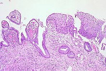
A precancerous condition is a condition, tumor or lesion involving abnormal cells which are associated with an increased risk of developing into cancer. Clinically, precancerous conditions encompass a variety of abnormal tissues with an increased risk of developing into cancer. Some of the most common precancerous conditions include certain colon polyps, which can progress into colon cancer, monoclonal gammopathy of undetermined significance, which can progress into multiple myeloma or myelodysplastic syndrome. and cervical dysplasia, which can progress into cervical cancer. Bronchial premalignant lesions can progress to squamous cell carcinoma of the lung.

<span class="mw-page-title-main">Koilocyte</span>

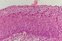
A koilocyte is a squamous epithelial cell that has undergone a number of structural changes, which occur as a result of infection of the cell by human papillomavirus (HPV). Identification of these cells by pathologists can be useful in diagnosing various HPV-associated lesions.

<span class="mw-page-title-main">Gardasil</span> Human papillomavirus vaccine

Gardasil is an HPV vaccine for use in the prevention of certain strains of human papillomavirus (HPV). It was developed by Merck & Co. High-risk human papilloma virus (hr-HPV) genital infection is the most common sexually transmitted infection among women. The HPV strains that Gardasil protects against are sexually transmitted, specifically HPV types 6, 11, 16 and 18. HPV types 16 and 18 cause an estimated 70% of cervical cancers, and are responsible for most HPV-induced anal, vulvar, vaginal, and penile cancer cases. HPV types 6 and 11 cause an estimated 90% of genital warts cases. HPV type 16 is responsible for almost 90% of HPV-positive oropharyngeal cancers, and the prevalence is higher in males than females. Though Gardasil does not treat existing infection, vaccination is still recommended for HPV-positive individuals, as it may protect against one or more different strains of the disease.

<span class="mw-page-title-main">Vulvar cancer</span> Cancer involving the vulva

Vulvar cancer is a cancer of the vulva, the outer portion of the female genitals. It most commonly affects the labia majora. Less often, the labia minora, clitoris, or vaginal glands are affected. Symptoms include a lump, itchiness, changes in the skin, or bleeding from the vulva.

Vaginal cancer is an extraordinarily rare form of cancer that develops in the tissue of the vagina. Primary vaginal cancer originates from the vaginal tissue – most frequently squamous cell carcinoma, but primary vaginal adenocarcinoma, sarcoma, and melanoma have also been reported – while secondary vaginal cancer involves the metastasis of a cancer that originated in a different part of the body. Secondary vaginal cancer is more common. Signs of vaginal cancer may include abnormal vaginal bleeding, dysuria, tenesmus, or pelvic pain, though as many as 20% of women diagnosed with vaginal cancer are asymptomatic at the time of diagnosis. Vaginal cancer occurs more frequently in women over age 50, and the mean age of diagnosis of vaginal cancer is 60 years. It often can be cured if found and treated in early stages. Surgery alone or surgery combined with pelvic radiation is typically used to treat vaginal cancer.

Vaginal intraepithelial neoplasia (VAIN) is a condition that describes premalignant histological findings in the vagina characterized by dysplastic changes.

HspE7 is an investigational therapeutic vaccine candidate being developed by Nventa Biopharmaceuticals for the treatment of precancerous and cancerous lesions caused by the human papillomavirus (HPV). HspE7 uses recombinant DNA technology to covalently fuse a heat shock protein (Hsp) to a target antigen, thereby stimulating cellular immune system responses to specific diseases. HspE7 is a patented construct consisting of the HPV Type 16 E7 protein and heat shock protein 65 (Hsp65) and is currently the only candidate using Hsp technology to target the over 20 million Americans already infected with HPV.

An anal Pap smear is the anal counterpart of the cervical Pap smear. It is used for the early detection of anal cancer. Some types of human papillomavirus (HPV) can cause anal cancer. Other HPV types cause anogenital warts. Cigarette smokers, men who have sex with men, individuals with a history of immunosuppression and women with a history of cervical, vaginal and vulval cancer are at increased risk of getting anal cancer. Vaccination against HPV before initial sexual exposure can reduce the risk of anal cancer.

The Bethesda system (TBS), officially called The Bethesda System for Reporting Cervical Cytology, is a system for reporting cervical or vaginal cytologic diagnoses, used for reporting Pap smear results. It was introduced in 1988 and revised in 1991, 2001, and 2014. The name comes from the location of the conference, sponsored by the National Institutes of Health, that established the system.

Cervicography is a diagnostic medical procedure in which a non-physician takes pictures of the cervix and submits them to a physician for interpretation. Other related procedures are speculoscopy and colposcopy. The procedure is considered a screening test for cervical cancer and is complementary to Pap smear. The technique was initially developed by Adolf Stafl, MD, of Medical College of Wisconsin in 1981.

Epithelial dysplasia, a term becoming increasingly referred to as intraepithelial neoplasia, is the sum of various disturbances of epithelial proliferation and differentiation as seen microscopically. Individual cellular features of dysplasia are called epithelial atypia.

Neuroendocrine carcinoma of the cervix is best defined separately:Neuroendocrine: Of, relating to, or involving the interaction between the nervous system and the hormones of the endocrine glands.Carcinoma: An invasive malignant tumor derived from epithelial tissue that tends to metastasize to other areas of the body.

<span class="mw-page-title-main">Cervical screening</span> Type of medical screening

Cervical cancer screening is a medical screening test designed to identify risk of cervical cancer. Cervical screening may involve looking for viral DNA, and/or to identify abnormal, potentially precancerous cells within the cervix as well as cells that have progressed to early stages of cervical cancer. One goal of cervical screening is to allow for intervention and treatment so abnormal lesions can be removed prior to progression to cancer. An additional goal is to decrease mortality from cervical cancer by identifying cancerous lesions in their early stages and providing treatment prior to progression to more invasive disease.

Pamela Toliman is a medical researcher from Papua New Guinea (PNG) who has researched areas such as sexually transmitted diseases, HIV/AIDS, cervical cancer and COVID-19.

References

  1. 1 2 3 4 5 6 Kumar V, Abbas AK, Fausto N, Mitchell RN (2007). Robbins Basic Pathology (8th ed.). Saunders Elsevier. pp. 718–721. ISBN   978-1-4160-2973-1.
  2. 1 2 "Colposcopy and treatment of cervical intraepithelial neoplasia: a beginners manual". screening.iarc.fr. Retrieved 2018-12-20.
  3. 1 2 3 Section 4 Gynecologic Oncology > Chapter 29. Preinvasive Lesions of the Lower Genital Tract > Cervical Intraepithelial Neoplasia in:Bradshaw KD, Schorge JO, Schaffer J, Halvorson LM, Hoffman BG (2008). Williams' Gynecology. McGraw-Hill Professional. ISBN   978-0-07-147257-9.
  4. "Human papillomavirus (HPV) and cervical cancer". www.who.int. Retrieved 2018-12-18.
  5. Boda D, Docea AO, Calina D, Ilie MA, Caruntu C, Zurac S, Neagu M, Constantin C, Branisteanu DE, Voiculescu V, Mamoulakis C, Tzanakakis G, Spandidos DA, Drakoulis N, Tsatsakis AM (March 2018). "Human papilloma virus: Apprehending the link with carcinogenesis and unveiling new research avenues (Review)". International Journal of Oncology. 52 (3): 637–655. doi:10.3892/ijo.2018.4256. PMC   5807043 . PMID   29393378.
  6. 1 2 Tainio K, Athanasiou A, Tikkinen KA, Aaltonen R, Cárdenas J, Glazer-Livson S, et al. (February 2018). "Clinical course of untreated cervical intraepithelial neoplasia grade 2 under active surveillance: systematic review and meta-analysis". BMJ. 360: k499. doi:10.1136/bmj.k499. PMC   5826010 . PMID   29487049.
  7. DiSaia PJ, Creasman WT (2007). "Invasive cervical cancer.". Clinical Gynecologic Oncology (7th ed.). Philadelphia: Mosby Elsevier. p. 55.
  8. Heard I (January 2009). "Prevention of cervical cancer in women with HIV". Current Opinion in HIV and AIDS. 4 (1): 68–73. doi:10.1097/COH.0b013e328319bcbe. PMID   19339941. S2CID   25047280.
  9. International Collaboration of Epidemiological Studies of Cervical Cancer (2006-09-01). "Cervical carcinoma and reproductive factors: collaborative reanalysis of individual data on 16,563 women with cervical carcinoma and 33,542 women without cervical carcinoma from 25 epidemiological studies". International Journal of Cancer. 119 (5): 1108–1124. doi:10.1002/ijc.21953. ISSN   0020-7136. PMID   16570271. S2CID   29433753.
  10. Beutner KR, Tyring S (May 1997). "Human papillomavirus and human disease". The American Journal of Medicine. 102 (5A): 9–15. doi:10.1016/s0002-9343(97)00178-2. PMID   9217657.
  11. de Villiers EM, Fauquet C, Broker TR, Bernard HU, zur Hausen H (June 2004). "Classification of papillomaviruses". Virology. 324 (1): 17–27. doi: 10.1016/j.virol.2004.03.033 . PMID   15183049.
  12. Soloman D (1989). "The 1988 Bethesda System for reporting cervical/vaginal cytologic diagnoses: developed and approved at the National Cancer Institute workshop in Bethesda, MD, December 12-13, 1988". Diagnostic Cytopathology. 5 (3): 331–4. doi:10.1002/dc.2840050318. PMID   2791840. S2CID   19684695.
  13. Solomon D, Davey D, Kurman R, Moriarty A, O'Connor D, Prey M, et al. (April 2002). "The 2001 Bethesda System: terminology for reporting results of cervical cytology". JAMA. 287 (16): 2114–9. doi:10.1001/jama.287.16.2114. PMID   11966386.
  14. Melnikow J, Nuovo J, Willan AR, Chan BK, Howell LP (October 1998). "Natural history of cervical squamous intraepithelial lesions: a meta-analysis". Obstetrics and Gynecology. 92 (4 Pt 2): 727–35. doi:10.1097/00006250-199810001-00046. PMID   9764690.
  15. Nagi CS, Schlosshauer PW (August 2006). "Endocervical glandular involvement is associated with high-grade SIL". Gynecologic Oncology. 102 (2): 240–3. doi:10.1016/j.ygyno.2005.12.029. PMID   16472847.
  16. Darragh TM, Colgan TJ, Thomas Cox J, Heller DS, Henry MR, Luff RD, et al. (January 2013). "The Lower Anogenital Squamous Terminology Standardization project for HPV-associated lesions: background and consensus recommendations from the College of American Pathologists and the American Society for Colposcopy and Cervical Pathology". International Journal of Gynecological Pathology. 32 (1): 76–115. doi:10.1097/PGP.0b013e31826916c7. PMID   23202792. S2CID   205943774.
  17. Gajjar K, Martin-Hirsch PP, Bryant A, Owens GL (July 2016). "Pain relief for women with cervical intraepithelial neoplasia undergoing colposcopy treatment". The Cochrane Database of Systematic Reviews. 7 (7): CD006120. doi:10.1002/14651858.cd006120.pub4. PMC   6457789 . PMID   27428114.
  18. Lowy DR, Schiller JT (May 2006). "Prophylactic human papillomavirus vaccines". The Journal of Clinical Investigation. 116 (5): 1167–73. doi: 10.1172/JCI28607 . PMC   1451224 . PMID   16670757.
  19. "FDA approves Gardasil 9 for prevention of certain cancers caused by five additional types of HPV". U.S. Food and Drug Administration (FDA) (press release). 10 December 2014. Archived from the original on 10 January 2015. Retrieved 28 February 2015.
  20. 1 2 American Congress of Obstetricians and Gynecologists, "Five Things Physicians and Patients Should Question", Choosing Wisely: an initiative of the ABIM Foundation , American Congress of Obstetricians and Gynecologists , retrieved August 1, 2013
  21. Martin-Hirsch, Pierre PL; Paraskevaidis, Evangelos; Bryant, Andrew; Dickinson, Heather O (2013-12-04). "Surgery for cervical intraepithelial neoplasia". Cochrane Database of Systematic Reviews. 2014 (12): CD001318. doi:10.1002/14651858.cd001318.pub3. ISSN   1465-1858. PMC   4170911 . PMID   24302546.
  22. 1 2 Athanasiou, Antonios; Veroniki, Areti Angeliki; Efthimiou, Orestis; Kalliala, Ilkka; Naci, Huseyin; Bowden, Sarah; Paraskevaidi, Maria; Arbyn, Marc; Lyons, Deirdre; Martin-Hirsch, Pierre; Bennett, Phillip; Paraskevaidis, Evangelos; Salanti, Georgia; Kyrgiou, Maria (2022-07-25). "Comparative effectiveness and risk of preterm birth of local treatments for cervical intraepithelial neoplasia and stage IA1 cervical cancer: a systematic review and network meta-analysis". The Lancet Oncology. 23 (8): 1097–1108. doi:10.1016/S1470-2045(22)00334-5. PMC   9630146 . PMID   35835138.
  23. 1 2 "Prevention of cervical cancer: what are the risks and benefits of different treatments?". NIHR Evidence (Plain English summary). National Institute for Health and Care Research. 2023-11-10. doi:10.3310/nihrevidence_60599.
  24. Kyrgiou M, Athanasiou A, Paraskevaidi M, Mitra A, Kalliala I, Martin-Hirsch P, et al. (July 2016). "Adverse obstetric outcomes after local treatment for cervical preinvasive and early invasive disease according to cone depth: systematic review and meta-analysis". BMJ. 354: i3633. doi:10.1136/bmj.i3633. PMC   4964801 . PMID   27469988.
  25. Kyrgiou M, Athanasiou A, Kalliala IE, Paraskevaidi M, Mitra A, Martin-Hirsch PP, et al. (November 2017). "Obstetric outcomes after conservative treatment for cervical intraepithelial lesions and early invasive disease". The Cochrane Database of Systematic Reviews. 11 (11): CD012847. doi:10.1002/14651858.cd012847. PMC   6486192 . PMID   29095502.
  26. Helm, C. William; Lorenz, Douglas J; Meyer, Nicholas J; Rising, William WR; Wulff, Judith L (2013-06-06). "Retinoids for preventing the progression of cervical intra-epithelial neoplasia". Cochrane Database of Systematic Reviews (6): CD003296. doi: 10.1002/14651858.cd003296.pub3 . ISSN   1465-1858. PMID   23740788.
  27. Kietpeerakool C, Chumworathayi B, Thinkhamrop J, Ussahgij B, Lumbiganon P (January 2017). "Antibiotics for infection prevention after excision of the cervical transformation zone". The Cochrane Database of Systematic Reviews. 1 (10): CD009957. doi:10.1002/14651858.cd009957.pub2. PMC   6464760 . PMID   28109160.
  28. Massad LS, Einstein MH, Huh WK, Katki HA, Kinney WK, Schiffman M, et al. (April 2013). "2012 updated consensus guidelines for the management of abnormal cervical cancer screening tests and cancer precursors". Journal of Lower Genital Tract Disease. 17 (5 Suppl 1): S1–S27. doi:10.1097/LGT.0b013e318287d329. PMID   23519301. S2CID   12551963.
  29. 1 2 Agorastos T, Miliaras D, Lambropoulos AF, Chrisafi S, Kotsis A, Manthos A, Bontis J (July 2005). "Detection and typing of human papillomavirus DNA in uterine cervices with coexistent grade I and grade III intraepithelial neoplasia: biologic progression or independent lesions?". European Journal of Obstetrics, Gynecology, and Reproductive Biology. 121 (1): 99–103. doi:10.1016/j.ejogrb.2004.11.024. PMID   15949888.
  30. Hillemanns P, Wang X, Staehle S, Michels W, Dannecker C (February 2006). "Evaluation of different treatment modalities for vulvar intraepithelial neoplasia (VIN): CO(2) laser vaporization, photodynamic therapy, excision and vulvectomy". Gynecologic Oncology. 100 (2): 271–5. doi:10.1016/j.ygyno.2005.08.012. PMID   16169064.
  31. Rapp L, Chen JJ (August 1998). "The papillomavirus E6 proteins". Biochimica et Biophysica Acta (BBA) - Reviews on Cancer. 1378 (1): F1-19. doi:10.1016/s0304-419x(98)00009-2. PMID   9739758.
  32. Bosch FX, Burchell AN, Schiffman M, Giuliano AR, de Sanjose S, Bruni L, Tortolero-Luna G, Kjaer SK, Muñoz N (August 2008). "Epidemiology and natural history of human papillomavirus infections and type-specific implications in cervical neoplasia". Vaccine. 26 Suppl 10 (Supplement 10): K1-16. doi:10.1016/j.vaccine.2008.05.064. PMID   18847553. 18847553.
  33. Monnier-Benoit S, Dalstein V, Riethmuller D, Lalaoui N, Mougin C, Prétet JL (March 2006). "Dynamics of HPV16 DNA load reflect the natural history of cervical HPV-associated lesions". Journal of Clinical Virology. 35 (3): 270–7. doi:10.1016/j.jcv.2005.09.001. PMID   16214397.
  34. Kyrgiou M, Mitra A, Arbyn M, Paraskevaidi M, Athanasiou A, Martin-Hirsch PP, et al. (September 2015). Cochrane Gynaecological, Neuro-oncology and Orphan Cancer Group (ed.). "Fertility and early pregnancy outcomes after conservative treatment for cervical intraepithelial neoplasia". The Cochrane Database of Systematic Reviews. 2016 (9): CD008478. doi:10.1002/14651858.CD008478.pub2. PMC   6457639 . PMID   26417855.
  35. Insinga RP, Glass AG, Rush BB (July 2004). "Diagnoses and outcomes in cervical cancer screening: a population-based study". American Journal of Obstetrics and Gynecology. 191 (1): 105–13. doi:10.1016/j.ajog.2004.01.043. PMID   15295350.