Fallopian tube cancer

Last updated
Primary fallopian tube cancer
Illu cervix.svg
Specialty Oncology, gynaecology
Symptoms Early: Vague, none [1] [2]
Later: Abnormal vaginal bleeding, abdominal distension, blood stained watery vaginal discharge, pelvic pain, loss of appetite, weight loss, feel full [1] [2]
Diagnostic method
Differential diagnosis Ovarian cancer, peritoneal cancer [1]

Primary fallopian tube cancer (PFTC), also known as tubal cancer, is a malignant neoplasm that originates from the fallopian tube. [1] [3] Along with primary ovarian and peritoneal carcinomas, it is grouped under epithelial ovarian cancers; cancers of the ovary that originate from a fallopian tube precursor. [4] [5]

Contents

Signs and symptoms

In the early stages, symptoms are typically vague. [2] Other symptoms may include abnormal vaginal bleeding, blood stained watery vaginal discharge, pelvic pain, or abdominal distension. [2] An affected person may feel full or have weight loss. [1]

Vaginal discharge in fallopian tube carcinoma results from intermittent hydrosalphinx, also known as hydrops tubae profluens. [6]

Pathology

The most common cancer type within this disease is adenocarcinoma; in the largest series of 3,051 cases as reported by Stewart et al. 88% of cases fell into this category. [7] According to their study, half of the cases were poorly differentiated, 89% unilateral, and the distribution showed a third each with local disease only, with regional disease only, and with distant extensions. Rarer forms of tubal neoplasm include leiomyosarcoma, and transitional cell carcinoma.

As the tumor is often enmeshed with the adjacent ovary, it may be the pathologist and not the surgeon who determines that the lesion is indeed tubal in origin.

Secondary tubal cancer usually originates from cancer of the ovaries, the endometrium, the GI tract, the peritoneum, and the breast.

Diagnosis

Diagnosis is by blood tests, medical imaging, and pathologic assessment of fallopian tissue. [1] Blood tests include Ca-125 and CBC. [1] Imaging includes transvaginal and abdominal ultrasound, CT scan, and MRI. [2] Pathologic assessment may include SEE-FIM Protocol. [1] A pelvic mass may be detected on a routine gynecologic examination. [2] It may be found at an early stage when removing the tubes and ovaries as a preventive measure. [1]

Differential

Ovarian and peritoneal cancers may present in a similar way. [1]

Staging

International Federation of Gynecology and Obstetrics (FIGO) staging [1]
StageDefinition
Stage 0Carcinoma in situ [1]
Stage IGrowth limited to fallopian tubes [1]
Stage IIGrowth involving one or both fallopian tubes with extension to pelvis [1]
Stage IIITumor involving one or both fallopian tubes with spread outside pelvis [1]
Stage IVGrowth involving one or more fallopian tubes with distant metastases [1]

Treatment

The initial approach to tubal cancer is generally surgical, and similar to that of ovarian cancer. As the lesion will spread first to the adjacent uterus and ovary, a total abdominal hysterectomy is an essential part of this approach, removing the ovaries, the tubes, and the uterus with the cervix. Also, peritoneal washings are taken, the omentum is removed, and pelvic and paraaortic lymph nodes are sampled. Staging at the time of surgery and pathological findings will determine further steps. In advanced cases when the cancer has spread to other organs and cannot be completely removed, cytoreductive surgery is used to lessen the tumor burden for subsequent treatments. Surgical treatments are typically followed by adjuvant, usually platinum-based, chemotherapy. [8] [9] Radiation therapy has been applied with some success to patients with tubal cancer for palliative or curative indications. [10]

Prognosis

Five-year survival rate is around 65%, though may range from 30% to 92% depending on stage at diagnosis and the amount of tumor remaining after surgery. [1] [4]

Frequency

Tubal cancer is thought to be a relatively rare primary cancer among women, accounting for 1 to 2 percent of all gynecologic cancers, [11] In the US, tubal cancer had an incidence of 0.41 per 100,000 women from 1998 to 2003. [7] Demographic distribution is similar to that of ovarian cancer, and the highest incidence is found in white, non-Hispanic women aged 60–79. [7] However, recent evidence suggests tubal cancer to be much more frequent. [1]

Evidence is accumulating that individuals with mutations of BRCA1 and BRCA2 are at higher risk for the development of PFTC. [12] [13]

History

The first descriptions were made by Renaud in 1847 and Ernst Gottlob Orthmann in 1888. [14]

Related Research Articles

<span class="mw-page-title-main">Krukenberg tumor</span> Medical condition

A Krukenberg tumor refers to a malignancy in the ovary that metastasized from a primary site, classically the gastrointestinal tract, although it can arise in other tissues such as the breast. Gastric adenocarcinoma, especially at the pylorus, is the most common source. Krukenberg tumors are often found in both ovaries, consistent with its metastatic nature.

<span class="mw-page-title-main">Ovarian cancer</span> Cancer originating in or on the ovary

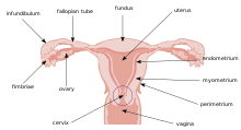
Ovarian cancer is a cancerous tumor of an ovary. It may originate from the ovary itself or more commonly from communicating nearby structures such as fallopian tubes or the inner lining of the abdomen. The ovary is made up of three different cell types including epithelial cells, germ cells, and stromal cells. When these cells become abnormal, they have the ability to divide and form tumors. These cells can also invade or spread to other parts of the body. When this process begins, there may be no or only vague symptoms. Symptoms become more noticeable as the cancer progresses. These symptoms may include bloating, vaginal bleeding, pelvic pain, abdominal swelling, constipation, and loss of appetite, among others. Common areas to which the cancer may spread include the lining of the abdomen, lymph nodes, lungs, and liver.

<span class="mw-page-title-main">Serous tumour</span> Medical condition

A serous tumour is a neoplasm that typically has papillary to solid formations of tumor cells with crowded nuclei, and which typically arises on the modified Müllerian-derived serous membranes that surround the ovaries in females. Such ovarian tumors are part of the surface epithelial-stromal tumour group of ovarian tumors. They are common neoplasms with a strong tendency to occur bilaterally, and they account for approximately a quarter of all ovarian tumors.

<span class="mw-page-title-main">Surface epithelial-stromal tumor</span> Medical condition

Surface epithelial-stromal tumors are a class of ovarian neoplasms that may be benign or malignant. Neoplasms in this group are thought to be derived from the ovarian surface epithelium or from ectopic endometrial or fallopian tube (tubal) tissue. Tumors of this type are also called ovarian adenocarcinoma. This group of tumors accounts for 90% to 95% of all cases of ovarian cancer; however is mainly only found in postmenopausal women with the exception of the United States where 7% of cases occur in women under the age of 40. Serum CA-125 is often elevated but is only 50% accurate so it is not a useful tumor marker to assess the progress of treatment. 75% of women with epithelial ovarian cancer are found within the advanced-stages; however younger patients are more likely to have better prognoses than older patients.

<span class="mw-page-title-main">Gynecologic ultrasonography</span> Application of medical ultrasonography to the female pelvic organs

Gynecologic ultrasonography or gynecologic sonography refers to the application of medical ultrasonography to the female pelvic organs as well as the bladder, the adnexa, and the recto-uterine pouch. The procedure may lead to other medically relevant findings in the pelvis.This technique is useful to detect myomas or mullerian malformations.

<span class="mw-page-title-main">Pseudomyxoma peritonei</span> Medical condition

Pseudomyxoma peritonei (PMP) is a clinical condition caused by cancerous cells that produce abundant mucin or gelatinous ascites. The tumors cause fibrosis of tissues and impede digestion or organ function, and if left untreated, the tumors and mucin they produce will fill the abdominal cavity. This will result in compression of organs and will destroy the function of the colon, small intestine, stomach, or other organs. Prognosis with treatment in many cases is optimistic, but the disease is lethal if untreated, with death occurring via cachexia, bowel obstruction, or other types of complications.

In medicine, Meigs's syndrome, also Meigs syndrome or Demons–Meigs syndrome, is the triad of ascites, pleural effusion, and benign ovarian tumor. Meigs syndrome resolves after the resection of the tumor. Because the transdiaphragmatic lymphatic channels are larger in diameter on the right, the pleural effusion is classically on the right side. The causes of the ascites and pleural effusion are poorly understood. Atypical Meigs syndrome, characterized by a benign pelvic mass with right-sided pleural effusion but without ascites, can also occur. As in typical Meigs syndrome, pleural effusion resolves after removal of the pelvic mass.

<span class="mw-page-title-main">Primary peritoneal carcinoma</span> Medical condition

Primary peritoneal cancer or carcinoma is also known as serous surface papillary carcinoma, primary peritoneal carcinoma, extra-ovarian serous carcinoma, primary serous papillary carcinoma, and psammomacarcinoma. It was historically classified under "carcinoma of unknown primary" (CUP). Primary peritoneal cancer is a cancer of the cells lining the peritoneum, or abdominal cavity. It usually affects women and is diagnosed after the age of 60; it very rarely affects men.

<span class="mw-page-title-main">Hydrosalpinx</span> Medical condition

A hydrosalpinx is a condition that occurs when a fallopian tube is blocked and fills with serous or clear fluid near the ovary. The blocked tube may become substantially distended giving the tube a characteristic sausage-like or retort-like shape. The condition is often bilateral and the affected tubes may reach several centimeters in diameter. The blocked tubes cause infertility. A fallopian tube filled with blood is a hematosalpinx, and with pus a pyosalpinx.

<span class="mw-page-title-main">Adnexal mass</span> Medical condition

An adnexal mass is a lump in the tissue of the adnexa of the uterus. Adnexal masses can be benign (noncancerous) or malignant (cancerous), and they can be categorized as simple or complex.

<span class="mw-page-title-main">Uterine serous carcinoma</span> Type of cancer of the uterus

Uterine serous carcinoma is a malignant form of serous tumor that originates in the uterus. It is an uncommon form of endometrial cancer that typically arises in postmenopausal women. It is typically diagnosed on endometrial biopsy, prompted by post-menopausal bleeding.

A borderline tumor, sometimes called low malignant potential (LMP) tumor, is a distinct but yet heterogeneous group of tumors defined by their histopathology as atypical epithelial proliferation without stromal invasion. It generally refers to such tumors in the ovary but borderline tumors may rarely occur at other locations as well.

<span class="mw-page-title-main">Endosalpingiosis</span> Medical condition

Endosalpingiosis is a condition in which fallopian tube-like epithelium is found outside the fallopian tube. It is unknown what causes this condition. It is generally accepted that the condition develops from transformation of coelomic tissue. It is often an incidental finding and is not usually associated with any pathology.

<span class="mw-page-title-main">Ovarian serous cystadenoma</span> Medical condition

Ovarian serous cystadenoma, also known as serous cystadenoma, is the most common ovarian neoplasm, representing 20% of ovarian neoplasms, and is benign.

<span class="mw-page-title-main">Adenosarcoma</span> Type of tumor

Adenosarcoma is a rare malignant tumor that occurs in women of all age groups, but most commonly post-menopause. Adenosarcoma arises from mesenchymal tissue and has a mixture of the tumoral components of an adenoma, a tumor of epithelial origin, and a sarcoma, a tumor originating from connective tissue. The adenoma, or epithelial component of the tumor, is benign, while the sarcomatous stroma is malignant. The most common site of adenosarcoma formation is the uterus, but it can also occur in the cervix and ovaries. It more rarely arises in the vagina and fallopian tubes as well as primary pelvic or peritoneal sites, such as the omentum, especially in those with a history of endometriosis. The rare cases of adenosarcoma outside the female genital tract usually occur in the liver, bladder, kidney, as well as the intestine and are typically associated with endometriosis.

<span class="mw-page-title-main">Paraovarian cyst</span> Medical condition

Paraovarian cysts or paratubal cysts are epithelium-lined fluid-filled cysts in the adnexa adjacent to the fallopian tube and ovary. The terms are used interchangeably, and depend on the location of the cyst.

<span class="mw-page-title-main">High-grade serous carcinoma</span> Medical condition

High-grade serous carcinoma (HGSC) is a type of tumour that arises from the serous epithelial layer in the abdominopelvic cavity and is mainly found in the ovary. HGSCs make up the majority of ovarian cancer cases and have the lowest survival rates. HGSC is distinct from low-grade serous carcinoma (LGSC) which arises from ovarian tissue, is less aggressive and is present in stage I ovarian cancer where tumours are localised to the ovary.

Ovarian germ cell tumors (OGCTs) are heterogeneous tumors that are derived from the primitive germ cells of the embryonic gonad, which accounts for about 2.6% of all ovarian malignancies. There are four main types of OGCTs, namely dysgerminomas, yolk sac tumor, teratoma, and choriocarcinoma.

<span class="mw-page-title-main">SEE-FIM Protocol</span> Pathology protocol to assess cancer risk

The SEE-FIM protocol is a pathology dissection protocol for Sectioning and Extensively Examining the Fimbria (SEE-FIM). This protocol is intended to provide for the optimal microscopic examination of the distal fallopian tube (fimbria) to identify either cancerous or precancerous conditions in this organ.

<span class="mw-page-title-main">Ovarian squamous cell carcinoma</span> Medical condition

Ovarian squamous cell carcinoma (oSCC) or squamous ovarian carcinoma (SOC) is a rare tumor that accounts for 1% of ovarian cancers. Included in the World Health Organization's classification of ovarian cancer, it mainly affects women above 45 years of age. Survival depends on how advanced the disease is and how different or similar the individual cancer cells are.

References

  1. 1 2 3 4 5 6 7 8 9 10 11 12 13 14 15 16 17 18 19 20 Ferri, Fred F. (2024). "Fallopian tube cancer". Ferri's Clinical Advisor 2024. Philadelphia: Elsevier. p. 568.e5. ISBN   978-0-323-75576-4.
  2. 1 2 3 4 5 6 7 "Fallopian tube cancer". www.cancerresearchuk.org. Retrieved 30 March 2024.
  3. WHO Classification of Tumours Editorial Board, ed. (2020). "4. Tumours of the fallopian tube: introduction". Female genital tumours: WHO Classification of Tumours. Vol. 4 (5th ed.). Lyon (France): International Agency for Research on Cancer. p. 216. ISBN   978-92-832-4504-9.
  4. 1 2 Stasenko, Marina; Fillipova, Olga; Tew, William P. (July 2019). "Fallopian Tube Carcinoma". Journal of Oncology Practice. 15 (7): 375–382. doi:10.1200/JOP.18.00662. ISSN   1554-7477.
  5. Rashid, Sameera; Arafah, Maria A.; Akhtar, Mohammed (1 May 2022). "The Many Faces of Serous Neoplasms and Related Lesions of the Female Pelvis: A Review". Advances in Anatomic Pathology. 29 (3): 154–167. doi:10.1097/PAP.0000000000000334. ISSN   1533-4031. PMC   8989637 . PMID   35180738.
  6. GOLDMAN JA, GANS B, ECKERLING B (November 1961). "Hydrops tubae profluens--symptom in tubal carcinoma". Obstet Gynecol. 18: 631–4. PMID   13899814.
  7. 1 2 3 Stewart SL, Wike JM, Foster SL, Michaud F (2007). "The incidence of primary fallopian tube cancer in the United States". Gynecol. Oncol. 107 (3): 392–7. doi:10.1016/j.ygyno.2007.09.018. PMID   17961642.
  8. Liapis A, Bakalianou K, Mpotsa E, Salakos N, Fotiou S, Kondi-Paffiti A (2008). "Fallopian tube malignancies: A retrospective clinical pathological study of 17 cases". J Obstet Gynaecol. 28 (1): 93–5. doi:10.1080/01443610701811894. PMID   18259909. S2CID   5351886.
  9. Takeshima N, Hasumi K (2000). "Treatment of fallopian tube cancer. Review of the literature". Arch Gynecol Obstet. 264 (1): 13–9. doi:10.1007/pl00007475. PMID   10985612. S2CID   34114333.
  10. Schray MF, Podratz KC, Malkasian GD (1987). "Fallopian tube cancer: the role of radiation therapy". Radiother. Oncol. 10 (4): 267–75. doi:10.1016/s0167-8140(87)80032-4. PMID   3444903.
  11. UCSF. "Gynecologic Cancer: Fallopian Tube Cancer". accessed 08-14-2008
  12. BRCA mutations link to tubal cancer, accessed 08-14-2008
  13. Piek, J. M. J. (2004). Hereditary serous ovarian carcinogenesis, a hypothesis (PhD thesis). Vrije Universiteit Amsterdam. hdl:1871/9013. ISBN   9064646406.
  14. Laury, Anna; Huang, Eric C.; Crum, Christopher P.; Hecht, Jonathan (2013). "7. Peritoneal and tubal serous carcinoma". In Deligdisch, Liane; Kase, Nathan G.; Cohen, Carmel J. (eds.). Altchek's Diagnosis and Management of Ovarian Disorders (3rd ed.). Cambridge University Press. pp. 111–120. ISBN   978-1-107-01281-3.