Kyasanur Forest disease

Last updated
Kyasanur forest disease
Other namesMonkey disease, monkey fever
Kyasanur Forest disease virus ecology.jpg
Virus ecology
Specialty Infectious disease

Kyasanur forest disease (KFD) is a tick-borne viral haemorrhagic fever endemic to South-western part of India. [1] The disease is caused by a virus belonging to the family Flaviviridae. KFDV is transmitted to humans through the bite of infected hard ticks ( Haemaphysalis spinigera ) which act as a reservoir of KFDV.

Contents

Signs and symptoms

The symptoms of the disease include a high fever with frontal headaches, chills, severe muscle pain, vomiting, and other gastrointestinal symptoms. Bleeding problems may occur 3–4 days after initial symptom onset. Patients may experience abnormally low blood pressure, and low platelet, red blood cell, and white blood cell count. After 1–2 weeks of symptoms, some patients recover without complication. However, the illness is biphasic for a subset of patients (10-20%) who experience a second wave of symptoms at the beginning of the third week. These symptoms include fever and signs of neurological manifestations, such as severe headache, mental disturbances, tremors, and vision deficits. [2] [3] [4] The convalescent period is typically very long, lasting several months. Muscle aches and weakness also occur during this period, and the patient is unable to engage in physical activities.

Cause

Virology

Kyasanur Forest disease virus
Ijms-20-04657-g002.webp
Flavivirus structure and genome
Virus classification OOjs UI icon edit-ltr.svg
(unranked): Virus
Realm: Riboviria
Kingdom: Orthornavirae
Phylum: Kitrinoviricota
Class: Flasuviricetes
Order: Amarillovirales
Family: Flaviviridae
Genus: Flavivirus
Species:
Kyasanur Forest disease virus
Synonyms [5]

Kyasanur Forest virus

The KFD virus is a typical flavivirus measuring about 40-60 nm in diameter. The genome of KFDV consists of 10,774 nucleotides of single-stranded, positive-sense RNA encoding a single polyprotein that is cleaved post-translationally into three structural (C, prM/M and E) and seven non-structural (NS1, NS2a, NS2b, NS3, NS4a, NS4b and NS5) proteins. [6] [7] [8] The genome of KFDV is very similar (>92% homologous) to that of Alkhurma Hemorrhagic Fever Virus which is primarily found in Saudi Arabia. These two species both belong to the family Flaviviridae and diverged over 700 years ago and have thus remained geographically separated. [9]

Transmission

A variety of animals are thought to be reservoir hosts for the disease, including porcupines, rats, squirrels, mice, and shrews. [2] Monkeys are the main amplifying hosts for KFD virus and they are also affected by the virus. The surili Presbytis entellus and the bonnet macaque are very susceptible to the KFD virus. They develop tremendous viremia and infect the ticks. The vector for disease transmission is Haemaphysalis spinigera , a forest tick. [10] Humans contract infection from the bite of nymphs of the tick. Man is a terminal host and there no human-to-human transmission because the human domestic environment does not sustain the ticks.

Pathology

The pathogenesis of KFDV is not completely understood. Research using mice models found that KFDV primarily replicated in the brain. [11] Other research has expanded on this by described neurological changes that occurred within infected organisms. This experiment was completed by using KFDV-infected mice and discovered that KFDV caused gliosis, inflammation, and cell death in the brain. They posited that KFDV could be primarily a neuropathic disease and other symptoms are due to this pathogenesis. [12]

Diagnosis

In earlier days suspected case were confirmed in a laboratory by serum inoculation into suckling mice (Swiss Albino mice) and subsequent death of mice was leveled as KFD Positive case. Other methods of diagnosis included hemagglutination inhibition (HI), complement fixation, neutralization tests. [13] However, new research has introduced more efficient molecular based methods to diagnose KFDV. These methods include: RT-PCR, nested RT-PCR, TaqMan-based real-time RT-PCR, Immunoglobin M antibodies and Immunoglobin G detection by ELISA. The two methods involving RT-PCR are able to function by attaching a primer to the NS-5 gene, which is highly conserved among the genus to which KFDV belongs. PCR positivity is limited to 8–10 days from the onset of symptoms. The ELISA based methods allows for the detections of anti-KFDV antibodies in patients typically from 5th day of onset of symptoms up to 3 months. [14]

Prevention and treatment

Prevention is by vaccination, as well as preventive measures such as protective clothing and tick population control. The vaccine for KFDV consists of formalin-inactivated KFDV. The vaccine has a 62.4% effectiveness rate for individuals who receive two doses. For individuals who receive an additional dose, the effectiveness increases to 82.9%. [15] Specific antiviral treatments are not available as of 2022. [16]

Risk factors and risk groups

The spill-over of Kyasanur forest disease happens at the crossroads of the animal-human interaction, especially villages adjoining forest areas and inter-state borders. [17] People who frequently visit the forest areas of the Western Ghats region such as forest guards and officials, range forest officer (RFO), forest watchers, shepherds, firewood collectors, dry leaf collectors, hunters, people who handle dead animal carcasses,  travelers who camp in the forest areas, tribal communities living inside the forest areas (Jenu kurubas and Betta kurubas), cashew nut workers especially those who engage in cleaning the dry leaves before the harvest season (seen in Pali and Mauxi outbreaks, North Goa), and areca nut farm workers working in infected tick areas will have a high risk of acquiring KFD infection. People who live in the KFD endemic areas and refuse to take KFD vaccination are at risk in contracting the infection.

History

The disease was first reported from Kattinakere village forest which is in the Kyasanur forest range of Karnataka in India in March 1957. When the officials visited the Kattinakere forest and discovered the diseases they noticed a sign board informing that this was the Kyasanur forest range. Hence the name. The disease first manifested as an epizootic outbreak among monkeys, killing several of them in the year 1957. Hence the disease is also locally known as "monkey disease" or "monkey fever". [18] The similarity with Russian spring-summer encephalitis was noted and the possibility of migratory birds carrying the disease was raised. [19] Studies began to look for the possible species that acted as reservoirs for the virus and the agents responsible for transmission. Subsequent studies failed to find any involvement of migratory birds, although the possibility of their role in initial establishment was not ruled out. The virus was found to be quite distinctive and not closely related to the Russian virus strains. Antigenic relatedness is, however, close to many other strains including the Omsk hemorrhagic fever (OHF) and birds from Siberia have been found to show an antigenic response to KFD virus. Sequence based studies note the distinctiveness of OHF. [20] Early studies in India were conducted in collaboration with the US Army Medical Research Unit and this led to controversy and conspiracy theories. [21] [22]

Subsequent studies based on sequencing found that the Alkhurma virus found in Saudi Arabia is closely related. [23] In 1989 a patient in Nanjianin, China was found with fever symptoms and in 2009 its viral gene sequence was found to exactly match with that of the KFD reference virus of 1957. This has been questioned, though, since the Indian virus shows variations in sequence over time and the exact match with the virus sequence of 1957 and the Chinese virus of 1989 is not expected. This study also found using immune response tests that birds and humans in the region appeared to have been exposed to the virus. [24] Another study has suggested that the virus is recent in origin dating the nearest common ancestor of it and related viruses to around 1942, based on the estimated rate of sequence substitutions. The study also raises the possibility of bird involvement in long-distance transfer. [25] It appears that these viruses diverged 700 years ago. [26]

A recent outbreak in 2020, claimed two lives in Siddapura, Karnataka. The peak season for this disease in Malnad is from March till May but has been observed to peak earlier in the year as well. There were a total of 55 reported cases in Shivamogga district, Karntaka. [27] [28]

Affected states in India

Disease distribution Kyasanur Forest disease distribution map.jpg
Disease distribution

The disease initially reported from Shimoga district of Karnataka which is a primitive sylvan territory in Western Ghats of India. The disease spread out to other districts of Karnataka involving districts of Chikkamagalore, Uttara Kannada, Dakshina Kannada, Udupi, Chamarajanagar (2012), Belagavi (2016). In 2013, KFDV was detected in monkey autopsies from Nilgiris district of Tamil Nadu state. Monkey deaths and human cases have now been reported from three neighbouring states bordering Karnataka, i.e., Wayanad (2013) and Malappuram districts of Kerala (2014), North Goa district of Goa state (2015), and Sindhudurg district of Maharashtra (2016). [29]

Serological evidence for KFD

There are reported serological evidence for KFD detected in humans in other parts of India, namely Kutch and Saurashtra regions of Gujarat state, Kingaon and Parbatpur of West Bengal state. [30] A seroprevalence study in Andaman and Nicobar islands in 2002 revealed a high prevalence of hemagglutination inhibition (HI) antibodies against KFDV. [31]

Epidemiology

The disease has a fatality rate of 3-10%, and it affects 400-500 people annually. [10] [14]

The disease was first noted at Kyasanur village near Sagar in Shivamogga district of Karnataka. The virus has been detected in monkeys in parts of Bandipur National Park (Chamarajnagar) and parts of the Nilgiris. Human infection occurred in Bandipur through handling of dead monkeys that were infected. A human carrier was also detected in Wayanad (Kerala). [32] The disease has shown its presence in the adjacent states of Karnataka including Kerala, Maharashtra, Goa, Tamil Nadu and Gujarat. [33] [34] [35]

Related Research Articles

<span class="mw-page-title-main">Lassa fever</span> Viral disease spread by a type of mouse

Lassa fever, also known as Lassa hemorrhagic fever, is a type of viral hemorrhagic fever caused by the Lassa virus. Many of those infected by the virus do not develop symptoms. When symptoms occur they typically include fever, weakness, headaches, vomiting, and muscle pains. Less commonly there may be bleeding from the mouth or gastrointestinal tract. The risk of death once infected is about one percent and frequently occurs within two weeks of the onset of symptoms. Of those who survive, about a quarter have hearing loss, which improves within three months in about half of these cases.

<span class="mw-page-title-main">Dengue fever</span> Tropical disease caused by the dengue virus, transmitted by mosquito

Dengue fever is a mosquito-borne tropical disease caused by the dengue virus. It is frequently asymptomatic; if symptoms appear they typically begin 3 to 14 days after infection. These may include a high fever, headache, vomiting, muscle and joint pains, and a characteristic skin itching and skin rash. Recovery generally takes two to seven days. In a small proportion of cases, the disease develops into a more severe dengue hemorrhagic fever, resulting in bleeding, low levels of blood platelets and blood plasma leakage, or into dengue shock syndrome, where dangerously low blood pressure occurs.

<span class="mw-page-title-main">Chikungunya</span> Infection caused by the Chikungunya virus

Chikungunya is an infection caused by the Chikungunya virus (CHIKV). The disease was first identified in 1952 in Tanzania and named based on the Kimakonde words for "to become contorted". Symptoms include fever and joint pain. These typically occur two to twelve days after exposure. Other symptoms may include headache, muscle pain, joint swelling, and a rash. Symptoms usually improve within a week; however, occasionally the joint pain may last for months or years. The risk of death is around 1 in 1,000. The very young, old, and those with other health problems are at risk of more severe disease.

Tick-borne diseases, which afflict humans and other animals, are caused by infectious agents transmitted by tick bites. They are caused by infection with a variety of pathogens, including rickettsia and other types of bacteria, viruses, and protozoa. The economic impact of tick-borne diseases is considered to be substantial in humans, and tick-borne diseases are estimated to affect ~80 % of cattle worldwide. Most of these pathogens require passage through vertebrate hosts as part of their life cycle. Tick-borne infections in humans, farm animals, and companion animals are primarily associated with wildlife animal reservoirs. many tick-borne infections in humans involve a complex cycle between wildlife animal reservoirs and tick vectors. The survival and transmission of these tick-borne viruses are closely linked to their interactions with tick vectors and host cells. These viruses are classified into different families, including Asfarviridae, Reoviridae, Rhabdoviridae, Orthomyxoviridae, Bunyaviridae, and Flaviviridae.

<i>Bunyavirales</i> Order of RNA viruses

Bunyavirales is an order of segmented negative-strand RNA viruses with mainly tripartite genomes. Member viruses infect arthropods, plants, protozoans, and vertebrates. It is the only order in the class Ellioviricetes. The name Bunyavirales derives from Bunyamwera, where the original type species Bunyamwera orthobunyavirus was first discovered. Ellioviricetes is named in honor of late virologist Richard M. Elliott for his early work on bunyaviruses.

<span class="mw-page-title-main">Viral hemorrhagic fever</span> Type of illnesses

Viral hemorrhagic fevers (VHFs) are a diverse group of animal and human illnesses. VHFs may be caused by five distinct families of RNA viruses: the families Filoviridae, Flaviviridae, Rhabdoviridae, and several member families of the Bunyavirales order such as Arenaviridae, and Hantaviridae. All types of VHF are characterized by fever and bleeding disorders and all can progress to high fever, shock and death in many cases. Some of the VHF agents cause relatively mild illnesses, such as the Scandinavian nephropathia epidemica, while others, such as Ebola virus, can cause severe, life-threatening disease.

<span class="mw-page-title-main">Tick-borne encephalitis</span> Medical condition

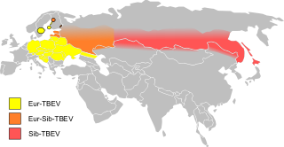
Tick-borne encephalitis (TBE) is a viral infectious disease involving the central nervous system. The disease most often manifests as meningitis, encephalitis or meningoencephalitis. Myelitis and spinal paralysis also occurs. In about one third of cases sequelae, predominantly cognitive dysfunction, persist for a year or more.

<span class="mw-page-title-main">Crimean–Congo hemorrhagic fever</span> Disease of humans and other animals

Crimean–Congo hemorrhagic fever (CCHF) is a viral disease. Symptoms of CCHF may include fever, muscle pains, headache, vomiting, diarrhea, and bleeding into the skin. Onset of symptoms is less than two weeks following exposure. Complications may include liver failure. Survivors generally recover around two weeks after onset.

<i>Thogotovirus</i> Genus of viruses

Thogotovirus is a genus of enveloped RNA viruses, one of seven genera in the virus family Orthomyxoviridae. Their single-stranded, negative-sense RNA genome has six or seven segments. Thogotoviruses are distinguished from most other orthomyxoviruses by being arboviruses – viruses that are transmitted by arthropods, in this case usually ticks. Thogotoviruses can replicate in both tick cells and vertebrate cells; one subtype has also been isolated from mosquitoes. A consequence of being transmitted by blood-sucking vectors is that the virus must spread systemically in the vertebrate host – unlike influenza viruses, which are transmitted by respiratory droplets and are usually confined to the respiratory system.

Alkhurma virus (ALKV) is a zoonotic virus of the Flaviviridae virus family. ALKV causes Alkhurma hemorrhagic fever (AHF), or alternatively termed as Alkhurma hemorrhagic fever virus, and is mainly based in Saudi Arabia.

Omsk hemorrhagic fever is a viral hemorrhagic fever caused by a Flavivirus.

Mayaro virus disease is a mosquito-borne zoonotic pathogen endemic to certain humid forests of tropical South America. Infection with Mayaro virus causes an acute, self-limited dengue-like illness of 3–5 days' duration. The causative virus, abbreviated MAYV, is in the family Togaviridae, and genus Alphavirus. It is closely related to other alphaviruses that produce a dengue-like illness accompanied by long-lasting arthralgia. It is only known to circulate in tropical South America.

Langat virus (LGTV) is a virus of the genus Flavivirus. The virus was first isolated in Malaysia in 1956 from a hard tick of the Ixodes genus. This virus is antigenically related to Omsk hemorrhagic fever virus, Kyasanur forest disease virus, Alkhurma virus, Louping ill virus and other viruses of the tick-borne encephalitis virus (TBEV) complex. The Langat virus does not pose a significant epidemiological threat in comparison with TBEV. There are no known cases of human diseases associated with LGTV. The Malaysian strain is naturally attenuated and induces neutralizing antibodies to tick-borne encephalitis virus (TBEV) and protection against other TBEV complex viruses in animals.

<span class="mw-page-title-main">Marburg virus</span> Species of filamentous virus responsible for hemorrhagic fever

Marburg virus (MARV) is a hemorrhagic fever virus of the Filoviridae family of viruses and a member of the species Marburg marburgvirus, genus Marburgvirus. It causes Marburg virus disease in primates, a form of viral hemorrhagic fever. The virus is considered to be extremely dangerous. The World Health Organization (WHO) rates it as a Risk Group 4 Pathogen. In the United States, the National Institute of Allergy and Infectious Diseases ranks it as a Category A Priority Pathogen and the Centers for Disease Control and Prevention lists it as a Category A Bioterrorism Agent. It is also listed as a biological agent for export control by the Australia Group.

In 1954 the Hazara orthonairovirus, one of the 34 tick-borne viruses of the genus Orthonairovirus, was discovered in Pakistan in the Ixodes tick native to that region. Today this virus is studied in mice in an attempt to develop treatments for the highly pathogenic Crimean-Congo Hemorrhagic Fever virus.

Batai orthobunyavirus (BATV) is a RNA virus belonging to order Bunyavirales, genus Orthobunyavirus.

Pylore Krishnaier Rajagopalan is an Indian vector control scientist, biologist and acarologist, known for his pioneering contributions to the control programmes against vector-borne diseases in India. He is a former director of the Indian Council of Medical Research managed Vector Control Research Centre, Pondicherry. He graduated in 1949 from the Banaras Hindu University and obtained a Masters in Zoology with University First Rank there itself in 1951. In 1952 he joined the fledgling Virus Research Centre in Pune, and worked under the supervision of some of the finest vector control specialists such as Dr T Ramachandra Rao. In recognition of his outstanding work as a young research scientist, in 1957 he was awarded a Fellowship by the Rockefeller Foundation to pursue a Master's program in Public Health from the University of California. He went on to secure a Diploma in Acarology from the University of Maryland at College Park.

<i>Ornithodoros savignyi</i> Species of tick

Ornithodoros savignyi, known as sand tampan, African eyed tampan or Kalahari sand tampan, is one of some 37 species in the genus Ornithodoros and is a soft tick with a leathery, mammillated integument, causing paralysis and tampan toxicosis, two unrelated conditions. The sand tampan is an ectoparasite on humans, their livestock and wild animals, including birds and bats. Occurring in semi-desert areas of Africa, Saudi Arabia and other parts of the Persian Gulf, India, Sri Lanka and into Asia, it is able to survive for lengthy periods without feeding, spending most of its life burrowed under sand or loose soil, often in wait for animals that rest or sleep under trees or in the lee of rocks, but also in places where people or their animals congregate such as marketplaces, places of worship, cattle kraals and village squares. The timing of its activity is geared to coincide with that of potential hosts, but hot sunny conditions are usually avoided. Because of its habit of feeding and dropping from its host, adult dispersal is limited, whereas larvae may remain attached to their hosts for several days. During its life cycle it will feed on multiple hosts between moults.

<span class="mw-page-title-main">MK-608</span> Chemical compound

MK-608 is an antiviral drug, an adenosine analog. It was originally developed by Merck & Co. as a treatment for hepatitis C, but despite promising results in animal studies, it was ultimately unsuccessful in clinical trials. Subsequently it has been widely used in antiviral research and has shown activity against a range of viruses, including Dengue fever, tick-borne encephalitis virus, poliovirus, and most recently Zika virus, in both in vitro and animal models. Since it has already failed in human clinical trials previously, it is unlikely MK-608 itself will be developed as an antiviral medication, but the continuing lack of treatment options for these emerging viral diseases means that much research continues using MK-608 and related antiviral drugs.

<span class="mw-page-title-main">NITD008</span> Chemical compound

NITD008 is an antiviral drug classified as an adenosine analog. It was developed as a potential treatment for flavivirus infections and shows broad spectrum antiviral activity against many related viruses such as dengue virus, West Nile virus, yellow fever virus, Powassan virus, hepatitis C virus, Kyasanur Forest disease virus, Omsk hemorrhagic fever virus, and Zika virus. However, NITD008 proved too toxic in pre-clinical animal testing to be suitable for human trials, but it continues to be used in research to find improved treatments for emerging viral diseases.

References

  1. EA Gould; T Solomon (February 9, 2008). "Pathogenic flaviviruses". The Lancet. 371 (961): 500–509. doi: 10.1016/S0140-6736(08)60238-X . ISSN   0140-6736. PMID   18262042. S2CID   205949828.
  2. 1 2 Gerhard Dobler (27 January 2010). "Zoonotic tick-borne flaviviruses". Veterinary Microbiology. 140 (3–4, Zoonoses: Advances and Perspectives): 221–228. doi:10.1016/j.vetmic.2009.08.024. ISSN   0378-1135. PMID   19765917.
  3. Dobler, Gerhard (2010). "Zoonotic tick-borne flaviviruses". Veterinary Microbiology. 140 (3/4): 221–228. doi:10.1016/j.vetmic.2009.08.024. PMID   19765917.
  4. Mourya, Devendra; Yadav, Pragya; Sandhya, V; Reddy, Shivanna (2013). "Spread of Kyasanur Forest Disease, Bandipur Tiger Reserve, India, 2012-2013". Emerging Infectious Diseases. 19 (9): 1540–1541. doi:10.3201/eid1909.121884. PMC   3810911 . PMID   23977946.
  5. ICTV 2nd Report Fenner, F (1976). "Classification and nomenclature of viruses. Second report of the International Committee on Taxonomy of Viruses" (PDF). Intervirology. 7 (1–2): 1–115. doi: 10.1159/000149938 . PMID   826499.
  6. Cook, Bradley W. M.; Cutts, Todd A.; Court, Deborah A.; Theriault, Steven (February 2012). "The generation of a reverse genetics system for Kyasanur Forest Disease Virus and the ability to antagonize the induction of the antiviral state in vitro". Virus Research. 163 (2): 431–438. doi:10.1016/j.virusres.2011.11.002. ISSN   1872-7492. PMID   22100401.
  7. Cook, Bradley; Ranadheera, Charlene; Nikiforuk, Aidan; Cutts, Todd; Kobasa, Darwyn; Court, Deborah; Theriault, Steven (2016). "Limited Effects of Type I Interferons on Kyasanur Forest Disease Virus in Cell Culture". PLOS Neglected Tropical Diseases. 10 (8): e0004871. doi: 10.1371/journal.pntd.0004871 . PMC   4968803 . PMID   27479197.
  8. Dodd, Kimberly; Bird, Brian; Khristova, Marina; Albariño, César; Carroll, Serena; Comer, James; Erickson, Bobbie; Rollin, Pierre; Nichol, Stuart (2011). "Ancient Ancestry of KFDV and AHFV Revealed by Complete Genome Analyses of Viruses Isolated from Ticks and Mammalian Hosts". PLOS Neglected Tropical Diseases. 5 (10): e1352. doi: 10.1371/journal.pntd.0001352 . PMC   3186760 . PMID   21991403.
  9. Dodd, Kimberly; Bird, Brian; Jones, Megan; Nichol, Stuart; Spiropoulou, Christina (2014). "Kyasanur Forest Disease Virus Infection in Mice Is Associated with Higher Morbidity and Mortality than Infection with the Closely Related Alkhurma Hemorrhagic Fever Virus". PLOS ONE. 9 (6): e100301. Bibcode:2014PLoSO...9j0301D. doi: 10.1371/journal.pone.0100301 . PMC   4065072 . PMID   24950196.
  10. 1 2 Holbrook, Michael (2012). "Kyasanur forest disease". Antiviral Research. 96 (3): 353–362. doi:10.1016/j.antiviral.2012.10.005. PMC   3513490 . PMID   23110991.
  11. Sawatsky, Bevan; McAuley, Alexander; Holbrook, Michael; Bente, Dennis (2014). "Comparative Pathogenesis of Alkhumra Hemorrhagic Fever and Kyasanur Forest Disease Viruses in a Mouse Model". PLOS Neglected Tropical Diseases. 8 (6): e2934. doi: 10.1371/journal.pntd.0002934 . PMC   4055546 . PMID   24922308.
  12. Basu, Atanu; Yadav, Pragya; Prasad, Sharda; Badole, Sachin; Patil, Dilip; Kohlapure, Rajendra; Mourya, Devendra (2016). "An Early Passage Human Isolate of Kyasanur Forest Disease Virus Shows Acute Neuropathology in Experimentally Infected CD-1 Mice". Vector Borne & Zoonotic Diseases. 16 (7): 496–498. doi:10.1089/vbz.2015.1917. PMID   27171207.
  13. Upadhyaya, S.; Narasimha Murthy, D. P.; Yashodhara Murthy, B. K. (July 1975). "Viraemia studies on the Kyasanur Forest Disease human cases of 1966". The Indian Journal of Medical Research. 63 (7): 950–953. ISSN   0971-5916. PMID   175006.
  14. 1 2 Mourya, Devendra; Yadav, Pragya; Mehla, Rajeev; Barde, Pradip; Yergolkar, Prasanna; Kumar, Sandeep; Thakare, Jyotsna; Mishra, Akhilesh (2012). "Diagnosis of Kyasanur forest disease by nested RT-PCR, real-time RT-PCR and IgM capture ELISA". Journal of Virological Methods. 186 (1/2): 49–54. doi:10.1016/j.jviromet.2012.07.019. PMID   22874757.
  15. Kasabi, Gudadappa; Murhekar, Manoj; Sandhya, Vijay; Raghunandan, Ramappa; Kiran, Shivani; Channabasappa, Gowdra; Mehendale, Sanjay (2013). "Coverage and Effectiveness of Kyasanur Forest Disease (KFD) Vaccine in Karnataka, South India, 2005-10". PLOS Neglected Tropical Diseases. 7 (1): e2025. doi: 10.1371/journal.pntd.0002025 . PMC   3554520 . PMID   23359421.
  16. Gupta N, Wilson W, Neumayr A, Saravu K. Kyasanur forest disease: a state-of-the-art review. QJM. 2022 Jun 7;115(6):351-358. doi : 10.1093/qjmed/hcaa310 PMID   33196834
  17. Davison, Catherine (16 March 2023). "Why areca nut plantations are driving 'monkey fever'". BBC. Retrieved 2023-03-17.
  18. Nichter, Mark (1987). "Kyasanur Forest Disease: An Ethnography of a Disease of Development". Medical Anthropology Quarterly. New Series. 1 (4): 406–423. doi:10.1525/maq.1987.1.4.02a00040.
  19. Work, Telford H.; Roderiguez, FR; Bhatt, PN (1959). "Virological Epidemiology of the 1958 Epidemic of Kyasanur Forest Disease" (PDF). American Journal of Public Health. 49 (7): 869–874. doi:10.2105/AJPH.49.7.869. PMC   1372906 . PMID   13661478.
  20. Lin D, Li L, Dick D, Shope RE, Feldmann H, Barrett AD, Holbrook MR (2003). "Analysis of the complete genome of the tick-borne flavivirus Omsk hemorrhagic fever virus". Virology. 313 (1): 81–90. doi: 10.1016/S0042-6822(03)00246-0 . PMID   12951023.
  21. Harry Hoogstraal; Makram N. Kaiser; Melvin A. Traylor; Ezzat Guindy; Sobhy Gaber (1963). "Ticks (Ixodidae) on birds migrating from Europe and Asia to Africa, 1959-61". Bull. World Health Organ. 28 (2): 235–262. PMC   2554471 . PMID   13961632.
  22. Lewis, Michael (2002). "Scientists or Spies? Ecology in a Climate of Cold War Suspicion". Economic and Political Weekly. 37 (24): 2324–32. JSTOR   4412243.
  23. Charrel RN, Zaki AM, Attoui H, Fakeeh M, Billoir F, Yousef AI, de Chesse R, De Micco P, Gould EA, de Lamballerie X (2001). "Complete coding sequence of the Alkhurma virus, a tick-borne Flavivirus causing severe hemorrhagic fever in humans in Saudi Arabia". Biochem. Biophys. Res. Commun. 287 (2): 455–61. doi:10.1006/bbrc.2001.5610. PMID   11554750.
  24. Jinglin Wang; Hailin Zhang; Shihong Fu; Huanyu Wang; Daxin Ni; Roger Nasci; Qing Tang; Guodong Liang (2009). "Isolation of Kyasanur Forest Disease Virus from Febrile Patient, Yunnan, China". Emerg. Infect. Dis. 15 (2): 326–328. doi:10.3201/eid1502.080979. PMC   2657630 . PMID   19193286.
  25. Rajeev Mehla; Sandeep R.P. Kumar; Pragya Yadav; Pradip V. Barde; Prasanna N. Yergolkar; Bobbie R. Erickson; Serena A. Carroll; Akhilesh C. Mishra; Stuart T. Nichol; Devendra T. Mourya (2009). "Recent Ancestry of Kyasanur Forest Disease Virus". Emerging Infectious Diseases. 15 (9): 1431–1437. doi:10.3201/eid1509.080759. PMC   2819879 . PMID   19788811.
  26. Dodd KA, Bird BH, Khristova ML, Albariño CG, Carroll SA, Comer JA, Erickson BR, Rollin PE, Nichol ST (2011). "Ancient ancestry of KFDV and AHFV revealed by complete genome analyses of viruses isolated from ticks and mammalian hosts". PLOS Negl Trop Dis. 5 (10): e1352. doi: 10.1371/journal.pntd.0001352 . PMC   3186760 . PMID   21991403.
  27. "Monkey Fever or Kyasanur Forest Disease". Educationphile. Retrieved 2020-03-02.
  28. "Monkey fever claims second victim in Karnataka". Deccan Herald. 2020-03-01. Retrieved 2020-03-02.
  29. Mourya, D. T.; Yadav, P. D. (2016-02-03). "Recent Scenario of Emergence of Kyasanur Forest Disease in India and Public Health Importance". Current Tropical Medicine Reports. 3 (1): 7–13. doi:10.1007/s40475-016-0067-1. ISSN   2196-3045. S2CID   87259702.
  30. Pattnaik, Priyabrata (May 2006). "Kyasanur forest disease: an epidemiological view in India". Reviews in Medical Virology. 16 (3): 151–165. doi:10.1002/rmv.495. ISSN   1052-9276. PMID   16710839. S2CID   32814428.
  31. Padbidri, V. S.; Wairagkar, N. S.; Joshi, G. D.; Umarani, U. B.; Risbud, A. R.; Gaikwad, D. L.; Bedekar, S. S.; Divekar, A. D.; Rodrigues, F. M. (December 2002). "A serological survey of arboviral diseases among the human population of the Andaman and Nicobar Islands, India". The Southeast Asian Journal of Tropical Medicine and Public Health. 33 (4): 794–800. ISSN   0125-1562. PMID   12757228.
  32. Mourya DT, Yadav PD, Sandhya VK, Reddy S (2013). "Spread of Kyasanur Forest disease, Bandipur Tiger Reserve, India, 2012–2013 [letter]". Emerging Infectious Diseases. 19 (9): 1540–1541. doi:10.3201/eid1909.121884. PMC   3810911 . PMID   23977946.
  33. Awate, P.; Yadav, P.; Patil, D.; Shete, A.; Kumar, V.; Kore, P.; Dolare, J.; Deshpande, M.; Bagde, S.; Sapkal, G.; Gurav, Y.; Mourya, D.T. (2016). "Outbreak of Kyasanur Forest disease (monkey fever) in Sindhudurg, Maharashtra State, India, 2016". Journal of Infection. 72 (6): 759–761. doi:10.1016/j.jinf.2016.03.006. PMID   26997635.
  34. Patil, D.Y.; Yadav, P.D.; Shete, A.M.; Nuchina, J.; Meti, R.; Bhattad, D.; Someshwar, S.; Mourya, D.T. (2017). "Occupational exposure of cashew nut workers to Kyasanur Forest disease in Goa, India". International Journal of Infectious Diseases. 61: 67–69. doi: 10.1016/j.ijid.2017.06.004 . PMID   28627428.
  35. Sadanandane, C.; Elango, A.; Marja, Noonu; Sasidharan, P.V; Raju, K.H.K; Jambulingam, P. (2017). "An outbreak of Kyasanur forest disease in the Wayanad and Malappuram districts of Kerala, India". Ticks and Tick-borne Diseases. 8 (1): 25–30. doi:10.1016/j.ttbdis.2016.09.010. PMID   27692988.