Viral hemorrhagic fevers (VHFs) are a diverse group of diseases. "Viral" means a health problem caused by infection from a virus, "hemorrhagic" means to bleed, and "fever" means an unusually high body temperature. Bleeding and fever are common signs of VHFs, which is how the group of infections got its common name.
Hemorrhage (bleeding) and sometimes bleeding diathesis (a person loses more blood than usual from an injury – for example, getting only a little cut, and losing a lot of blood)
The severity of symptoms varies with the type of virus. The "VHF syndrome" causes bleeding diathesis, capillary leak, and circulatory shock. It happens to most people who have Filoviridae infections (such as Ebola virus or Marburg virus), Crimean–Congo hemorrhagic fever (CCHF), or the South American hemorrhagic fevers (which are caused by Arenaviridae). VHF syndrome only happens to a small minority of people who have dengue fever or Rift Valley fever.
Causes
Five families of RNA viruses have been recognized as being able to cause hemorrhagic fevers.[citation needed]
The family Rhabdoviridae (order Mononegavirales). In September 2012 scientists writing in the journal PLOS Pathogens reported the isolation of a member of the Rhabdoviridae responsible for two fatal and two non-fatal cases of hemorrhagic fever in the Bas-Congo district of the Democratic Republic of Congo. The virus was named Bas-Congo virus. The non-fatal cases occurred in healthcare workers involved in the treatment of the other two, suggesting the possibility of person-to-person transmission.[1] This virus is related to the Ephemerovirus and Tibrovirus genera.
The pathogen that caused the cocoliztli epidemics in Mexico of 1545 and 1576 is still unknown, and the 1545 epidemic may have been bacterial rather than viral.[2][3]
Pathophysiology
Different hemorrhagic fever viruses act on the body differently, resulting in variable symptoms. In most VHFs, several mechanisms likely contribute to symptoms, including liver damage, disseminated intravascular coagulation (DIC), and bone marrow dysfunction. In DIC, small blood clots form in blood vessels throughout the body, removing platelets necessary for clotting from the bloodstream and reducing clotting ability. DIC is thought to cause bleeding in Rift Valley, Marburg, and Ebola fevers. For filoviral hemorrhagic fevers, there are four general mechanisms of pathogenesis. The first mechanism is the dissemination of the virus due to suppressed responses by macrophages and dendritic cell (antigen-presenting cells). The second mechanism is prevention of antigen specific immune response. The third mechanism is apoptosis of lymphocytes. The fourth mechanism is when infected macrophages interact with toxic cytokines, leading to diapedesis and coagulation deficiency. From the vascular perspective, the virus will infect vascular endothelial cells, leading to the reorganization of the VE-cadherin catenin complex (a protein important in cell adhesion). This reorganization creates intercellular gaps in endothelial cells. The gaps lead to increased endothelial permeability and allow blood to escape from the vascular circulatory system.[citation needed]
The reasons for variation among patients infected with the same virus are unknown but stem from a complex system of virus-host interactions. Dengue fever becomes more virulent during a second infection by means of antibody-dependent enhancement. After the first infection, macrophages display antibodies on their cell membranes specific to the dengue virus. By attaching to these antibodies, dengue viruses from a second infection are better able to infect the macrophages, thus reducing the immune system's ability to fight off infection.[citation needed]
Diagnosis
Definitive diagnosis is usually made at a reference laboratory with advanced biocontainment capabilities. The findings of laboratory investigation vary somewhat between the viruses but in general, there is a decrease in the total white cell count (particularly the lymphocytes), a decrease in the platelet count, an increase in the blood serum liver enzymes, and reduced blood clotting ability measured as an increase in both the prothrombin (PT) and activated partial thromboplastin times (PTT). The hematocrit may be elevated. The serum urea and creatine may be raised but this is dependent on the hydration status of the patient. The bleeding time tends to be prolonged.[citation needed]
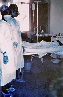
Any person who is taking care of a patient with any VHF (except dengue fever) should take multiple precautions against exposure and infection. The precautions include hand hygiene, double gloves, gowns, shoe and leg coverings, and face shields or goggles. Lassa, CCHF, Ebola, and Marburg viruses may be particularly prone to nosocomial (hospital-based) spread. Airborne precautions should be utilized including, at a minimum, a fit-tested, HEPA filter-equipped respirator (such as an N95 mask), a battery-powered, air-purifying respirator, or a positive pressure supplied air respirator to be worn by personnel coming within 1.8 meters (six feet) of a VHF patient. Groups of patients should be cohorted (sequestered) to a separate building or a ward with an isolated air-handling system. Environmental decontamination is typically accomplished with hypochlorite (e.g. bleach) or phenolic disinfectants.[6]
Management
Medical management of VHF patients may require intensive supportive care. Antiviral therapy with intravenous ribavirin may be useful in Bunyaviridae and Arenaviridae infections (specifically Lassa fever, RVF, CCHF, and HFRS due to Old World Hantavirus infection) and can be used only under an experimental protocol as IND approved by the U.S. Food and Drug Administration (FDA). Interferon may be effective in Argentine or Bolivian hemorrhagic fevers (also available only as IND).[citation needed]
Potential therapies
A potential novel treatment, the NMT inhibitor, has been shown to completely inhibit Lassa (LAS) and Junín (JUN) viral infections in cells based assays.[7]
Uíge Province in Angola was the site of another outbreak of Marburg virus disease in 2005, the largest one to date of this disease.[12]
A VHF outbreak in the village of Mweka, Democratic Republic of the Congo (DRC) that started in August 2007, and that has killed 103 people (100 adults and three children), has been shown to be caused (at least partially) by Ebola virus.
The VHF viruses are spread in a variety of ways. Some may be transmitted to humans through a respiratory route.[citation needed] The viruses are considered by military medical planners to have a potential for aerosol dissemination, weaponization, or likelihood for confusion with similar agents that might be weaponized.[16][17]
↑ Scott, Susan and Duncan, Christopher. (2004). Return of the Black Death: The World's Greatest Serial Killer West Sussex; John Wiley and Sons. ISBN0-470-09000-6.
This page is based on this Wikipedia article Text is available under the CC BY-SA 4.0 license; additional terms may apply. Images, videos and audio are available under their respective licenses.