Meralgia paraesthetica

Last updated
Meralgia paresthetica
Other names Bernhardt-Roth syndrome [1]
Gray826-LFC.png
Innervation of lateral cutaneous nerve of the thigh (shaded area) on the right leg.
Specialty Neurology
Symptoms Pain, dysthesias, paresthesias, and hyperesthesia on the anterolateral thigh [2]
CausesAnything that compresses the lateral femoral cutaneous nerve, such as tight fitting clothes at the waist and injuries. [3]
Risk factors Obesity, pregnancy, diabetes, middle-age [3]
Diagnostic method Clinical examination and patient history [2]
Differential diagnosis Lumbar L2/L3 pathology, lumbar plexopathy, another entrapped peripheral nerve, and pelvic tumors [3] [4]
TreatmentSteroid injections, nerve decompression, and neurectomy [3]

Meralgia paresthetica or meralgia paraesthetica is numbness or pain in the outer thigh not caused by injury to the thigh, but by injury to a nerve that extends from the spinal column to the thigh.

Contents

This chronic nerve entrapment disorder [5] involves a single nerve—the lateral cutaneous nerve of the thigh, which is also called the lateral femoral cutaneous nerve (and hence the syndrome lateral femoral cutaneous neuropathy). [6] The term "meralgia paraesthetica" combines four Greek roots to mean "thigh pain with anomalous perception". The disorder has also been nicknamed bikini brief syndrome [7] and skinny pants syndrome, [8] because it can be caused by wearing tight clothing.

It is also known as Bernhardt-Roth syndrome, after Martin Bernhardt and Vladimir Karlovich Roth who first described it.

Anatomy

The lateral femoral cutaneous nerve (LFCN) originates from the lumbar plexus and contains fibers from lumbar nerves L2 and L3. [9] [4] [2] [3] The LFCN then traverses to the lateral border of the psoas major muscle, crosses the iliacus muscle, and continues to the anterior superior iliac spine (bony landmark). The nerve then passes between inguinal ligament (passes under) and sartorius muscle (passes over). It then divides into an anterior and posterior branch as it travels into the thigh. [5]

Signs and symptoms

The lateral femoral cutaneous nerve (LFCN) is a purely sensory nerve [3] [2] , and consequently the symptoms are also sensory [4] . Symptoms are typically unilateral, seen in about 78% of cases, but may be bilateral. [4] [2]

The most common symptom is pain, paresthesias, or dysthesias on the anterolateral surface of the thigh that extends just above the knee. [3] [10] [5] [2] [11] [4] Examples of paresthesias (abnormal sensations but not unpleasant) and dysthesias (abnormal sensations that are unpleasant) are burning, tingling, numbness, stinging, hypersensitivity, and itching. The symptom intensity can be mild to severe in a way that limits function due to pain. [5]

Activity and position can affect the symptoms. For example, symptoms may be exacerbated by standing or prolonged hip extension, with relief when sitting or flexing the hip. [4] [3] Sometimes strenuous exercise may exacerbate symptoms. [9] [5]

Causes

Lateral cutaneous nerve of thigh and other structures passing between the left inguinal ligament and ilium, frontolateral view of the right side of the pelvis. Gray546-LFC.png
Lateral cutaneous nerve of thigh and other structures passing between the left inguinal ligament and ilium, frontolateral view of the right side of the pelvis.

The cause of Meralgia Paresthetica (MP) is nerve compression or injury to the lateral femoral cutaneous nerve (LFCN). [11] [9] [4] [10] [3]

The site of compression is often at or near the inguinal ligament as the LFCN passes between the upper front hip bone (ilium) and the inguinal ligament near the attachment at the anterior superior iliac spine (the upper point of the hip bone). [2] [3]

Causes of compression can be due to tight fitting clothing at the waist, tight belts, or tight seat belts placing pressure on the LFCN. [4] [2] [3] It can also be due to conditions which increase intra-abdominal volume and consequently pressure on the LFCN such as ascites, obesity [9] , and pregnancy [4] . [2] Less commonly, anything that directly presses on the LFCN anywhere along its course, such a tumor or cyst, can also be a cause. [9] Meralgia Paresthetica can also be caused by accidents that cause injury to the LFCN such as a seat belt injury from a car accident. [9] Iatrogenic injuries to the LFCN are also possible and can happen due to peri-operative positioning [3] or spinal, pelvic, and abdominal operations. [2] [12] In cases where MP is elicited by strenuous physical activity, it's thought that anatomic variations may predispose the LFCN to compression with certain limb movements. [13]

Pathophysiology

Anatomic variations in the course of the lateral femoral cutaneous nerve (LFCN) through the inguinal ligament, its branching level, and course through the thigh is thought to predispose the LFCN to nerve damage through injury or nerve compression. [2]

Diagnosis

Diagnosis is generally based on clinical examination and patient history. [4] [2] [11] The clinical examination may include checking the region that the patient is reporting paresthetias/dysthesias as well as sensory differences between each leg if the symptoms are unilateral. Patient history might include relevant details about recent surgeries, injuries, lifestyle activities that could irritate the nerve.

In typical cases a nerve block is performed. [3] In a nerve block, an anesthetic injection near the nerve numbs the entire nerve to confirm lateral femoral cutaneous nerve (LFCN) involvement and also to distinguish it from lumbosacral root pain. [5] [2] [4] The nerve block test of the LFCN is considered positive if the patient has immediate symptom relief lasting 30-40 minutes after the injection. [5]

Atypical presentations involve an extended workup that involves imaging, electrophysiology testing, neurosonography to rule out similar conditions with overlapping symptoms (differential diagnoses). [3] Some differential diagnoses include L2/L3 lumbar radiculopathy [9] [3] [4] [5] , another entrapped peripheral nerve (e.g. femoral neuropathy) [3] [2] , lumbar plexopathy [2] , abdominal masses [4] , and pelvic tumors [4] .

A lumbar MRI can rule out lumbar radiculopathy. [11] Imaging like MRI/CT/x-ray can be used to rule out mass lesions (e.g. tumors) that could compress the LFCN. [2] [4] Magnetic resonance neurography (MRN) can be used to assess signal alterations along the LFCN. [14] Neurophysiological testing can include sensory nerve conduction studies or somatosensory evoked potentials. [11] [2] [5] Image-guided nerve blocks can be used to rule out the involvement of other nerves. [4] [3]

Treatment

Meralgia Paresthetica is typically a self-limiting condition with spontaneous remission. [4] [2] In one study evaluating the natural history of Meralgia Paresthetica, 62% of patients with no treatment had complete symptom resolution after 2 years. [15] The limitation in existing treatment data is that there are no randomized control trials, much of the data is observational, and it is difficult to distinguish the effectiveness of a treatment from the natural history of the disease (how the disease progresses without treatment). [2]

Conservative

Conservative therapy seeks to reduce aggravating factors. This includes eliminating tight fitting clothing and tight belts, losing weight, avoiding activities that aggravate symptoms, and physical therapy to stretch the muscles and tendons present along the course of the lateral femoral cutaneous nerve. [3] [5] [4]

Medications can be used for symptomatic treatment. [5] Examples include NSAIDs, topical capsaicin, and lidocaine gel/patches for epidermal hypersensitivity or dysesthesia. [4]

Nerve blocks can also be used therapeutically especially if combined with corticosteroids. [3] [10] [2] The relief from steroid injections is highly variable, with long-term, complete pain relief of approximately 22%. [10] The revision rate for injections is approximately 81% and a revision is likely to either be a repeat injection or a nerve decompression. [10]

An emerging non-surgical treatment is radiofrequency ablation to destroy the lateral femoral cutaneous nerve. [10]

Surgical

If repeated nerve blocks don't lead to symptom improvement, then surgery is recommended. [3]

The main surgical treatments for Meralgia Paresthetica are nerve decompression and neurectomy. [3] [5] The main difference between the decompression and neurectomy is that the decompression will remove material around the nerve which might compress it, while the neurectomy removes the nerve itself. By keeping the nerve intact, a decompression preserves sensation. [10] With a neuroectomy, there will be permanent numbness distal to the site of transaction because the nerve is transected and removed. [3] [4] [5] However, in studies measuring patient quality of life, patients seem to be rarely bothered by the loss of sensation. [10]

While both procedures are effective, the neurectomy has better outcomes on complete pain relief. [4] [10] [5] In a systematic review, complete pain relief was seen in 85% of neurectomy cases and 63% of decompression cases. [10] A Cochrane review found that complete pain relief in 85-100% of neurectomy cases and 60-99% of decompression cases. [2] The revision rate for nerve decompression is 12% and a revision was either a repeat decompression or neurectomy. The revision rate for neurectomy is approximately 0%. [10]

One systematic review found that the complication rate after neurectomy is 0%, and after neurolysis is 5%. [10] The most common complications after either surgery is hematomas, subcutaneous effusions, wound healing disorders, and wound infections. [3] [10]

Epidemiology

The incidence is 32-43 cases per 100,000 people per year. [3] [16] There is an association between Meralgia Paresthetica (MP) and age, body mass index (BMI), and diabetes. [3] [5] In 68% of cases MP occurs in middle-aged men. [2]

History

Meralgia Paresthetica (MP) was first described in 1885 by Martin Bernhardt and named by Vladimir Karlovich Roth in 1895. [5] Roth noticed the syndrome in cavalrymen who wore their belts too tightly. [17] Consequently, Meralgia Paresthetica is also known as Bernhardt-Roth syndrome. [4] [5]

See also

Related Research Articles

<span class="mw-page-title-main">Sciatic nerve</span> Large nerve in humans and other animals

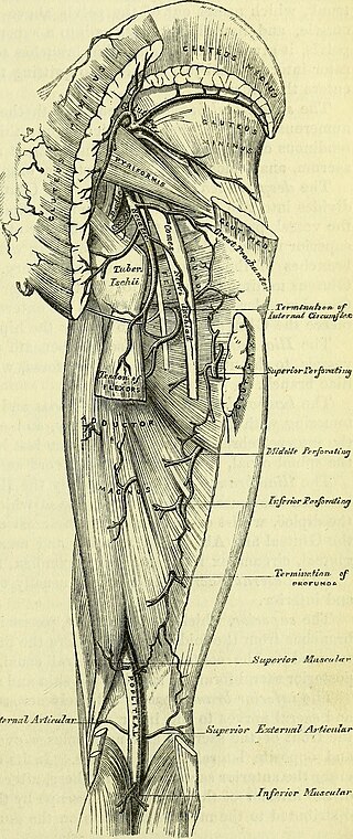
The sciatic nerve, also called the ischiadic nerve, is a large nerve in humans and other vertebrate animals which is the largest branch of the sacral plexus and runs alongside the hip joint and down the lower limb. It is the longest and widest single nerve in the human body, going from the top of the leg to the foot on the posterior aspect. The sciatic nerve has no cutaneous branches for the thigh. This nerve provides the connection to the nervous system for the skin of the lateral leg and the whole foot, the muscles of the back of the thigh, and those of the leg and foot. It is derived from spinal nerves L4 to S3. It contains fibers from both the anterior and posterior divisions of the lumbosacral plexus.

<span class="mw-page-title-main">Spondylosis</span> Degeneration of the vertebral column

Spondylosis is the degeneration of the vertebral column from any cause. In the more narrow sense it refers to spinal osteoarthritis, the age-related degeneration of the spinal column, which is the most common cause of spondylosis. The degenerative process in osteoarthritis chiefly affects the vertebral bodies, the neural foramina and the facet joints. If severe, it may cause pressure on the spinal cord or nerve roots with subsequent sensory or motor disturbances, such as pain, paresthesia, imbalance, and muscle weakness in the limbs.

<span class="mw-page-title-main">Cauda equina syndrome</span> Nerve damage at the end of the spinal cord

Cauda equina syndrome (CES) is a condition that occurs when the bundle of nerves below the end of the spinal cord known as the cauda equina is damaged. Signs and symptoms include low back pain, pain that radiates down the leg, numbness around the anus, and loss of bowel or bladder control. Onset may be rapid or gradual.

<span class="mw-page-title-main">Nerve block</span> Deliberate inhibition of nerve impulses

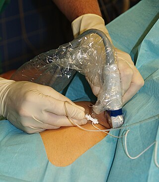
Nerve block or regional nerve blockade is any deliberate interruption of signals traveling along a nerve, often for the purpose of pain relief. Local anesthetic nerve block is a short-term block, usually lasting hours or days, involving the injection of an anesthetic, a corticosteroid, and other agents onto or near a nerve. Neurolytic block, the deliberate temporary degeneration of nerve fibers through the application of chemicals, heat, or freezing, produces a block that may persist for weeks, months, or indefinitely. Neurectomy, the cutting through or removal of a nerve or a section of a nerve, usually produces a permanent block. Because neurectomy of a sensory nerve is often followed, months later, by the emergence of new, more intense pain, sensory nerve neurectomy is rarely performed.

<span class="mw-page-title-main">Iliopsoas</span> Joined psoas and the iliacus muscles

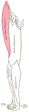
The iliopsoas muscle refers to the joined psoas major and the iliacus muscles. The two muscles are separate in the abdomen, but usually merge in the thigh. They are usually given the common name iliopsoas. The iliopsoas muscle joins to the femur at the lesser trochanter. It acts as the strongest flexor of the hip.

<span class="mw-page-title-main">Femoral nerve</span> Long nerve down the thigh and inner leg

The femoral nerve is a nerve in the thigh that supplies skin on the upper thigh and inner leg, and the muscles that extend the knee. It is the largest branch of the lumbar plexus.

<span class="mw-page-title-main">Lateral cutaneous nerve of thigh</span> Nerve of the thigh

The lateral cutaneous nerve of the thigh is a cutaneous nerve of the thigh. It originates from the dorsal divisions of the second and third lumbar nerves from the lumbar plexus. It passes under the inguinal ligament to reach the thigh. It supplies sensation to the skin on the lateral part of the thigh by an anterior branch and a posterior branch.

A neurectomy, or nerve resection is a neurosurgical procedure in which a peripheral nerve is cut or removed to alleviate neuropathic pain or permanently disable some function of a nerve. The nerve is not intended to grow back. For chronic pain it may be an alternative to a failed nerve decompression when the target nerve has no motor function and numbness is acceptable. Neurectomies have also been used to permanently block autonomic function, and special sensory function not related to pain.

<span class="mw-page-title-main">Spinal disc herniation</span> Injury to the connective tissue between spinal vertebrae

A spinal disc herniation is an injury to the intervertebral disc between two spinal vertebrae, usually caused by excessive strain or trauma to the spine. It may result in back pain, pain or sensation in different parts of the body, and physical disability. The most conclusive diagnostic tool for disc herniation is MRI, and treatment may range from painkillers to surgery. Protection from disc herniation is best provided by core strength and an awareness of body mechanics including good posture.

The saphenous nerve is the largest cutaneous branch of the femoral nerve. It is derived from the lumbar plexus (L3-L4). It is a strictly sensory nerve, and has no motor function. It commences in the proximal (upper) thigh and travels along the adductor canal. Upon exiting the adductor canal, the saphenous nerve terminates by splitting into two terminal branches: the sartorial nerve, and the infrapatellar nerve. The saphenous nerve is responsible for providing sensory innervation to the skin of the anteromedial leg.

<span class="mw-page-title-main">Vladimir Karlovich Roth</span>

Vladimir Karlovich Roth, sometimes Rot was a Russian Empire neuropathologist.

<span class="mw-page-title-main">Martin Bernhardt</span> German neuropathologist

Martin Bernhardt was a German neuropathologist.

In medicine, Carnett's sign is a finding on clinical examination in which (acute) abdominal pain remains unchanged or increases when the muscles of the abdominal wall are tensed. For this part of the abdominal examination, the patient can be asked to lift the head and shoulders from the examination table to tense the abdominal muscles. An alternative is to ask the patient to raise both legs with straight knees.

<span class="mw-page-title-main">Nerve compression syndrome</span> Human disease

Nerve compression syndrome, or compression neuropathy, or nerve entrapment syndrome, is a medical condition caused by chronic, direct pressure on a peripheral nerve. It is known colloquially as a trapped nerve, though this may also refer to nerve root compression. Its symptoms include pain, tingling, numbness and muscle weakness. The symptoms affect just one particular part of the body, depending on which nerve is affected. The diagnosis is largely clinical and can be confirmed with diagnostic nerve blocks. Occasionally imaging and electrophysiology studies aid in the diagnosis. Timely diagnosis is important as untreated chronic nerve compression may cause permanent damage. A surgical nerve decompression can relieve pressure on the nerve but cannot always reverse the physiological changes that occurred before treatment. Nerve injury by a single episode of physical trauma is in one sense an acute compression neuropathy but is not usually included under this heading, as chronic compression takes a unique pathophysiological course.

<span class="mw-page-title-main">Spinal stenosis</span> Disease of the bony spine that results in narrowing of the spinal canal

Spinal stenosis is an abnormal narrowing of the spinal canal or neural foramen that results in pressure on the spinal cord or nerve roots. Symptoms may include pain, numbness, or weakness in the arms or legs. Symptoms are typically gradual in onset and improve with leaning forward. Severe symptoms may include loss of bladder control, loss of bowel control, or sexual dysfunction.

<span class="mw-page-title-main">Anterior cutaneous nerve entrapment syndrome</span> Medical condition

Anterior cutaneous nerve entrapment syndrome (ACNES) is a nerve entrapment condition that causes chronic pain of the abdominal wall. It occurs when nerve endings of the lower thoracic intercostal nerves (7–12) are 'entrapped' in abdominal muscles, causing a severe localized nerve (neuropathic) pain that is usually experienced at the front of the abdomen.

Femoral nerve dysfunction, also known as femoral neuropathy, is a rare type of peripheral nervous system disorder that arises from damage to nerves, specifically the femoral nerve. Given the location of the femoral nerve, indications of dysfunction are centered around the lack of mobility and sensation in lower parts of the legs. The causes of such neuropathy can stem from both direct and indirect injuries, pressures and diseases. Physical examinations are usually first carried out, depending on the high severity of the injury. In the cases of patients with hemorrhage, imaging techniques are used before any physical examination. Another diagnostic method, electrodiagnostic studies, are recognized as the gold standard that is used to confirm the injury of the femoral nerve. After diagnosis, different treatment methods are provided to the patients depending upon their symptoms in order to effectively target the underlying causes. Currently, femoral neuropathy is highly underdiagnosed and its precedent medical history is not well documented worldwide.

A nerve decompression is a neurosurgical procedure to relieve chronic, direct pressure on a nerve to treat nerve entrapment, a pain syndrome characterized by severe chronic pain and muscle weakness. In this way a nerve decompression targets the underlying pathophysiology of the syndrome and is considered a first-line surgical treatment option for peripheral nerve pain. Despite treating the underlying cause of the disease, the symptoms may not be fully reversible as delays in diagnosis can allow permanent damage to occur to the nerve and surrounding microvasculature. Traditionally only nerves accessible with open surgery have been good candidates, however innovations in laparoscopy and nerve-sparing techniques made nearly all nerves in the body good candidates, as surgical access is no longer a barrier.

<span class="mw-page-title-main">Deep gluteal syndrome</span> Medical condition

Deep gluteal syndrome describes the non-discogenic extrapelvic entrapment of the sciatic nerve in the deep gluteal space. It is an extension of non-discogenic sciatic nerve entrapment beyond the traditional model of piriformis syndrome. Symptoms are pain or dysthesias the buttocks, hip, and posterior thigh with or without radiating leg pain. Patients often report pain when sitting. The two most common causes are piriformis syndrome and fibrovascular bands, but many other causes exist. Diagnosis is usually done through physical examination, magnetic resonance imaging, magnetic resonance neurography, and diagnostic nerve blocks. Surgical treatment is an endoscopic sciatic nerve decompression.

<span class="mw-page-title-main">Pathophysiology of nerve entrapment</span>

Nerve entrapment involves a cascade of physiological changes caused by compression and tension. Some of these changes are irreversible. The magnitude and duration of the forces determines the extent of injury. In the acute form, mechanical injury and metabolic blocks impede nerve function. In the chronic form, there is a sequence of changes starting with a breakdown of the blood-nerve-barrier, followed by edema with connective tissue changes, followed by diffuse demyelination, and finally followed by axonmetesis. The injury will often be a mixed lesion where mild/moderate compression is a combination of a metabolic block and neuropraxia, while severe compression combines elements of neuropraxia and axonmetesis.

References

  1. Pearce JM (2006). "Meralgia paraesthetica (Bernhardt-Roth syndrome)". Journal of Neurology, Neurosurgery & Psychiatry. 77 (1): 84. doi:10.1136/jnnp.2005.072363. PMC   2117390 . PMID   16361600.
  2. 1 2 3 4 5 6 7 8 9 10 11 12 13 14 15 16 17 18 19 20 21 22 Khalil N, Nicotra A, Rakowicz W (December 2012). "Treatment for meralgia paraesthetica". Cochrane Database Syst Rev. 12 (12): CD004159. doi:10.1002/14651858.CD004159.pub3. PMC   7197425 . PMID   23235604.
  3. 1 2 3 4 5 6 7 8 9 10 11 12 13 14 15 16 17 18 19 20 21 22 23 24 25 Scholz C, Hohenhaus M, Pedro MT, Uerschels AK, Dengler NF (September 2023). "Meralgia Paresthetica: Relevance, Diagnosis, and Treatment". Dtsch Arztebl Int. 120 (39): 655–661. doi:10.3238/arztebl.m2023.0170. PMC   10622057 . PMID   37534445.
  4. 1 2 3 4 5 6 7 8 9 10 11 12 13 14 15 16 17 18 19 20 21 22 Coffey R, Gupta V. Meralgia Paresthetica. [Updated 2023 May 1]. In: StatPearls [Internet]. Treasure Island (FL): StatPearls Publishing; 2024 Jan-. Available from: https://www.ncbi.nlm.nih.gov/books/NBK557735/
  5. 1 2 3 4 5 6 7 8 9 10 11 12 13 14 15 16 17 Cheatham SW, Kolber MJ, Salamh PA (December 2013). "Meralgia paresthetica: a review of the literature". Int J Sports Phys Ther. 8 (6): 883–93. PMC   3867081 . PMID   24377074.
  6. IASP, XXXI: LOCAL SYNDROMES IN THE LEG OR FOOT: PAIN OF NEUROLOGICAL ORIGIN Archived 2012-12-19 at the Wayback Machine , 2012
  7. Lambert HW, Wineski LE (July 2010). Lippincott's Illustrated Q&A Review of Anatomy and Embryology. Lippincott Williams & Wilkins. ISBN   978-1-60547-315-4.
  8. "Make your pencil jeans less dangerous to your health". Vanguard. March 8, 2012.
  9. 1 2 3 4 5 6 7 de Ruiter GC, Oosterhuis JW, Vissers TF, Kloet A (May 2023). "Unusual causes for meralgia paresthetica: systematic review of the literature and single center experience". Neurosurg Rev. 46 (1): 107. doi:10.1007/s10143-023-02023-2. PMC   10162905 . PMID   37148363.
  10. 1 2 3 4 5 6 7 8 9 10 11 12 13 Lu VM, Burks SS, Heath RN, Wolde T, Spinner RJ, Levi AD (January 2021). "Meralgia paresthetica treated by injection, decompression, and neurectomy: a systematic review and meta-analysis of pain and operative outcomes". J Neurosurg. 135 (3): 912–922. doi:10.3171/2020.7.JNS202191. PMID   33450741.
  11. 1 2 3 4 5 Schönberg B, Pigorsch M, Huscher D, Baruchi S, Reinsch J, Zdunczyk A, Scholz C, Uerschels AK, Dengler NF (February 2023). "Diagnosis and treatment of meralgia paresthetica between 2005 and 2018: a national cohort study". Neurosurg Rev. 46 (1): 54. doi:10.1007/s10143-023-01962-0. PMC   9925535 . PMID   36781569.
  12. Ivins GK (2000). "Meralgia Paresthetica, the Elusive Diagnosis". Annals of Surgery. 232 (2): 281–6. doi:10.1097/00000658-200008000-00019. PMC   1421141 . PMID   10903608.
  13. Kho KH, Blijham PJ, Zwarts MJ (June 2005). "Meralgia paresthetica after strenuous exercise". Muscle Nerve. 31 (6): 761–3. doi:10.1002/mus.20271. PMID   15654691.
  14. Chhabra A, Del Grande F, Soldatos T, Chalian M, Belzberg AJ, Williams EH, Jalali FS, Thawait GK, Eng J, Carrino JA (June 2013). "Meralgia paresthetica: 3-Tesla magnetic resonance neurography". Skeletal Radiol. 42 (6): 803–8. doi:10.1007/s00256-012-1557-4. PMID   23306718.
  15. ECKER AD, WOLTMAN HW. MERALGIA PARAESTHETICA: A REPORT OF ONE HUNDRED AND FIFTY CASES. JAMA. 1938;110(20):1650–1652. doi:10.1001/jama.1938.02790200018006
  16. van Slobbe AM, Bohnen AM, Bernsen RM, Koes BW, Bierma-Zeinstra SM (March 2004). "Incidence rates and determinants in meralgia paresthetica in general practice". J Neurol. 251 (3): 294–7. doi:10.1007/s00415-004-0310-x. PMID   15015008.
  17. Pearce JM (January 2006). "Meralgia paraesthetica (Bernhardt-Roth syndrome)". J Neurol Neurosurg Psychiatry. 77 (1): 84. doi:10.1136/jnnp.2005.072363. PMC   2117390 . PMID   16361600.