Purtscher's retinopathy

Last updated
Purtscher's retinopathy
Human eye cross-sectional view grayscale.png
Anatomy of eye(Retina on top)
Specialty Ophthalmology

Purtscher's retinopathy is a disease where part of the eye (retina) is damaged. Usually associated with severe head injuries, it may also occur with other types of trauma, such as long bone fractures, or with several non-traumatic systemic diseases. However, the exact cause of the disease is not well understood. There are no treatments specific for Purtscher's retinopathy, and the prognosis varies. The disease can threaten vision, sometimes causing temporary or permanent blindness.

Contents

It is named for the Austrian ophthalmologist, Othmar Purtscher (1852–1927), who detected it in 1910 and described it fully in 1912.

Presentation

Associated diseases

Pathophysiology

Purtscher's retinopathy likely involves complex pathophysiology, with several contributing factors, including complement-mediated aggregates, fat, air, fibrin clots and platelet clumps. [1] The diseases leads to the formation of cotton wool spots in the retina, a finding observed in several other diseases, and atrophy of the optic nerve. [2]

Diagnosis

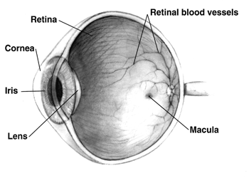
Where trauma is involved, only a funduscopic examination of the back of the eye (retina) is necessary to make the diagnosis. [2] Fluoroscein angiography may show a decrease in blood flow to the areas of whiteness in the retina.

Treatment

It may be treated with triamcinolone in some cases. [3] However, in general, there are no treatments for Purtscher's retinopathy. If it is caused by a systemic disease or emboli, then those conditions should be treated.

Prognosis

Purtscher's retinopathy can lead to loss of vision, [4] and recovery of vision may occur very little. [2] However, vision recovery does occur in some cases, and reports have varied on the long-term prognosis. [5]

History

Purtscher's retinopathy was first characterized in 1910 and 1912 as a syndrome of sudden blindness after head trauma, [6] with patches of hemorrhage and whitening of the retina in both eyes. [2] Later, it was discovered to occur after other types of trauma, such as chest trauma, and is associated with several non-traumatic systemic diseases. [2] Purtscher's retinopathy may also be associated with acute pancreatitis, vasculitis, embolization of such materials as fat and amniotic fluid, [7] systemic lupus erythematosus, thrombotic thrombocytopenic purpura, and chronic kidney failure. [2] Purtscher's retinopathy may be caused by extensive fractures of the long bones. [8]

Related Research Articles

<span class="mw-page-title-main">Retinopathy</span> Medical condition

Retinopathy is any damage to the retina of the eyes, which may cause vision impairment. Retinopathy often refers to retinal vascular disease, or damage to the retina caused by abnormal blood flow. Age-related macular degeneration is technically included under the umbrella term retinopathy but is often discussed as a separate entity. Retinopathy, or retinal vascular disease, can be broadly categorized into proliferative and non-proliferative types. Frequently, retinopathy is an ocular manifestation of systemic disease as seen in diabetes or hypertension. Diabetes is the most common cause of retinopathy in the U.S. as of 2008. Diabetic retinopathy is the leading cause of blindness in working-aged people. It accounts for about 5% of blindness worldwide and is designated a priority eye disease by the World Health Organization.

<span class="mw-page-title-main">Diabetic retinopathy</span> Medical condition

Diabetic retinopathy, is a medical condition in which damage occurs to the retina due to diabetes mellitus. It is a leading cause of blindness in developed countries.

<span class="mw-page-title-main">Hypertensive retinopathy</span> Medical condition

Hypertensive retinopathy is damage to the retina and retinal circulation due to high blood pressure.

<span class="mw-page-title-main">Thrombotic thrombocytopenic purpura</span> Medical condition with blood clots

Thrombotic thrombocytopenic purpura (TTP) is a blood disorder that results in blood clots forming in small blood vessels throughout the body. This results in a low platelet count, low red blood cells due to their breakdown, and often kidney, heart, and brain dysfunction. Symptoms may include large bruises, fever, weakness, shortness of breath, confusion, and headache. Repeated episodes may occur.

Microangiopathic hemolytic anemia (MAHA) is a microangiopathic subgroup of hemolytic anemia caused by factors in the small blood vessels. It is identified by the finding of anemia and schistocytes on microscopy of the blood film.

<span class="mw-page-title-main">Thrombocytopenia</span> Medical condition

Thrombocytopenia is a condition characterized by abnormally low levels of platelets, also known as thrombocytes, in the blood. Low levels of platelets in turn may lead to prolonged or excessive bleeding. It is the most common coagulation disorder among intensive care patients and is seen in a fifth of medical patients and a third of surgical patients.

<span class="mw-page-title-main">Panniculitis</span> Medical condition

Panniculitis is a group of diseases whose hallmark is inflammation of subcutaneous adipose tissue. Symptoms include tender skin nodules, and systemic signs such as weight loss and fatigue.

<span class="mw-page-title-main">Amaurosis fugax</span> Medical condition

Amaurosis fugax is a painless temporary loss of vision in one or both eyes.

Libman–Sacks endocarditis is a form of non-bacterial endocarditis that is seen in association with systemic lupus erythematosus, antiphospholipid syndrome, and malignancies. It is one of the most common cardiac manifestations of lupus.

<span class="mw-page-title-main">Petechia</span> Medical condition

A petechia is a small red or purple spot that can appear on the skin, conjunctiva, retina, and mucous membranes which is caused by haemorrhage of capillaries. The word is derived from Italian petecchia, 'freckle,' of obscure origin. It refers to one of the three descriptive types of hematoma differentiated by size, the other two being ecchymosis and purpura. The term is always used in the plural (petechiae), since a single petechia is seldom noticed or significant.

<span class="mw-page-title-main">Methylprednisolone</span> Corticosteroid medication

Methylprednisolone is a synthetic glucocorticoid, primarily prescribed for its anti-inflammatory and immunosuppressive effects. It is either used at low doses for chronic illnesses or used concomitantly at high doses during acute flares. Methylprednisolone and its derivatives can be administered orally or parenterally.

<span class="mw-page-title-main">ADAMTS13</span> Metalloprotease enzyme

ADAMTS13 —also known as von Willebrand factor-cleaving protease (VWFCP)—is a zinc-containing metalloprotease enzyme that cleaves von Willebrand factor (vWf), a large protein involved in blood clotting. It is secreted into the blood and degrades large vWf multimers, decreasing their activity.

<span class="mw-page-title-main">Fat embolism syndrome</span> Medical condition

Fat embolism syndrome occurs when fat enters the blood stream and results in symptoms. Symptoms generally begin within a day. This may include a petechial rash, decreased level of consciousness, and shortness of breath. Other symptoms may include fever and decreased urine output. The risk of death is about 10%.

<span class="mw-page-title-main">Intraocular hemorrhage</span> Medical condition

Intraocular hemorrhage is bleeding inside the eye. Bleeding can occur from any structure of the eye where there is vasculature or blood flow, including the anterior chamber, vitreous cavity, retina, choroid, suprachoroidal space, or optic disc.

<span class="mw-page-title-main">Lupus</span> Human autoimmune disease

Lupus, technically known as systemic lupus erythematosus (SLE), is an autoimmune disease in which the body's immune system mistakenly attacks healthy tissue in many parts of the body. Symptoms vary among people and may be mild to severe. Common symptoms include painful and swollen joints, fever, chest pain, hair loss, mouth ulcers, swollen lymph nodes, feeling tired, and a red rash which is most commonly on the face. Often there are periods of illness, called flares, and periods of remission during which there are few symptoms.

<span class="mw-page-title-main">Retinal vasculitis</span> Medical condition

Retinal vasculitis is inflammation of the vascular branches of the retinal artery, caused either by primary ocular disease processes, or as a specific presentation of any systemic form of vasculitis such as Behçet's disease, sarcoidosis, multiple sclerosis, or any form of systemic necrotizing vasculitis such as temporal arteritis, polyarteritis nodosa, and granulomatosis with polyangiitis, or due to lupus erythematosus, or rheumatoid arthritis. Eales disease, pars planitis, birdshot retinochoroidopathy, and Fuchs heterochromic iridocyclitis (FHI) can also cause retinal vasculitis. Infectious pathogens such as Mycobacterium tuberculosis, visceral larva migrans can also cause retinal vasculitis. Drug-induced vasculitis may involve retina as well, as seen in methamphetamine induced vasculitis.

Atypical hemolytic uremic syndrome (aHUS), also known as complement-mediated hemolytic uremic syndrome, is an extremely rare, life-threatening, progressive disease that frequently has a genetic component. In most cases it can be effectively controlled by interruption of the complement cascade. Particular monoclonal antibodies, discussed later in the article, have proven efficacy in many cases.

Autoimmune optic neuropathy (AON), sometimes called autoimmune optic neuritis, may be a forme fruste of systemic lupus erythematosus (SLE) associated optic neuropathy. AON is more than the presence of any optic neuritis in a patient with an autoimmune process, as it describes a relatively specific clinical syndrome. AON is characterized by chronically progressive or recurrent vision loss associated with serological evidence of autoimmunity. Specifically, this term has been suggested for cases of optic neuritis with serological evidence of vasculitis by positive ANA, despite the lack of meeting criteria for SLE. The clinical manifestations include progressive vision loss that tends to be steroid-responsive and steroid dependent.

Sickle cell retinopathy can be defined as retinal changes due to blood vessel damage in the eye of a person with a background of sickle cell disease. It can likely progress to loss of vision in late stages due to vitreous hemorrhage or retinal detachment. Sickle cell disease is a structural red blood cell disorder leading to consequences in multiple systems. It is characterized by chronic red blood cell destruction, vascular injury, and tissue ischemia causing damage to the brain, eyes, heart, lungs, kidneys, spleen, and musculoskeletal system.

References

  1. Buckley, Sally A.; James, B (July 1996). "Purtscher's retinopathy". Postgraduate Medical Journal. 72 (849): 409–12. doi:10.1136/pgmj.72.849.409. PMC   2398519 . PMID   8935600.
  2. 1 2 3 4 5 6 Yanoff, Myron; Duker, Jay S. (2008). Ophthalmology (3rd ed.). Edinburgh: Mosby. pp. 750–754. ISBN   978-0323057516.
  3. Olson J, Rouhani B, Mandava N (March 2008). "Sub-Tenon's triamcinolone for post-partum Purtscher's-like retinopathy". Clinical Ophthalmology. 2 (1): 195–8. doi:10.2147/opth.s2080. PMC   2698687 . PMID   19668404.
  4. Agrawal A, McKibbin M (Nov 2007). "Purtscher's retinopathy: epidemiology, clinical features, and outcome". British Journal of Ophthalmology. 91 (11): 1456–9. doi:10.1136/bjo.2007.117408. PMC   2095457 . PMID   17556428.
  5. Schmidt, D; Otto, T (June 2004). "Prognosis and differential diagnosis of Purtscher's retinopathy". Der Ophthalmologe: Zeitschrift der Deutschen Ophthalmologischen Gesellschaft. 101 (6): 576–83. doi:10.1007/s00347-003-0952-6. PMID   15197574.
  6. Purtscher O (1912). "Angiopathia retinae traumatica. Lymphorrhagien des Augengrundes". Albrecht von Græfe's Archiv für Ophthalmologie. 82 (2): 347–71. doi:10.1007/bf01929449. S2CID   40420476.
  7. Retinopathy, Purtscher's at eMedicine
  8. Chuang, EL; Miller FS, 3rd; Kalina, RE (March 1985). "Retinal lesions following long bone fractures". Ophthalmology. 92 (3): 370–4. doi:10.1016/s0161-6420(85)34023-x. PMID   3991127.