Septo-optic dysplasia

Last updated
Septo-optic dysplasia
Other namesde Morsier syndrome [1] [2]
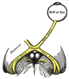
Gray773.png
The optic nerve is underdeveloped in this condition.
Specialty Ophthalmology
Diagnostic method congenital hypopituitarism, holoprosencephaly [3]

Septo-optic dysplasia (SOD), known also as de Morsier syndrome, is a rare congenital malformation syndrome that features a combination of the underdevelopment of the optic nerve, pituitary gland dysfunction, and absence of the septum pellucidum (a midline part of the brain). Two or more of these features need to be present for a clinical diagnosis—only 30% of patients have all three. [4] French-Swiss doctor Georges de Morsier first recognized the relation of a rudimentary or absent septum pellucidum with hypoplasia of the optic nerves and chiasm in 1956. [5]

Contents

Signs and symptoms

The symptoms of SOD can be divided into those related to optic nerve underdevelopment, pituitary hormone abnormalities, and mid-line brain abnormalities. Symptoms may vary greatly in their severity. [6]

Optic nerve underdevelopment

About one quarter of people with SOD have significant visual impairment in one or both eyes, as a result of optic nerve underdevelopment. Developmental delays are more common in children with bilateral optic nerve hypoplasia than those with unilateral optic nerve hypoplasia. [6] Bilateral optic nerve hypoplasia is also associated with a more severe disease course. [7]

There may be nystagmus (involuntary eye movements, often side-to-side). [6] In cases of bilateral optic nerve hypoplasia this can usually be detected within the first three months of life. It may be followed by strabismus developing in the first year. [7]

Pituitary hormone abnormalities

Underdevelopment of the pituitary gland in SOD leads to hypopituitarism, most commonly in the form of growth hormone deficiency. [6] In severe cases panhypopituitarism may occur. [8]

Mid-line brain abnormalities

In SOD, mid-line brain structures such as the corpus callosum and the septum pellucidum may fail to develop normally, leading to neurological problems such as seizures or developmental delay. [8] Patients with seizures are more likely to show additional neurological abnormalities such as cortical dysplasia, polymicrogyria and schizencephaly. Such abnormalities are always identified when spastic quadriplegia is present.[ citation needed ]

Neurological symptoms are typically considered late onset manifestations of SOD. Common initial presentations include epilepsy, development delays and limb weakness. Intellectual abilities vary widely from normal to severe intellectual disability. [7] Early studies indicated intellectual disability occurs in 71% of cases, cerebral palsy occurs in 57%, and behavioral problems occur in 20%, but further research has indicated that these symptoms may be less common and caused by additional neurological abnormalities. [4]

Causes

SOD results from an abnormality in the development of the embryonic forebrain at 4–6 weeks of pregnancy. [6] There is no known single cause of SOD, but it is thought that both genetic and environmental factors may be involved. [8]

Genetic

Rare familial recurrence has been reported, suggesting at least one genetic form (HESX1). [9] Five homozygous and eight heterozygous pathogenic HESX1 mutations have been discovered. Patients with homozygous mutations present with a typical SOD phenotype while those with heterozygous mutations are mildly affected. [6] In addition to HESX1, mutations in OTX2, SOX2 and PAX6 have been implicated in SOD. [8] SOX2 mutations in SOD patients are associated with severe bilateral ocular anomalies such as microphthalmia and anophthalmia. Additional features associated with SOX2 mutations include developmental delay, oesophageal atresia, short stature and sensorineural hearing loss. Genetic abnormalities are identified in fewer than one percent of patients. [6]

Diagnosis

A diagnosis of SOD is made when at least two of the following triad are present: optic nerve underdevelopment; pituitary hormone abnormalities; and mid-line brain abnormalities. Diagnosis is usually made at birth or during childhood, and a clinical diagnosis can be confirmed by MRI scans. [6]

Treatment

There is no cure for SOD. [6] Treatment aims to manage symptoms and may require a multidisciplinary team of specialists including neurologists, ophthalmologists and endocrinologists. Hormone deficiencies may be treated with HRT but vision impairments are not usually treatable. [3]

Epidemiology

A European survey put the prevalence of SOD at somewhere in the region of 1.9 to 2.5 per 100,000 live births, with the United Kingdom having a particularly high rate and with increased risk for younger mothers. [10]

History

In 1941 Dr. David Reeves at the Children's Hospital Los Angeles described an association between underdevelopment of the optic nerve with an absent septum pellucidum. Fifteen years later French doctor Georges de Morsier reported his theory that the two abnormalities were connected and coined the term septo-optic dysplasia. In 1970 American doctor William Hoyt made the connection between the three features of SOD and named the syndrome after de Morsier. [11]

British model and television personality Katie Price's son, Harvey, has this condition. [12]

Related Research Articles

<span class="mw-page-title-main">Dwarfism</span> Small size of an organism, caused by growth deficiency or genetic mutations

Dwarfism is a condition wherein an organism is exceptionally small, and mostly occurs in the animal kingdom. In humans, it is sometimes defined as an adult height of less than 147 centimetres, regardless of sex; the average adult height among people with dwarfism is 120 centimetres (4 ft). Disproportionate dwarfism is characterized by either short limbs or a short torso. In cases of proportionate dwarfism, both the limbs and torso are unusually small. Intelligence is usually normal, and most have a nearly normal life expectancy. People with dwarfism can usually bear children, though there are additional risks to the mother and child depending upon the underlying condition.

<span class="mw-page-title-main">Megalencephaly</span> Medical condition

Megalencephaly is a growth development disorder in which the brain is abnormally large. It is characterized by a brain with an average weight that is 2.5 standard deviations above the mean of the general population. Approximately 1 out of 50 children (2%) are said to have the characteristics of megalencephaly in the general population.

<span class="mw-page-title-main">Macrocephaly</span> Abnormally large head size

Macrocephaly is a condition in which circumference of the human head is abnormally large. It may be pathological or harmless, and can be a familial genetic characteristic. People diagnosed with macrocephaly will receive further medical tests to determine whether the syndrome is accompanied by particular disorders. Those with benign or familial macrocephaly are considered to have megalencephaly.

Optic nerve hypoplasia (ONH) is a medical condition arising from the underdevelopment of the optic nerve(s). This condition is the most common congenital optic nerve anomaly. The optic disc appears abnormally small, because not all the optic nerve axons have developed properly. It is often associated with endocrinopathies, developmental delay, and brain malformations. The optic nerve, which is responsible for transmitting visual signals from the retina to the brain, has approximately 1.2 million nerve fibers in the average person. In those diagnosed with ONH, however, there are noticeably fewer nerves.

<span class="mw-page-title-main">Septum pellucidum</span> Thin membrane between the lateral ventricles of the brain

The septum pellucidum is a thin, triangular, vertical double membrane separating the anterior horns of the left and right lateral ventricles of the brain. It runs as a sheet from the corpus callosum down to the fornix.

<span class="mw-page-title-main">Coloboma</span> Hole in one of the structures of the eye

A coloboma is a hole in one of the structures of the eye, such as the iris, retina, choroid, or optic disc. The hole is present from birth and can be caused when a gap called the choroid fissure, which is present during early stages of prenatal development, fails to close up completely before a child is born. Ocular coloboma is relatively uncommon, affecting less than one in every 10,000 births.

Bilateral frontoparietal polymicrogyria is a genetic disorder with autosomal recessive inheritance that causes a cortical malformation. Our brain has folds in the cortex to increase surface area called gyri and patients with polymicrogyria have an increase number of folds and smaller folds than usual. Polymicrogyria is defined as a cerebral malformation of cortical development in which the normal gyral pattern of the surface of the brain is replaced by an excessive number of small, fused gyri separated by shallow sulci and abnormal cortical lamination. From ongoing research, mutation in GPR56, a member of the adhesion G protein-coupled receptor (GPCR) family, results in BFPP. These mutations are located in different regions of the protein without any evidence of a relationship between the position of the mutation and phenotypic severity. It is also found that GPR56 plays a role in cortical pattering.

Agenesis of the corpus callosum (ACC) is a rare birth defect in which there is a complete or partial absence of the corpus callosum. It occurs when the development of the corpus callosum, the band of white matter connecting the two hemispheres in the brain, in the embryo is disrupted. The result of this is that the fibers that would otherwise form the corpus callosum are instead longitudinally oriented along the ipsilateral ventricular wall and form structures called Probst bundles.

Aplasia is a birth defect where an organ or tissue is wholly or largely absent. It is caused by a defect in a developmental process.
<span class="mw-page-title-main">Duane-radial ray syndrome</span> Medical condition

Duane-radial ray syndrome, also known as Okihiro Syndrome, is a rare autosomal dominant disorder that primarily affects the eyes and causes abnormalities of bones in the arms and hands. This disorder is considered to be a SALL4-related disorder due to the SALL4 gene mutations leading to these abnormalities. It is diagnosed by clinical findings on a physical exam as well as genetic testing and imaging. After being diagnosed, there are other evaluations that one may go through in order to determine the extent of the disease. There are various treatments for the symptoms of this disorder.
<span class="mw-page-title-main">1p36 deletion syndrome</span> Medical condition

1p36 deletion syndrome is a congenital genetic disorder characterized by moderate to severe intellectual disability, delayed growth, hypotonia, seizures, limited speech ability, malformations, hearing and vision impairment, and distinct facial features. The symptoms may vary, depending on the exact location of the chromosomal deletion.

<span class="mw-page-title-main">Papillorenal syndrome</span> Medical condition

Papillorenal syndrome is an autosomal dominant genetic disorder marked by underdevelopment (hypoplasia) of the kidney and colobomas of the optic nerve.

<span class="mw-page-title-main">3C syndrome</span> Medical condition

3C syndrome is a rare condition whose symptoms include heart defects, cerebellar hypoplasia, and cranial dysmorphism. It was first described in the medical literature in 1987 by Ritscher and Schinzel, for whom the disorder is sometimes named.

<span class="mw-page-title-main">HESX1</span> Protein-coding gene in the species Homo sapiens

Homeobox expressed in ES cells 1, also known as homeobox protein ANF, is a homeobox protein that in humans is encoded by the HESX1 gene.

<span class="mw-page-title-main">Antley–Bixler syndrome</span> Medical condition

Antley–Bixler syndrome is a rare, severe autosomal recessive congenital disorder characterized by malformations and deformities affecting the majority of the skeleton and other areas of the body.

<span class="mw-page-title-main">Roberts syndrome</span> Medical condition

Roberts syndrome, or sometimes called pseudothalidomide syndrome, is an extremely rare autosomal recessive genetic disorder that is characterized by mild to severe prenatal retardation or disruption of cell division, leading to malformation of the bones in the skull, face, arms, and legs.

Gonadotropin-releasing hormone (GnRH) insensitivity also known as Isolated gonadotropin-releasing hormone (GnRH)deficiency (IGD) is a rare autosomal recessive genetic and endocrine syndrome which is characterized by inactivating mutations of the gonadotropin-releasing hormone receptor (GnRHR) and thus an insensitivity of the receptor to gonadotropin-releasing hormone (GnRH), resulting in a partial or complete loss of the ability of the gonads to synthesize the sex hormones. The condition manifests itself as isolated hypogonadotropic hypogonadism (IHH), presenting with symptoms such as delayed, reduced, or absent puberty, low or complete lack of libido, and infertility, and is the predominant cause of IHH when it does not present alongside anosmia.

Georges de Morsier was a Swiss neurologist.

<span class="mw-page-title-main">Strømme syndrome</span> Rare genetic condition involving intestinal atresia, eye abnormalities and microcephaly

Strømme syndrome is a very rare autosomal recessive genetic condition characterised by intestinal atresia, eye abnormalities and microcephaly. The intestinal atresia is of the "apple-peel" type, in which the remaining intestine is twisted around its main artery. The front third of the eye is typically underdeveloped, and there is usually moderate developmental delay. Less common features include an atrial septal defect, increased muscle tone or skeletal abnormalities. Physical features may include short stature, large, low-set ears, a small jaw, a large mouth, epicanthic folds, or fine, sparse hair.

<span class="mw-page-title-main">Muscle–eye–brain disease</span> Medical condition

Muscle–eye–brain (MEB) disease, also known as muscular dystrophy-dystroglycanopathy congenital with brain and eye anomalies A3 (MDDGA3), is a kind of rare congenital muscular dystrophy (CMD), largely characterized by hypotonia at birth. Patients have muscular dystrophy, central nervous system abnormalities and ocular abnormalities. The condition is degenerative.
References

  1. synd/2548 at Who Named It?
  2. de Morsier G (1956). "Études sur les dysraphies, crânioencéphaliques. III. Agénésie du septum palludicum avec malformation du tractus optique. La dysplasie septo-optique" [Studies on dysraphias, cranioencephalic. III. Agenesis of the septum palludicum with malformation of the optic tract. Septo-optic dysplasia.]. Schweizer Archiv für Neurologie und Psychiatrie (in French). Zurich. 77: 267–292.
  3. 1 2 "Septo-Optic Dysplasia Spectrum". Genetic and Rare Diseases Information Center (GARD). Retrieved 5 August 2021.
  4. 1 2 Gleason, CA; Devascar, S (5 October 2011). "Congenital malformations of the Central Nervous System". Avery's Diseases of the Newborn (9 ed.). Saunders. p.  857. ISBN   978-1437701340.
  5. Daroff RB, Jankovic J, Mazziotta JC, Pomeroy SL (2015-10-25). Bradley's neurology in clinical practice (Seventh ed.). London. ISBN   9780323339162. OCLC   932031625.{{cite book}}: CS1 maint: location missing publisher (link)
  6. 1 2 3 4 5 6 7 8 9 Webb EA, Dattani MT (April 2010). "Septo-optic dysplasia". European Journal of Human Genetics. 18 (4): 393–7. doi: 10.1038/ejhg.2009.125 . PMC   2987262 . PMID   19623216.
  7. 1 2 3 Ganau M, Huet S, Syrmos N, Meloni M, Jayamohan J (2019). "Neuro-Ophthalmological Manifestations Of Septo-Optic Dysplasia: Current Perspectives". Eye and Brain. 11 (11): 37–47. doi: 10.2147/EB.S186307 . PMC   6805786 . PMID   31695544.
  8. 1 2 3 4 "Septo-Optic Dysplasia". Genetics Home Reference. U.S. National Library of Medicine. Retrieved 16 July 2015.
  9. Dattani MT, Martinez-Barbera JP, Thomas PQ, Brickman JM, Gupta R, Mårtensson IL, et al. (June 1998). "Mutations in the homeobox gene HESX1/Hesx1 associated with septo-optic dysplasia in human and mouse". Nature Genetics. 19 (2): 125–33. doi:10.1038/477. PMID   9620767. S2CID   28880292.
  10. Garne E, Rissmann A, Addor MC, Barisic I, Bergman J, Braz P, et al. (September 2018). "Epidemiology of septo-optic dysplasia with focus on prevalence and maternal age - A EUROCAT study". European Journal of Medical Genetics. 61 (9): 483–488. doi:10.1016/j.ejmg.2018.05.010. PMID   29753093. S2CID   21673637.
  11. Borchert M (March 2012). "Reappraisal of the optic nerve hypoplasia syndrome". Journal of Neuro-Ophthalmology. 32 (1): 58–67. doi: 10.1097/WNO.0b013e31824442b8 . PMID   22330852. S2CID   12131899.
  12. Singh A (25 January 2021). "Katie Price's dilemma over what's best for disabled son will be familiar to many". The Telegraph.