Temporal lobe epilepsy

Last updated

Temporal lobe epilepsy
Lobes of the brain NL.svg
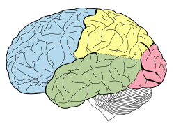
Lobes of the brain. Temporal lobe in green
Specialty Neurology, Psychiatry

In the field of neurology, temporal lobe epilepsy is an enduring brain disorder that causes unprovoked seizures from the temporal lobe. Temporal lobe epilepsy is the most common type of focal onset epilepsy among adults. [1] Seizure symptoms and behavior distinguish seizures arising from the medial temporal lobe from seizures arising from the lateral (neocortical) temporal lobe . [2] Memory and psychiatric comorbidities may occur. Diagnosis relies on electroencephalographic (EEG) and neuroimaging studies. [3] [4] Anticonvulsant medications, epilepsy surgery and dietary treatments may improve seizure control. [5] [6] [6] [7] [8]

Contents

Types

Under the International League Against Epilepsy (ILAE) 2017 classification of the epilepsies, focal onset epilepsy occurs from seizures arising from a biological neural network within a single cerebral hemisphere. [9] [10] Temporal lobe epilepsy occurs from seizures arising within the temporal lobe. [10] Temporal lobe epilepsy is the most common focal onset epilepsy, and 80% of temporal lobe epilepsy is mesial (medial) temporal lobe epilepsy, temporal lobe epilepsy arising from the inner (medial) part of the temporal lobe that may involve the hippocampus, parahippocampal gyrus or amygdala. [2] [11] The less common lateral temporal lobe or neocortical temporal lobe seizures arise from the outer (lateral) temporal lobe. [2] The ILAE 2017 classification distinguishes focal aware from focal impaired seizures. [10] A focal aware temporal lobe seizure occurs if a person remains aware of what occurs during the entire seizure; awareness may be retained even if impaired responsiveness occurs during the seizure. [10] A focal impaired awareness temporal lobe seizure occurs if a person becomes unaware during any part of the seizure. [10]

Symptoms and behavior

Medial temporal lobe epilepsy

During a temporal lobe seizure, a person may experience a seizure aura; an aura is an autonomic, cognitive, emotional or sensory experience that commonly occurs during the beginning part of a seizure. [10] [2] The common medial temporal lobe seizure auras include a rising epigastric feeling, abdominal discomfort, taste (gustatory), smell (olfactory), tingling (somatosensory), fear, déjà vu, jamais vu, flushing, or rapid heart rate (tachycardia). [2] A person may then stare blankly, appear motionless (behavioral arrest) and lose awareness. [2] Repeated stereotyped motor behaviors ( automatisms ) may occur such as repeated swallowing, lip smacking, picking, fumbling, patting or vocalizations. [2] Dystonic posture is an unnatural stiffening of one arm occurring during a seizure. [12] A dystonic posture on one side of the body commonly indicates seizure onset from the opposite side of the brain e.g. right arm dystonic posture arising from a left temporal lobe seizure. [12] Impaired language function (dysphasia) during or soon following a seizure is more likely to occur when seizures arise from the language dominant side of the brain. [12]

Lateral temporal lobe epilepsy

The common auras from seizures arising from primary auditory cortex include vertigo, humming sound, ringing sound, buzzing sound, hearing a song, hearing voices or altered hearing sensation. [2] Lateral temporal lobe seizures arising from the temporal-parietal lobe junction may cause complex visual hallucinations. [2] In comparison to mesial temporal lobe seizures, lateral temporal lobe seizures are briefer duration seizures, occur with earlier loss of awareness, and are more likely become a focal to bilateral tonic-clonic seizure. [2] Impaired language function (dysphasia) during or soon following a seizure is more likely to occur when seizures arise from the language dominant side of the brain. [12]

Comorbidities

Memory

The major cognitive impairment in mesial temporal lobe epilepsy is a progressive memory impairment. [13] :71 This involves declarative memory impairment, including episodic memory and semantic memory, and is worse when medications fail to control seizures. [14] [15] [13] :71 Mesial temporal lobe epilepsy arising from the language dominant hemisphere impairs verbal memory, and mesial temporal lobe epilepsy arising from the language non-dominant hemisphere impairs nonverbal memory. [13] :71 [15]

Psychiatric comorbidities

Psychiatric disorders are more common among those with epilepsy, and the highest prevalence occurs among those with temporal lobe epilepsy. [16] The most common psychiatric comorbidity is major depressive disorder. [16] Other disorders include post-traumatic stress disorder, general anxiety disorder, psychosis, obsessive-compulsive disorder, schizophrenia, bipolar disorder, substance use disorder and a ~9% prevalence of suicide. [16]

Personality

Geschwind syndrome is a syndrome of altered sexuality (most often hyposexuality), religiosity, and compulsive or extensive writing and drawing occurring in persons with temporal lobe epilepsy. [17] :347–348 However, subsequent studies did not support the association of these behavioral traits with temporal lobe epilepsy. [17] :347–348 There are reports of religious behaviors occurring in persons with temporal lobe epilepsy. [18] [19] [20] [21] [22]

Causes

Hippocampal sclerosis, brain tumor, traumatic brain injury, cerebral vascular malformation, neuronal migration disorders, infections such as encephalitis and meningitis, autoimmune disease (limbic encephalitis) and genetic disorders may cause temporal lobe epilepsy. [23]

Risk factors

Many persons with uncontrolled temporal lobe epilepsy had childhood febrile seizures. [24] A brief febrile seizure only slighty increases the risk for developing afebrile seizures. [25] However, the prolonged seizure of febrile status epilepticus leads to a 9% risk for developing epilepsy. [25] There is no clear relationship between febrile seizures and development of hippocampal sclerosis. [25]

Mechanisms

The neural circuit of the hippocampus shows dentate gyrus (DG). subiculum (SB). entorhinal cortex (EC), CA1 sector and CA3 sector CajalHippocampus (modified).png
The neural circuit of the hippocampus shows dentate gyrus (DG). subiculum (SB). entorhinal cortex (EC), CA1 sector and CA3 sector
Scalp electrodes are placed to record an electroencephalogram EEG Recording Cap.jpg
Scalp electrodes are placed to record an electroencephalogram
Brain MRI with hippocampus identified by cross hairs. Hippocampus-mri.jpg
Brain MRI with hippocampus identified by cross hairs.
Brain positron emission tomography (PET) scan PET Normal brain.jpg
Brain positron emission tomography (PET) scan

Neuronal loss

Hippocampal sclerosis occurs with severe CA1 and less severe CA3 and CA4 neuronal loss. [26] Experimental research has shown that N-methyl-d-aspartate (NMDA) receptor activation causes neuronal cell loss, and electrical stimulation-induced animal models of temporal lobe epilepsy duplicate the cell loss pattern of temporal lobe epilepsy in humans. [26] Repetitive seizures irreversibly damage interneurons leading to persistent loss of recurrent inhibition. [26] Damage of GABAergic interneurons lead to loss of inhibition, uncontrolled neuronal firing, leading to seizures. [26] The secondary epileptogenesis hypothesis is that repetitive seizures lead to interneuron loss, loss of glutamatergic principal neurons, axonal sprouting, and formation of new recurrent glutamatergic excitatory circuits leading to a more severe epilepsy. [27] Mechanisms related to neuronal loss incompletely account for temporal lobe epilepsy as temporal lobe epilepsy may occur with only minimal neuronal cell loss. [26]

Neuron-specific type 2 K+/Cl− cotransporter (KCC2) mutation

This KCC2 mutation prevents subicular neurons from potassium and chloride ion extrusion, leading to intracellular chloride accumulation, and positive γ-Aminobutyric acid (GABA) mediated currents. [26] Accumulated chloride efflux through GABA receptors leads to neuronal depolarization, increased neuronal excitability and ultimately seizures. [26] Persons with this mutation have mesial temporal lobe epilepsy with hippocampal sclerosis. [26]

Granule cell dispersion

Dentate gyrus granule cell dispersion refers to a granule cell layer that is widened, poorly demarcated, or accompanied by granule cells outside the layer (ectopic granule cells). [28] :1318 In the normal brain, dentate granule cells block seizure spread from entorhinal cortex to the hippocampus. [26] A hypothesis is that granule cell dispersion may disrupt the normal mossy fiber pathway connecting granule cells and CA3 pyramidal cells leading to mossy fiber sprouting and new excitatory networks capable of generating seizures. [26] However, a study has shown that a similar pattern of granule cell dispersion may occur in persons without epilepsy. [29]

Cortical developmental malformations

Focal cortical dysplasia is a brain malformation that may cause temporal lobe epilepsy. [26] This malformation may cause abnormal cortical layers (dyslamination ), occur with abnormal neurons ( dysmorphic neurons, balloon cells ) and may occur with a brain tumor or vascular malformation. [26] An abnormality of the MTOR pathway leads to hyperexcitable glutamate mediated neurons leading to seizures. [26]

Diagnosis

A surgeon performs epilepsy brain surgery Epilepsy-surgery-in-progress.jpg
A surgeon performs epilepsy brain surgery
Vagus nerve stimulation Vagus nerve stimulation.jpg
Vagus nerve stimulation
Responsive neurostimulation device m`rkt RNS.jpg
Responsive neurostimulation device
Surgeon implants a brain electrode for deep brain stimulation Peds DBS.jpg
Surgeon implants a brain electrode for deep brain stimulation

Electroencephalogram

The temporal lobe epileptiform discharge is a pattern seen on the electroencephalgram (EEG) test; temporal lobe epileptiform discharges occur between seizures and confirm the diagnosis of temporal lobe epilepsy. [3] Long-term video-EEG monitoring may record the behavior and EEG during a seizure. [3] Magnetoencephalography may diagnose temporal lobe epilepsy by recording epileptiform discharges or seizure patterns arising from the magnetic fields of neural electrical currents. [3]

Neuroimaging

Neuroimaging tests may identify the cause for seizures and the seizure focus, the brain location where seizures begin. [4] In newly diagnosed epilepsy, magnetic resonance imaging (MRI) can detect brain lesion in up to 12 to 14% of persons with epilepsy. [30] However, for those with chronic epilepsy, MRI can detect brain lesion in 80% of the persons with epilepsy. [30] 3-Tesla MRI scan is advised for those with evidence of focal epilepsy such as temporal lobe epilepsy. [4] Abnormalities identified by MRI scan include hippocampal sclerosis, focal cortical dysplasia, other cortical developmental brain malformations, developmental and low-grade tumors, cavernous hemangioma, hypoxic-ischemic brain injury, traumatic brain injury and encephalitis. [4]

18F-fluorodeoxyglucose (18F-FDG) brain positron emission tomography (PET) may show a brain region of decreased glucose metabolism at a time between seizures; this hypometabolic region may correspond to the seizure focus, and PET scan is more sensitive for temporal lobe seizure focus localization compared to epilepsy arising from other brain lobes. [4] Single-photon emission computed tomography (SPECT) may show a region of decreased blood flow occurring 40-60 seconds after injection during the seizure; this reduced blood flow region may correspond to the seizure focus. [4]

Computed tomography (CT) scan is less sensitive than MRI scan for identifying small tumors, vascular malformations, cortical developmental brain malformations, and abnormalities in the medial temporal lobe. [30] CT scan is advised in emergencies when the suspected cause of epilepsy may be intracerebral hemorrhage, brain abscess, large cerebral infarction or subdural empyema. [4] [30] A person who requires neuroimaging but cannot have an MRI scan due to implanted devices such as a cardiac pacemaker, defibrillator or cochlear implant may receive a CT scan. CT scan may better demonstrate calcium containing brain abnormalities causing epilepsy such as in tuberous sclerosis and Sturge–Weber syndrome. [4] [30]

Treatment

Medical treatment

Anticonvulsant oral medications control seizures in about two-thirds of persons with epilepsy, and control commonly occurs with one or two medications. [31]

Surgical treatment

Those with uncontrolled seizures despite treatment with multiple anticonvulsant medications have pharmacoresistant epilepsy, and they may require epilepsy surgery to achieve seizure control. [9] [31]

Penfield and Flanigan first described anterior temporal lobectomy, partial surgical removal of the temporal lobe, for treatment of mesial temporal lobe epilepsy in 1950. [32] In a prospective randomized controlled trial comparing anterior temporal lobectomy to medical therapy for pharmacoresistant temporal lobe epilepsy, surgery was more effective than medical therapy with 1-year seizure free outcome occurring in 58% of persons with anterior temporal lobectomy compared to 8% of persons with drug treatment. [5] Among those with intractable mesial temporal lobe epilepsy and hippocampal sclerosis, about 70% become seizure-free after epilepsy surgery. [33] :751 Studies show that language dominant anterior temporal lobectomy may lead to verbal memory decline. [15] However, study outcomes are more variable on language non-dominant anterior temporal lobectomy leading to nonverbal memory decline. [15]

Magnetic resonance-guided laser interstitial thermal therapy, stereotactic radiosurgery , and stereotactic radiofrequency ablation are surgical methods that treat epilepsy by destroying the abnormal brain tissue that causes seizures. [34] [35] [36]

Neurostimulation may also improve seizure control. [6] The vagus nerve stimulator (VNS) is surgically implanted in the chest, and delivers programmed electrical stimulation to the vagus nerve in the neck. [37] The responsive neurostimulation device is implanted in the skull, monitors electrical brain activity for seizures, and responds to seizures with programmed electrical stimulation to one or two brain areas. [38] Programmed deep brain stimulation of the anterior thalamic nucleus may treat seizures arising from more than 2 brain areas. [6]

Dietary treatment

The ketogenic diet and modified Atkins diet are additional temporal lobe epilepsy treatment options. [7] [8]

Remission

Among those who develop childhood temporal lobe epilepsy, epilepsy remits in about one-third of children. [39] Remission was more likely among those without hippocampal sclerosis, brain tumor, or focal cortical dysplasia on MRI scan. [39]

See also

Notes

  1. Muhlhofer et al. 2017.
  2. 1 2 3 4 5 6 7 8 9 10 Chowdhury et al. 2021.
  3. 1 2 3 4 Javidan 2012.
  4. 1 2 3 4 5 6 7 8 Duncan 2019.
  5. 1 2 Wiebe et al. 2001.
  6. 1 2 3 4 Touma et al. 2022.
  7. 1 2 Freeman, Kossoff & Hartman 2007.
  8. 1 2 Rezaei et al. 2019.
  9. 1 2 Scheffer et al. 2017.
  10. 1 2 3 4 5 6 Fisher et al. 2017.
  11. Tatum 2012.
  12. 1 2 3 4 Rusu et al. 2005.
  13. 1 2 3 Zeman, Kapur & Jones-Gotman 2012.
  14. Quiroga 2012.
  15. 1 2 3 4 Bauman, Devinsky & Liu 2019.
  16. 1 2 3 Lu et al. 2021.
  17. 1 2 David et al. 2012.
  18. Tedrus et al. 2015.
  19. d'Orsi & Tinuper 2006.
  20. Sirven, Drazkowski & Noe 2007.
  21. Arzy & Schurr 2016.
  22. Tedrus, Fonseca & Höehr 2013.
  23. Vadlamudi 2003.
  24. Patterson, Baram & Shinnar 2014.
  25. 1 2 3 Mewasingh, Chin & Scott 2020.
  26. 1 2 3 4 5 6 7 8 9 10 11 12 13 Ong 2019.
  27. Ben-Ari & Dudek 2010.
  28. Blümcke et al. 2013.
  29. Roy, Millen & Kapur 2020.
  30. 1 2 3 4 5 Salmenpera & Duncan 2005.
  31. 1 2 Kwan & Brodie 2000.
  32. Penfield & Flanigan 1950.
  33. Lamberink et al. 2020.
  34. Chen et al. 2023.
  35. Kerezoudis et al. 2022.
  36. Mustafa & Zaben 2022.
  37. Goggins, Mitani & Tanaka 2022.
  38. Geller 2018.
  39. 1 2 Spooner et al. 2006.

Related Research Articles

<span class="mw-page-title-main">Epilepsy</span> Group of neurological disorders causing seizures

Epilepsy is a group of non-communicable neurological disorders characterized by recurrent epileptic seizures. An epileptic seizure is the clinical manifestation of an abnormal, excessive, and synchronized electrical discharge in the brain cells called neurons. The occurrence of two or more unprovoked seizures defines epilepsy. The occurrence of just one seizure may warrant the definition in a more clinical usage where recurrence may be able to be prejudged. Epileptic seizures can vary from brief and nearly undetectable periods to long periods of vigorous shaking due to abnormal electrical activity in the brain. These episodes can result in physical injuries, either directly such as broken bones or through causing accidents. In epilepsy, seizures tend to recur and may have no detectable underlying cause. Isolated seizures that are provoked by a specific cause such as poisoning are not deemed to represent epilepsy. People with epilepsy may be treated differently in various areas of the world and experience varying degrees of social stigma due to the alarming nature of their symptoms.

<span class="mw-page-title-main">Seizure</span> Period of symptoms due to excessive or synchronous neuronal brain activity

A seizure is a period of symptoms due to abnormally excessive or synchronous neuronal activity in the brain. Outward effects vary from uncontrolled shaking movements involving much of the body with loss of consciousness, to shaking movements involving only part of the body with variable levels of consciousness, to a subtle momentary loss of awareness. These episodes usually last less than two minutes and it takes some time to return to normal. Loss of bladder control may occur.

<span class="mw-page-title-main">Hippocampus</span> Vertebrate brain region involved in memory consolidation

The hippocampus is a major component of the brain of humans and other vertebrates. Humans and other mammals have two hippocampi, one in each side of the brain. The hippocampus is part of the limbic system, and plays important roles in the consolidation of information from short-term memory to long-term memory, and in spatial memory that enables navigation. The hippocampus is located in the allocortex, with neural projections into the neocortex, in humans as well as other primates. The hippocampus, as the medial pallium, is a structure found in all vertebrates. In humans, it contains two main interlocking parts: the hippocampus proper, and the dentate gyrus.

<span class="mw-page-title-main">Focal cortical dysplasia</span> Medical condition

Focal cortical dysplasia (FCD) is a congenital abnormality of brain development where the neurons in an area of the brain failed to migrate in the proper formation in utero. Focal means that it is limited to a focal zone in any lobe. Focal cortical dysplasia is a common cause of intractable epilepsy in children and is a frequent cause of epilepsy in adults. There are three types of FCD with subtypes, including type 1a, 1b, 1c, 2a, 2b, 3a, 3b, 3c, and 3d, each with distinct histopathological features. All forms of focal cortical dysplasia lead to disorganization of the normal structure of the cerebral cortex :

<span class="mw-page-title-main">Hippocampal sclerosis</span> Medical condition

Hippocampal sclerosis (HS) or mesial temporal sclerosis (MTS) is a neuropathological condition with severe neuronal cell loss and gliosis in the hippocampus. Neuroimaging tests such as magnetic resonance imaging (MRI) and positron emission tomography (PET) may identify individuals with hippocampal sclerosis. Hippocampal sclerosis occurs in 3 distinct settings: mesial temporal lobe epilepsy, adult neurodegenerative disease and acute brain injury.

Focal seizures are seizures which affect initially only one hemisphere of the brain. The brain is divided into two hemispheres, each consisting of four lobes – the frontal, temporal, parietal and occipital lobes. A focal seizure is generated in and affects just one part of the brain – a whole hemisphere or part of a lobe. Symptoms will vary according to where the seizure occurs. When seizures occur in the frontal lobe the patient may experience a wave-like sensation in the head. When seizures occur in the temporal lobe, a feeling of déjà vu may be experienced. When seizures are localized to the parietal lobe, a numbness or tingling may occur. With seizures occurring in the occipital lobe, visual disturbances or hallucinations have been reported.

Frontal lobe epilepsy (FLE) is a neurological disorder that is characterized by brief, recurring seizures arising in the frontal lobes of the brain, that often occur during sleep. It is the second most common type of epilepsy after temporal lobe epilepsy (TLE), and is related to the temporal form in that both forms are characterized by partial (focal) seizures.

Anterior temporal lobectomy (ATL) is the complete or partial removal of the anterior portion of the temporal lobe of the brain. The exact boundaries for removal can vary slightly in practice and between neurosurgeons. It is a treatment option for temporal lobe epilepsy for those in whom anticonvulsant medications do not control epileptic seizures, and who have frequent seizures, and who additionally qualify based on a WADA test to localize the dominant hemisphere for language module.

In the field of neurology, seizure types are categories of seizures defined by seizure behavior, symptoms, and diagnostic tests. The International League Against Epilepsy (ILAE) 2017 classification of seizures is the internationally recognized standard for identifying seizure types. The ILAE 2017 classification of seizures is a revision of the prior ILAE 1981 classification of seizures. Distinguishing between seizure types is important since different types of seizures may have different causes, outcomes, and treatments.

Epilepsy surgery involves a neurosurgical procedure where an area of the brain involved in seizures is either resected, ablated, disconnected or stimulated. The goal is to eliminate seizures or significantly reduce seizure burden. Approximately 60% of all people with epilepsy have focal epilepsy syndromes. In 15% to 20% of these patients, the condition is not adequately controlled with anticonvulsive drugs. Such patients are potential candidates for surgical epilepsy treatment.

<span class="mw-page-title-main">Rolandic epilepsy</span> Most common epilepsy syndrome in childhood, usually subsiding with age

Benign Rolandic epilepsy or self-limited epilepsy with centrotemporal spikes is the most common epilepsy syndrome in childhood. Most children will outgrow the syndrome, hence the label benign. The seizures, sometimes referred to as sylvian seizures, start around the central sulcus of the brain.

Sleep-related hypermotor epilepsy (SHE), previously known as nocturnal frontal lobe epilepsy, is a form of focal epilepsy characterized by seizures which arise during sleep. The seizures are most typically characterized by complex motor behaviors. It is a relatively uncommon form of epilepsy that constitutes approximately 9-13% of cases. This disorder is associated with cognitive impairment in at least half of patients as well as excessive daytime sleepiness due to poor sleep quality. This disorder is sometimes misdiagnosed as a non-epileptic sleep disorder. There are many potential causes of SHE including genetic, acquired injuries and structural abnormalities.

<span class="mw-page-title-main">Epilepsy in children</span>

Epilepsy is a neurological condition of recurrent episodes of unprovoked epileptic seizures. A seizure is an abnormal neuronal brain activity that can cause intellectual, emotional, and social consequences. Epilepsy affects children and adults of all ages and races, and is one of the most common neurological disorders of the nervous system. Epilepsy is more common among children than adults, affecting about 6 out of 1000 US children that are between the age of 0 to 5 years old. The epileptic seizures can be of different types depending on the part of the brain that was affected, seizures are classified in 2 main types partial seizure or genralized seizure.

Febrile infection-related epilepsy syndrome (FIRES), is onset of severe seizures following a febrile illness in someone who was previously healthy. The seizures may initially be focal; however, often become tonic-clonic. Complications often include intellectual disability, behavioral problems, and ongoing seizures.

Granule cell dispersion is one of the abnormal structural changes that has been shown in brains of patients with temporal lobe epilepsy. It has also been shown in different animal models, such as the kainic acid model, pilocarpine model, and kindling model. But granule cell dispersion was not found by using perforant pathway stimulation.

An epilepsy syndrome is defined as "a characteristic cluster of clinical and EEG features, often supported by specific etiological findings ."

The hippocampus participates in the encoding, consolidation, and retrieval of memories. The hippocampus is located in the medial temporal lobe (subcortical), and is an infolding of the medial temporal cortex. The hippocampus plays an important role in the transfer of information from short-term memory to long-term memory during encoding and retrieval stages. These stages do not need to occur successively, but are, as studies seem to indicate, and they are broadly divided in the neuronal mechanisms that they require or even in the hippocampal areas that they seem to activate. According to Gazzaniga, "encoding is the processing of incoming information that creates memory traces to be stored." There are two steps to the encoding process: "acquisition" and "consolidation". During the acquisition process, stimuli are committed to short term memory. Then, consolidation is where the hippocampus along with other cortical structures stabilize an object within long term memory, which strengthens over time, and is a process for which a number of theories have arisen to explain the underlying mechanism. After encoding, the hippocampus is capable of going through the retrieval process. The retrieval process consists of accessing stored information; this allows learned behaviors to experience conscious depiction and execution. Encoding and retrieval are both affected by neurodegenerative and anxiety disorders and epilepsy.

Drug-resistant epilepsy (DRE), also known as refractory epilepsy, intractable epilepsy, or pharmacoresistant epilepsy, is diagnosed following a failure of adequate trials of two tolerated and appropriately chosen and used antiepileptic drugs (AEDs) to achieve sustained seizure freedom. The probability that the next medication will achieve seizure freedom drops with every failed AED. For example, after two failed AEDs, the probability that the third will achieve seizure freedom is around 4%. Drug-resistant epilepsy is commonly diagnosed after several years of uncontrolled seizures, however, in most cases, it is evident much earlier. Approximately 30% of people with epilepsy have a drug-resistant form.

Christine Kilpatrick is an Australian neurologist and the chief executive of Royal Melbourne Health. She has held this position since 2017. Previously, she was the chief executive of the Royal Children's Hospital from 2008 to 2017 and the executive director of Medical Services, Melbourne Health and executive director of the Royal Melbourne Hospital from 2004 to 2008. Before she held these positions, she worked as a neurologist at Royal Melbourne Health and engaged in extensive neurological research, especially epilepsy.

Malignant migrating partial seizures of infancy (MMPSI) is a rare epileptic syndrome that onsets before 6 months of age, commonly in the first few weeks of life. Once seizures start, the site of seizure activity repeatedly migrates from one area of the brain to another, with few periods of remission in between. These seizures are 'focal' (updated term for 'partial'), meaning they do not affect both sides of the brain at the same time. These continuous seizures cause damage to the brain, hence the descriptor 'malignant.'

References