Band 3 anion transport protein

Last updated
SLC4A1
Alpha Intercalated Cell Cartoon.svg
Available structures
PDB Ortholog search: PDBe RCSB
Identifiers
Aliases SLC4A1 , solute carrier family 4 (anion exchanger), member 1 (Diego blood group), AE1, BND3, CD233, DI, EMPB3, EPB3, FR, RTA1A, SW, WD, WD1, WR, CHC, SAO, SPH4, solute carrier family 4 member 1 (Diego blood group)
External IDs OMIM: 109270 MGI: 109393 HomoloGene: 133556 GeneCards: SLC4A1
Orthologs
SpeciesHumanMouse
Entrez
Ensembl
UniProt
RefSeq (mRNA)

NM_000342

NM_011403

RefSeq (protein)

NP_000333

NP_035533

Location (UCSC) Chr 17: 44.25 – 44.27 Mb Chr 11: 102.24 – 102.26 Mb
PubMed search [3] [4]
Wikidata
View/Edit Human View/Edit Mouse
solute carrier family 4 (anion exchanger), member 1, adapter protein
Alpha Intercalated Cell Cartoon.svg
Identifiers
SymbolSLC4A1AP
NCBI gene 22950
HGNC 13813
OMIM 602655
RefSeq NM_018158
UniProt P02730
Other data
Locus Chr. 2 p23.3
Search for
Structures Swiss-model
Domains InterPro

Band 3 anion transport protein, also known as anion exchanger 1 (AE1) or band 3 or solute carrier family 4 member 1 (SLC4A1), is a protein that is encoded by the SLC4A1 gene in humans.

Band 3 anion transport protein is a phylogenetically-preserved transport protein responsible for mediating the exchange of chloride (Cl) with bicarbonate (HCO3) across plasma membranes. Functionally similar members of the AE clade are AE2 and AE3. [5]

Function

Band 3 is present in the basolateral face of the α-intercalated cells of the collecting ducts of the nephron, which are the main acid-secreting cells of the kidney. They generate hydrogen ions and bicarbonate ions from carbon dioxide and water – a reaction catalysed by carbonic anhydrase. The hydrogen ions are pumped into the collecting duct tubule by vacuolar H+ ATPase, the apical proton pump, which thus excretes acid into the urine. kAE1, the kidney isoform of AE1, exchanges bicarbonate for chloride on the basolateral surface, essentially returning bicarbonate to the blood. Here it performs two functions:[ citation needed ]

Distribution

It is ubiquitous throughout the vertebrates. In mammals, it is present in two specific sites:[ citation needed ]

Gene products

The erythrocyte and kidney forms are different isoforms of the same protein. [6]

The erythrocyte isoform of AE1, known as eAE1, is composed of 911 amino acids. eAE1 is an important structural component of the erythrocyte cell membrane, making up to 25% of the cell membrane surface. Each red cell contains approximately one million copies of eAE1.[ citation needed ]

The kidney isoform of AE1, known as kAE1 (which is 65 amino acids shorter than erythroid AE1) is found in the basolateral membrane of alpha-intercalated cells in the cortical collecting duct of the kidney.[ citation needed ]

Clinical significance

Mutations of kidney AE1 cause distal (type 1) renal tubular acidosis, which is an inability to acidify the urine, even if the blood is too acidic. These mutations are disease causing as they cause mistargetting of the mutant band 3 proteins so that they are retained within the cell or occasionally addressed to the wrong (i.e. apical) surface.[ citation needed ]

Mutations of erythroid AE1 affecting the extracellular domains of the molecule may cause alterations in the individual's blood group, as band 3 determines the Diego antigen system (blood group).[ citation needed ]

More importantly erythroid AE1 mutations cause 15–25% of cases of hereditary spherocytosis (a disorder associated with progressive red cell membrane loss), and also cause the hereditary conditions of hereditary stomatocytosis [7] and Southeast Asian ovalocytosis. [8]

Interactions

Band 3 has been shown to interact with CA2 [9] [10] [11] [12] and CA4. [13]

Discovery

AE1 was discovered following SDS-PAGE (sodium dodecyl sulfate polyacrylamide gel electrophoresis) of erythrocyte cell membrane. The large 'third' band on the electrophoresis gel represented AE1, which was thus initially termed 'Band 3'. [14]

See also

Related Research Articles

<span class="mw-page-title-main">Red blood cell</span> Oxygen-delivering blood cell and the most common type of blood cell

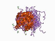
Red blood cells (RBCs), scientific name erythrocytes (from ancient Greek erythros 'red' and kytos 'hollow vessel', with -cyte translated as 'cell' in modern usage), also known as red cells, erythroid cells, and rarely haematids, are the most common type of blood cell and the vertebrate's principal means of delivering oxygen (O2) to the body tissues—via blood flow through the circulatory system. Erythrocytes take up oxygen in the lungs, or in fish the gills, and release it into tissues while squeezing through the body's capillaries.

<span class="mw-page-title-main">Acetazolamide</span> Chemical compound

Acetazolamide, sold under the trade name Diamox among others, is a medication used to treat glaucoma, epilepsy, acute mountain sickness, periodic paralysis, idiopathic intracranial hypertension, heart failure and to alkalinize urine. It may be used long term for the treatment of open angle glaucoma and short term for acute angle closure glaucoma until surgery can be carried out. It is taken by mouth or injection into a vein. Acetazolamide is a first generation carbonic anhydrase inhibitor and it decreases the ocular fluid and osmolality in the eye to decrease intraocular pressure.

<span class="mw-page-title-main">Renal physiology</span> Study of the physiology of the kidney

Renal physiology is the study of the physiology of the kidney. This encompasses all functions of the kidney, including maintenance of acid-base balance; regulation of fluid balance; regulation of sodium, potassium, and other electrolytes; clearance of toxins; absorption of glucose, amino acids, and other small molecules; regulation of blood pressure; production of various hormones, such as erythropoietin; and activation of vitamin D.

<span class="mw-page-title-main">Antiporter</span> Class of transmembrane transporter protein

An antiporter is an integral membrane protein involved in secondary active transport. It is a type of cotransporter, which means that uses the movement of one In the case of an antiporter, two or more different molecules or ions are moved across a phospholipid membrane, such as the plasma membrane, in opposite directions, one into the cell and one out of the cell. This is in contrast to symporters, which are another type of cotransporter that moves two or more ions in the same direction.

<span class="mw-page-title-main">Cotransporter</span> Type of membrane transport proteins

Cotransporters are a subcategory of membrane transport proteins (transporters) that couple the favorable movement of one molecule with its concentration gradient and unfavorable movement of another molecule against its concentration gradient. They enable coupled or cotransport and include antiporters and symporters. In general, cotransporters consist of two out of the three classes of integral membrane proteins known as transporters that move molecules and ions across biomembranes. Uniporters are also transporters but move only one type of molecule down its concentration gradient and are not classified as cotransporters.

The Na–K–Cl cotransporter (NKCC) is a transport protein that aids in the secondary active transport of sodium, potassium, and chloride into cells. In humans there are two isoforms of this membrane transport protein, NKCC1 and NKCC2, encoded by two different genes. Two isoforms of the NKCC1/Slc12a2 gene result from keeping or skipping exon 21 in the final gene product.

Alkaline tide (mal del puerco) refers to a condition, normally encountered after eating a meal, where during the production of hydrochloric acid by the parietal cells in the stomach, the parietal cells secrete bicarbonate ions across their basolateral membranes and into the blood, causing a temporary increase in blood pH.

Pendrin is an anion exchange protein that in humans is encoded by the SLC26A4 gene . Pendrin was initially identified as a sodium-independent chloride-iodide exchanger with subsequent studies showing that it also accepts formate and bicarbonate as substrates. Pendrin is similar to the Band 3 transport protein found in red blood cells. Pendrin is the protein which is mutated in Pendred syndrome, which is an autosomal recessive disorder characterized by sensorineural hearing loss, goiter and a partial organification problem detectable by a positive perchlorate test.

<span class="mw-page-title-main">Protein 4.1</span> Protein-coding gene in the species Homo sapiens

Protein 4.1,, is a protein associated with the cytoskeleton that in humans is encoded by the EPB41 gene. Protein 4.1 is a major structural element of the erythrocyte membrane skeleton. It plays a key role in regulating membrane physical properties of mechanical stability and deformability by stabilizing spectrin-actin interaction. Protein 4.1 interacts with spectrin and short actin filaments to form the erythrocyte membrane skeleton. Mutations of spectrin and protein 4.1 are associated with elliptocytosis or spherocytosis and anemia of varying severity.

<span class="mw-page-title-main">Chloride shift</span> Transfer of ions into red blood cells

Chloride shift (also known as the Hamburger phenomenon or lineas phenomenon, named after Hartog Jakob Hamburger) is a process which occurs in a cardiovascular system and refers to the exchange of bicarbonate (HCO3) and chloride (Cl) across the membrane of red blood cells (RBCs).

<span class="mw-page-title-main">Carbonic anhydrase II</span> Enzyme found in humans

Carbonic anhydrase II is one of sixteen forms of human α carbonic anhydrases. Carbonic anhydrase catalyzes reversible hydration of carbon dioxide. Defects in this enzyme are associated with osteopetrosis and renal tubular acidosis. Renal carbonic anhydrase allows the reabsorption of bicarbonate ions in the proximal tubule. Loss of carbonic anhydrase activity in bones impairs the ability of osteoclasts to promote bone resorption, leading to osteopetrosis.

<span class="mw-page-title-main">Electrogenic sodium bicarbonate cotransporter 1</span> Protein-coding gene in the species Homo sapiens

Electrogenic sodium bicarbonate cotransporter 1, sodium bicarbonate cotransporter is a membrane transport protein that in humans is encoded by the SLC4A4 gene.

<span class="mw-page-title-main">Anion exchange protein 2</span> Protein-coding gene in the species Homo sapiens

Anion exchange protein 2 (AE2) is a membrane transport protein that in humans is encoded by the SLC4A2 gene. AE2 is functionally similar to the Band 3 Cl/HCO3 exchange protein.

<span class="mw-page-title-main">Carbonic anhydrase 4</span> Enzyme found in humans

Carbonic anhydrase 4 is an enzyme that in humans is encoded by the CA4 gene.

<span class="mw-page-title-main">Chloride anion exchanger</span> Protein-coding gene in the species Homo sapiens

Chloride anion exchanger, also known as down-regulated in adenoma, is a protein that in humans is encoded by the SLC26A3 gene.

<span class="mw-page-title-main">Carbonic anhydrase 12</span> Enzyme found in humans

Carbonic anhydrase 12 is an enzyme that in humans is encoded by the CA12 gene.

<span class="mw-page-title-main">Anion exchange protein 3</span> Protein-coding gene in the species Homo sapiens

Anion exchange protein 3 is a membrane transport protein that in humans is encoded by the SLC4A3 gene. AE3 is functionally similar to the Band 3 Cl/HCO3 exchange protein but it is expressed primarily in brain neurons and in the heart. Like AE2 its activity is sensitive to pH. AE3 mutations have been linked to seizures.

<span class="mw-page-title-main">Anion exchange transporter</span> Protein-coding gene in the species Homo sapiens

Anion exchange transporter is a protein that in humans is encoded by the SLC26A7 gene.

<span class="mw-page-title-main">Carbonic anhydrase</span> Class of enzymes

The carbonic anhydrases form a family of enzymes that catalyze the interconversion between carbon dioxide and water and the dissociated ions of carbonic acid. The active site of most carbonic anhydrases contains a zinc ion. They are therefore classified as metalloenzymes. The enzyme maintains acid-base balance and helps transport carbon dioxide.

The anion exchanger family is a member of the large APC superfamily of secondary carriers. Members of the AE family are generally responsible for the transport of anions across cellular barriers, although their functions may vary. All of them exchange bicarbonate. Characterized protein members of the AE family are found in plants, animals, insects and yeast. Uncharacterized AE homologues may be present in bacteria. Animal AE proteins consist of homodimeric complexes of integral membrane proteins that vary in size from about 900 amino acyl residues to about 1250 residues. Their N-terminal hydrophilic domains may interact with cytoskeletal proteins and therefore play a cell structural role. Some of the currently characterized members of the AE family can be found in the Transporter Classification Database.

References

  1. 1 2 3 GRCh38: Ensembl release 89: ENSG00000004939 Ensembl, May 2017
  2. 1 2 3 GRCm38: Ensembl release 89: ENSMUSG00000006574 Ensembl, May 2017
  3. "Human PubMed Reference:". National Center for Biotechnology Information, U.S. National Library of Medicine.
  4. "Mouse PubMed Reference:". National Center for Biotechnology Information, U.S. National Library of Medicine.
  5. Alper SL (2009). "Molecular physiology and genetics of Na+-independent SLC4 anion exchangers". Journal of Experimental Biology. 212 (11): 1672–1683. doi:10.1242/jeb.029454. PMC   2683012 . PMID   19448077.
  6. Schlüter K, Drenckhahn D (August 1986). "Co-clustering of denatured hemoglobin with band 3: its role in binding of autoantibodies against band 3 to abnormal and aged erythrocytes". Proc. Natl. Acad. Sci. U.S.A. 83 (16): 6137–41. Bibcode:1986PNAS...83.6137S. doi: 10.1073/pnas.83.16.6137 . PMC   386454 . PMID   3461480.
  7. Bruce LJ, Robinson HC, Guizouarn H, Borgese F, Harrison P, King MJ, Goede JS, Coles SE, Gore DM, Lutz HU, Ficarella R, Layton DM, Iolascon A, Ellory JC, Stewart GW (2005). "Monovalent cation leaks in human red cells caused by single amino-acid substitutions in the transport domain of the band 3 chloride-bicarbonate exchanger, AE1". Nat. Genet. 37 (11): 1258–63. doi:10.1038/ng1656. PMID   16227998. S2CID   23554234.
  8. Jarolim P, Palek J, Amato D, Hassan K, Sapak P, Nurse GT, Rubin HL, Zhai S, Sahr KE, Liu SC (1991). "Deletion in erythrocyte band 3 gene in malaria-resistant Southeast Asian ovalocytosis". Proc. Natl. Acad. Sci. U.S.A. 88 (24): 11022–6. Bibcode:1991PNAS...8811022J. doi: 10.1073/pnas.88.24.11022 . PMC   53065 . PMID   1722314.
  9. Sterling D, Reithmeier RA, Casey JR (Dec 2001). "A transport metabolon. Functional interaction of carbonic anhydrase II and chloride/bicarbonate exchangers". J. Biol. Chem. 276 (51): 47886–94. doi: 10.1074/jbc.M105959200 . PMID   11606574.
  10. Vince JW, Reithmeier RA (October 1998). "Carbonic anhydrase II binds to the carboxyl terminus of human band 3, the erythrocyte C1-/HCO3- exchanger". J. Biol. Chem. 273 (43): 28430–7. doi: 10.1074/jbc.273.43.28430 . PMID   9774471.
  11. Vince JW, Carlsson U, Reithmeier RA (November 2000). "Localization of the Cl-/HCO3- anion exchanger binding site to the amino-terminal region of carbonic anhydrase II". Biochemistry. 39 (44): 13344–9. doi:10.1021/bi0015111. PMID   11063570.
  12. Vince JW, Reithmeier RA (May 2000). "Identification of the carbonic anhydrase II binding site in the Cl(-)/HCO(3)(-) anion exchanger AE1". Biochemistry. 39 (18): 5527–33. doi:10.1021/bi992564p. PMID   10820026.
  13. Sterling D, Alvarez BV, Casey JR (July 2002). "The extracellular component of a transport metabolon. Extracellular loop 4 of the human AE1 Cl-/HCO3- exchanger binds carbonic anhydrase IV". J. Biol. Chem. 277 (28): 25239–46. doi: 10.1074/jbc.M202562200 . PMID   11994299.
  14. Alberts, Bruce; Johnson, Alexander; Lewis, Julian; Raff, Martin; Roberts, Keith; Walter, Peter. Molecular Biology of the Cell (Fourth ed.). Garland Science. p. 604. ISBN   0815332181.

Further reading