Fonsecaea compacta

Last updated

Fonsecaea compacta
Fonsecaea compacta.jpg
Microscope image of Fonsecaea compacta at 1000X magnification
Scientific classification OOjs UI icon edit-ltr.svg
Domain: Eukaryota
Kingdom: Fungi
Division: Ascomycota
Class: Eurotiomycetes
Order: Chaetothyriales
Family: Herpotrichiellaceae
Genus: Fonsecaea
Species:
F. compacta
Binomial name
Fonsecaea compacta
(Carrion) Carrion (1940)
Synonyms
  • Hormodendrum compactumCarrion (1935)
  • Phialoconidiophora compactaCarrion (1936)
  • Fonsecaea compactum(Carrion) Carrion (1940)
  • Phialophora compacta(Carrion) Redaelli & Cif. (1942)
  • Rhinocladiella compacta (Carrion) Schol-Schwarz (1968)
  • Rhinocladiella compacta Carrion ex de Hoog (1977)
  • Hormodendrum pedrosoiBrumpt (1922)
  • Hormodendrum algerienseMontpell (1927)
  • Hormodendrum rossicumJacz. & Merlin (1929)
  • Phialoconidiophora guggenheimiaM. Moore & F. P. Almeida (1936)
  • Fonsecaea pedrosoivar. communis Carrion (1940)

Fonsecaea compacta is a saprophytic fungal species found in the family Herpotrichiellaceae. [1] It is a rare etiological agent of chromoblastomycosis, with low rates of correspondence observed from reports. [2] The main active components of F. compacta are glycolipids, yet very little is known about its composition. [3] F. compacta is widely regarded as a dysplastic variety of Fonsecaea pedrosoi , its morphological precursor. [4] [5] The genus Fonsecaea presently contains two species, F. pedrosoi and F. compacta. [1] Over 100 strains of F. pedrosoi have been isolated but only two of F. compacta. [6]

Contents

History

Fonsecaea compacta was first proposed by Carrion in 1935. [7] This proposal was considered invalid because a Latin diagnosis was not provided at the time. [7] The name F. compacta was later validated in 1940 when Carrion provided the required Latin diagnosis. [7] [8] Carrion & Emmons reported the presence of phialides in F. compacta, which were described as being typical of those formed by Phialophora verrucosa . [7] Owing to this observation, Redaelli & Ciferri transferred F. compacta to the genus Phialophora in 1942. [7] Given that the generic name Fonsecaea is feminine, the species epithet "compacta" rather than "compactum" is used for gender agreement. [1]

Classification

There is some disagreement concerning the nomenclature, such as whether the genus Fonsecaea is suitable. [9] This is largely due to discrepancy among medical mycologists as to which characteristics should be used to identify them. [1] At one time or another, F. compacta had been placed in other genera, including, Phialophora, Hormodendrum , Acrotheca, Phialoconidiophora, Rhinocladiella or Trichosporium. [10] The two more common ones are Rhinocladiella and Phialophora. [1] Confusion surrounding F. pedrosoi and F. compacta has resulted from their polymorphic nature, in that they may form more than one type of conidia arrangement within a single culture. [9] Evaluation of different isolates confirms the genus Fonsecaea is most logical, as characterized by their complex heads of conidia. [9] In 2004, it was reported that based on sequences of the internal transcribed spacer (ITS) region, 39 strains of Fonsecaea spp. and related species could be classified into three groups: Group A, including F. pedrosoi and F. compacta; Group B, including F. monophora and Group C, a heterogeneous collection containing Fonsecaea sp. and Cladophialophora spp. [11]

Taxonomic debate

The taxonomic status of F. compacta is uncertain. [10] The debate whether or not F. compacta is a distinct species of Fonsecaea has persisted for years, essentially since it was discovered. [1] Some authors maintain that F. compacta and F. pedrosoi are separate species given small differences in morphology of conidiophores and conidia. [4] [5] F. compacta and F. pedrosoi are readily distinguishable from each other. [1] F. compacta is characterized by its compact conidial heads, blunt scars and subglobose to ovoid conidia, while F. pedrosoi has loose conidial heads, prominent scars, and elongated conidia. [1] It was once thought that the two can not be combined into a single species considering there are base substitutions in 48 positions. [5] The two were also found to have identical D1/D2 sequences, a 600 nucleotide domain in a subunit of rDNA. [12] RAPD and RFLP methods were used to investigate genetic variations between these species, however no variations were found. [12] In 2004, scientists from the University of Chiba in Japan found that there is no difference in subunit ribosomal DNA D1/D2 domain sequence between F. pedrosoi and F. compacta, which may indicate that the latter is merely a morphological variation of the first. [13] More recently, several molecular investigations such as restriction fragment length polymorphism (RFLP) of mitochondrial DNA, ribosomal RNA (rRNA), ITS sequence, random amplified polymorphic DNA (RAPD), large subunit (LSU) rRNA D1/D2 domain sequence, and RFLP of small subunit (SSU) rRNA and ITS regions have revealed that F. pedrosoi and F. compacta have few distinctions at the molecular level. [14] and accordingly F. compacta has been considered a morphological variant of F. pedrosoi. [14]

Growth and morphology

The morphological forms of F. compacta are referred to as RhinocIadiella-like, Cladosporium -Iike, and Phialophora-like. [9] The Rhinocladiella-like and Phialophora-like types of development are best referred to as additional anamorphs of Fonsecaea. [1] Some isolates of Fonsecaea may form phialides with collarettes that are typical of the genus Phialophora. [1] When fungi produce more than one morphologic form in culture, such as the case with F. compacta and F. pedrosoi, the most stable, distinct, and unique form that is produced under standard conditions are used for identifying the fungus. [9] Colonies on potato dextrose agar are slow growing, velvety to woolly, and olive to olivaceous black in color. [9] Isolates of F. compacta may produce up to four different types of conidiophores. [9] The diagnostic form consists of densely clustered, one-celled, pale brown, primary conidia, up to 4 × 8 μm that develop irregularly upon pegs at the terminus of erect, dark, irregularly swollen, club shaped, conidiophores. [5] [9] The primary conidia give rise to one-celled, 3 × 3.5 μm, secondary conidia in a like manner. [9] The secondary conidia may in turn give rise to tertiary conidia. [9] The conidia are rounded and form compact heads. [6] Conidiophores bearing one-celled conidia like those produced by Rhinocladiella, branched chains of one-celled conidia arising from erect conidiophores like those produced by Cladosporium, and flask-shaped phialides having flared collarettes and balls of one-celled conidia like those produced by Phialophora may also be present. [9] On average, sizes range from 5 to 20 μm in diameter. [15]

Habitat and ecology

F. compacta is predominantly found in humid conditions such as Latin America and Asia, although it has also been seen in Europe.[ citation needed ] A large number of cases have been reported from Madagascar in Africa, Brazil and Japan.[ citation needed ] Its natural habitat consists of soil and woody plant material. [1] It is a saprotroph, commonly associated with forest litter decomposition. [5] [1]

Disease in humans

Fonsecaea compacta has the ability to cause a disease called Chromoblastomycosis. [2] The five main causal fungi of chromoblastomycosis are F. compacta, F. pedrosoi , Phialophora verrucosa, Exophiala dermatitidis and Cladophialophora carrionii. [6] F. compacta is a rare etiological agent of chromoblastomycosis in humans, as it has only been reported in a few instances.[ citation needed ] A Puerto Rican case in which the disease was confined to an upper limb and the lesions consisted of extensive, diffuse, even areas of infiltration with some papillomata on the hand and without tumors or nodules was confirmed to be caused by F. compacta. [16]

Epidemiology

Chromoblastomycosis is distributed worldwide, although it is more common in tropical and subtropical countries.[ citation needed ] Large numbers of cases have been reported from Madagascar in Africa, Brazil and Japan.[ citation needed ] Several studies have shown that it is prevalent in several other countries as well like Thailand, Korea, Pakistan.[ citation needed ] The five types of lesions described by Carrion in chromoblastomycosis are nodules, tumors, plaques, warty lesions.[ citation needed ]F. compacta is a very rare species, known only from a few clinical collections. [1] A few of these instances include five cases in India from which F. compacta was isolated. [17] One study of F. compacta in India produced an isolation rate of 15%. [17] Another study from Sri Lanka reported isolation of 2 cases of F. compacta. [2] Infection occurs more commonly in males than females, and typically between the ages of 30-50.[ citation needed ] It is less commonly seen in adolescence, with onset occurring before the age of 20 in 24% of cases. [17] [18]

Transmission

Infection caused by F. compacta is thought to be acquired through the same mechanisms as other more common agents of chromoblastomycosis, such as through puncture wounds caused by wooden splinters or thorny plants which allow the fungus to gain entry. [19] Increased cases are seen in agricultural workers such as adult male farmers and laborers, whose occupation brings them into close contact with soil, are mainly affected. [18] Poverty and malnutrition in Indian children may be responsible for the early development of clinical infection. [17] The Fonsecaea species have been reported to be recoverable from environmental sources and therefore the disease is considered to be of traumatic origin. [4] Nevertheless, the precise natural niche of both F. compacta has remained uncertain and hence it is unclear where and how symptomatic patients have acquired their infection. [4]

Treatment

Good hygiene and adequate nutrition may help the individual abort a potential infection. [17] Early stages of treatment for minor chromoblastomycosis cases involve surgical excision, electrodesiccation.[ citation needed ] cryosurgery, physical therapy, using liquid nitrogen for localized lesions is very effective and can be applied in combination with antifungal therapies. [19] More advanced cases require systemic antifungals treatment for extended periods of time. [19] Severe lesions tend to respond slowly or even become non-responding to antifungal drugs. [19] Presently, the most useful antifungals against chromoblastomycosis include itraconazole and terbinafine, which are highly expensive and often used in combination. [19] Cure rates observed with antifungal drugs vary from 15 to 80%. [19] In severe forms cure rates are particularly low and relapse rates are high. [19] F. compacta and F. pedrosoi are less susceptible to antifungal treatments so cure rates are lower compared to other agents of the disease. [19]

Related Research Articles

<span class="mw-page-title-main">Chromoblastomycosis</span> Medical condition

Chromoblastomycosis is a long-term fungal infection of the skin and subcutaneous tissue.

<i>Setosphaeria rostrata</i> Pathogenic fungus

Setosphaeria rostrata is a heat tolerant fungus with an asexual reproductive form (anamorph) known as Exserohilum rostratum. This fungus is a common plant pathogen, causing leaf spots as well as crown rot and root rot in grasses. It is also found in soils and on textiles in subtropical and tropical regions. Exserohilum rostratum is one of the 35 Exserohilum species implicated uncommonly as opportunistic pathogens of humans where it is an etiologic agent of sinusitis, keratitis, skin lesions and an often fatal meningoencephalitis. Infections caused by this species are most often seen in regions with hot climates like Israel, India and the southern USA.

<i>Acrophialophora fusispora</i> Species of ascomycete fungus found in soil, air and various plants

Acrophialophora fusispora is a poorly studied ascomycete fungus found in soil, air and various plants. A. fusispora is morphologically similar to the genera Paecilomyces and Masonia, but differ in the presence of pigmented conidiophores, verticillate phialides, and frequent sympodial proliferation. Moreover, A. fusispora is distinguished by its pigmented spindle-shaped conidia, covered with spiral bands. The fungus is naturally found in soils of tropical to temperate regions. The fungus has been identified as a plant and animal pathogen, and has recently been recognized as an emerging opportunistic human pathogen. A. fusispora infection in human is rare and has few documented clinical cases, but due to the rarity of the fungus and potential misidentification, the infections may be underdiagnosed. Clinical cases of A. fusispora include cases of keratitis, pulmonary colonization and infection, and cerebral infections. The fungus also has two documented cases of infection in dogs.

Exophiala jeanselmei is a saprotrophic fungus in the family Herpotrichiellaceae. Four varieties have been discovered: Exophiala jeanselmei var. heteromorpha, E. jeanselmei var. lecanii-corni, E. jeanselmei var. jeanselmei, and E. jeanselmei var. castellanii. Other species in the genus Exophiala such as E. dermatitidis and E. spinifera have been reported to have similar annellidic conidiogenesis and may therefore be difficult to differentiate.

<i>Phialophora</i> Genus of fungi

Phialophora is a form genus of fungus with short conidiophores, sometimes reduced to phialides; their conidia are unicellular. They may be parasites, or saprophytic.

<i>Fonsecaea pedrosoi</i> Species of fungus

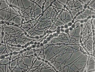
Fonsecaea pedrosoi is a fungal species in the family Herpotrichiellaceae, and the major causative agent of chromoblastomycosis. This species is commonly found in tropical and sub-tropical regions, especially in South America, where it grows as a soil saprotroph. Farming activities in the endemic zone are a risk factor for the development of chromoblastomycosis.

<i>Exophiala dermatitidis</i> Species of fungus

Exophiala dermatitidis is a thermophilic black yeast, and a member of the Herpotrichiellaceae. While the species is only found at low abundance in nature, metabolically active strains are commonly isolated in saunas, steam baths, and dish washers. Exophiala dermatitidis only rarely causes infection in humans, however cases have been reported around the world. In East Asia, the species has caused lethal brain infections in young and otherwise healthy individuals. The fungus has been known to cause cutaneous and subcutaneous phaeohyphomycosis, and as a lung colonist in people with cystic fibrosis in Europe. In 2002, an outbreak of systemic E. dermatitidis infection occurred in women who had received contaminated steroid injections at North Carolina hospitals.

Coniochaeta hoffmannii, also known as Lecythophora hoffmannii, is an ascomycete fungus that grows commonly in soil. It has also been categorized as a soft-rot fungus capable of bringing the surface layer of timber into a state of decay, even when safeguarded with preservatives. Additionally, it has pathogenic properties, although it causes serious infection only in rare cases. A plant pathogen lacking a known sexual state, C. hoffmannii has been classified as a "dematiaceous fungus" despite its contradictory lack of pigmentation; both in vivo and in vitro, there is no correlation between its appearance and its classification.

Phaeohyphomycosis is a diverse group of fungal infections, caused by dematiaceous fungi whose morphologic characteristics in tissue include hyphae, yeast-like cells, or a combination of these. It can be associated with an array of melanistic filamentous fungi including Alternaria species, Exophiala jeanselmei, and Rhinocladiella mackenziei.

Phialemonium curvatum is a pathogenic fungus in the phylum Ascomycota. The genus was created to accommodate taxa intermediate to Acremonium and Phialophora. This genus is characterized by its abundance of adelophialides and few discrete phialides with no signs of collarettes. Specifically, P. curvatum is characterized by its grayish white colonies and its allantoid conidia. Phialemonium curvatum is typically found in a variety of environments including air, soil, industrial water and sewage. Furthermore, P. curvatum affects mainly immunocompromised and is rarely seen in immunocompetent people. The species has been known to cause peritonitis, endocarditis, endovascular infections, osteomyelitis as well as cutaneous infections of wounds and burns.

<i>Madurella mycetomatis</i> Species of fungus

Madurella mycetomatis is a fungus primarily reported in Central Africa as a cause of mycetoma in humans. It has been misclassified for many years, but with improvement of molecular techniques, its phylogenetic classification has been established. Many methods exist to identify M. mycetomatis, both in lesions and in culture. Histological examination is especially useful, as it has many unique morphological features. Strain-level differences in response to antifungal agents is informative for treatment and laboratory isolation of cultures.

<i>Aspergillus alabamensis</i> Species of fungus

Aspergillus alabamensis is a soil fungus in the division Ascomycota first described in 2009 as a segregated taxon of A. terreus. Originally thought to be a variant of A. terreus, A. alabamensis is situated in a distinctive clade identified by genetic analysis. While A. alabamensis has been found to be morphologically similar to Aspergillus terreus by morphological studies, the two differ significantly in active metabolic pathways, with A. alabamensis producing the mycotoxins citrinin and citreoviridin but lacking mevinolin.

<i>Phialemonium obovatum</i> Species of fungus

Phialemonium obovatum is a saprotrophic filamentous fungus able to cause opportunistic infections in humans with weakened immune systems. P. obovatum is widespread throughout the environment, occurring commonly in sewage, soil, air and water. Walter Gams and Michael McGinnis described the genus Phialemonium to accommodate species intermediate between the genera Acremonium and Phialophora. Currently, three species of Phialemonium are recognized of which P. obovatum is the only one to produce greenish colonies and obovate conidia. It has been investigated as one of several microfungi with potential use in the accelerated aging of wine.

<i>Rhinocladiella mackenziei</i> Species of fungus

Rhinocladiella mackenziei is a deeply pigmented mold that is a common cause of human cerebral phaeohyphomycosis. Rhinocladiella mackenziei was believed to be endemic solely to the Middle East, due to the first cases of infection being limited to the region. However, cases of R. mackenziei infection are increasingly reported from regions outside the Middle East. This pathogen is unique in that the majority of cases have been reported from immunologically normal people.

<i>Cladophialophora carrionii</i> Species of fungus

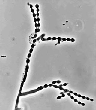
Cladophialophora carrionii is a melanized fungus in the genus Cladophialophora that is associated with decaying plant material like cacti and wood. It is one of the most frequent species of Cladophialophora implicated in human disease. Cladophialophora carrionii is a causative agent of chromoblastomycosis, a subcutaneous infection that occurs in sub-tropical areas such as Madagascar, Australia and northwestern Venezuela. Transmission occurs through traumatic implantation of plant material colonized by C. carrionii, mainly infecting rural workers. When C. carrionii infects its host, it transforms from a mycelial state to a muriform state to better tolerate the extreme conditions in the host's body.

<i>Phialophora verrucosa</i> Species of fungus

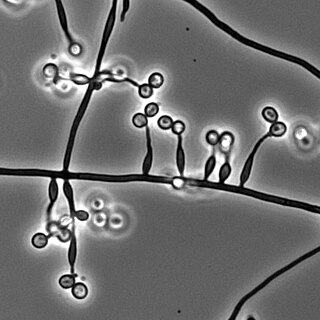
Phialophora verrucosa is a pathogenic, dematiaceous fungus that is a common cause of chromoblastomycosis. It has also been reported to cause subcutaneous phaeohyphomycosis and mycetoma in very rare cases. In the natural environment, it can be found in rotting wood, soil, wasp nests, and plant debris. P. verrucosa is sometimes referred to as Phialophora americana, a closely related environmental species which, along with P. verrucosa, is also categorized in the P. carrionii clade.

<i>Metarhizium granulomatis</i> Species of fungus

Metarhizium granulomatis is a fungus in the family Clavicipitaceae associated with systemic mycosis in veiled chameleons. The genus Metarhizium is known to infect arthropods, and collectively are referred to green-spored asexual pathogenic fungi. This species grows near the roots of plants and has been reported as an agent of disease in captive veiled chameleons. The etymology of the species epithet, "granulomatis" refers to the ability of the fungus to cause granulomatous disease in susceptible reptiles.

Sarocladium kiliense is a saprobic fungus that is occasionally encountered as a opportunistic pathogen of humans, particularly immunocompromised and individuals. The fungus is frequently found in soil and has been linked with skin and systemic infections. This species is also known to cause disease in the green alga, Cladophora glomerata as well as various fruit and vegetable crops grown in warmer climates.

Aspergillus giganteus is a species of fungus in the genus Aspergillus that grows as a mold. It was first described in 1901 by Wehmer, and is one of six Aspergillus species from the Clavati section of the subgenus Fumigati. Its closest taxonomic relatives are Aspergillus rhizopodus and Aspergillus longivescia.

Scytalidium hyalinum is an ascomycete fungus currently in the genus Scytalidium. It causes dermatomycosis and systemic infections in humans and it is widespread throughout the world.

References

  1. 1 2 3 4 5 6 7 8 9 10 11 12 13 McGinnis, Michael R. (2012). Laboratory Handbook of Medical Mycology. Elsevier. ISBN   978-0323138864.
  2. 1 2 3 Attapattu, Maya Chandrani (1997). "Chromoblastomycosis – A clinical and mycological study of 71 cases from Sri Lanka". Mycopathologia. 137 (3): 145–151. doi:10.1023/A:1006819530825. ISSN   0301-486X. PMID   9368408. S2CID   26091759.
  3. Santos, André L. S.; Palmeira, Vanila F.; Rozental, Sonia; Kneipp, Lucimar F.; Nimrichter, Leonardo; Alviano, Daniela S.; Rodrigues, Marcio L.; Alviano, Celuta S. (2007-09-01). "Biology and pathogenesis of Fonsecaea pedrosoi, the major etiologic agent of chromoblastomycosis". FEMS Microbiology Reviews. 31 (5): 570–591. doi:10.1111/j.1574-6976.2007.00077.x. ISSN   1574-6976. PMID   17645522.
  4. 1 2 3 4 De Hoog, G. S.; Attili-Angelis, D.; Vicente, V. A.; Van Den Ende, A. H. G. Gerrits; Queiroz-Telles, F. (2004). "Molecular ecology and pathogenic potential of Fonsecaea species". Medical Mycology. 42 (5): 405–416. doi: 10.1080/13693780410001661464 . ISSN   1369-3786. PMID   15552642.
  5. 1 2 3 4 5 Attili, D. S.; Hoog, G. S. de; Pizzirani-Kleiner, A. A. (1998). "rDNA-RFLP and ITSI sequencing of species of the genus Fonsecaea, agents of chromoblastomycosis". Medical Mycology. 36 (4): 219–225. doi: 10.1080/02681219880000331 . ISSN   1369-3786. PMID   9776838.
  6. 1 2 3 Vollum, Dorothy I. (1977). "Chromomycosis: a review". British Journal of Dermatology. 96 (4): 454–458. doi:10.1111/j.1365-2133.1977.tb07145.x. ISSN   1365-2133. PMID   861185. S2CID   34940097.
  7. 1 2 3 4 5 McGinnis, M R (1980). "Recent Taxonomic Developments and Changes in Medical Mycology". Annual Review of Microbiology. 34 (1): 109–135. doi:10.1146/annurev.mi.34.100180.000545. ISSN   0066-4227. PMID   7002021.
  8. Lupi, Omar; Tyring, Stephen K.; McGinnis, Michael R. (2005). "Tropical dermatology: Fungal tropical diseases". Journal of the American Academy of Dermatology. 53 (6): 931–951. doi:10.1016/j.jaad.2004.10.883. PMID   16310053.
  9. 1 2 3 4 5 6 7 8 9 10 11 McGinnis, Michael R. (1983). "Chromoblastomycosis and phaeohyphomycosis: New concepts, diagnosis, and mycology". Journal of the American Academy of Dermatology. 8 (1): 1–16. doi:10.1016/s0190-9622(83)70001-0. PMID   6826791.
  10. 1 2 Murphy, Juneann W.; Friedman, Herman; Bendinelli, Mauro (2013). Fungal Infections and Immune Responses. Springer Science & Business Media. ISBN   978-1489924001.
  11. Yaguchi, Takashi; Tanaka, Reiko; Nishimura, Kazuko; Udagawa, Shun-ichi (2007). "Molecular phylogenetics of strains morphologically identified as Fonsecaea pedrosoi from clinical specimens". Mycoses. 50 (4): 255–260. doi:10.1111/j.1439-0507.2007.01383.x. ISSN   1439-0507. PMID   17576315. S2CID   35086036.
  12. 1 2 Abliz, Paride; Fukushima, Kazutaka; Takizawa, Kayoko; Nishimura, Kazuko (2004). "Identification of pathogenic dematiaceous fungi and related taxa based on large subunit ribosomal DNA D1/D2 domain sequence analysis". FEMS Immunology & Medical Microbiology. 40 (1): 41–49. doi: 10.1016/S0928-8244(03)00275-X . ISSN   0928-8244. PMID   14734185.
  13. Krzyściak, Paweł M.; Pindycka-Piaszczyńska, Małgorzata; Piaszczyński, Michał (2014). "Chromoblastomycosis". Advances in Dermatology and Allergology. 31 (5): 310–321. doi:10.5114/pdia.2014.40949. ISSN   1642-395X. PMC   4221348 . PMID   25395928.
  14. 1 2 Liu, Dongyou (2011). Molecular Detection of Human Fungal Pathogens. CRC Press. ISBN   978-1439812419.
  15. Tille, Patricia (2013). Bailey & Scott's Diagnostic Microbiology (13 ed.). Elsevier Health Sciences. ISBN   978-0323277426.
  16. Carrión, A. L. (1942). "Chromoblastomycosis". Mycologia. 34 (4): 424–441. doi:10.2307/3754985. JSTOR   3754985.
  17. 1 2 3 4 5 Sharma, N. L.; Sharma, R. C.; Grover, P. S.; Gupta, M. L.; Sharma, A. K.; Mahajan, V. K. (1999). "Chromoblastomycosis in India". International Journal of Dermatology. 38 (11): 846–851. doi:10.1046/j.1365-4362.1999.00820.x. ISSN   0011-9059. PMID   10583618. S2CID   755756.
  18. 1 2 Ameen, M. (2009). "Chromoblastomycosis: clinical presentation and management". Clinical and Experimental Dermatology. 34 (8): 849–854. doi:10.1111/j.1365-2230.2009.03415.x. ISSN   1365-2230. PMID   19575735. S2CID   205278413.
  19. 1 2 3 4 5 6 7 8 Queiroz-Telles, Flavio; Esterre, Phillippe; Perez-Blanco, Maigualida; Vitale, Roxana G.; Salgado, Claudio Guedes; Bonifaz, Alexandro (2009). "Chromoblastomycosis: an overview of clinical manifestations, diagnosis and treatment". Medical Mycology. 47 (1): 3–15. doi:10.1080/13693780802538001. ISSN   1369-3786. PMID   19085206.