Sporothrix schenckii

Last updated

Sporothrix schenckii
Conidiophores and conidia of the fungus Sporothrix schenckii PHIL 4208 lores.jpg
Conidiophores and conidia
Scientific classification OOjs UI icon edit-ltr.svg
Domain: Eukaryota
Kingdom: Fungi
Division: Ascomycota
Class: Sordariomycetes
Order: Ophiostomatales
Family: Ophiostomataceae
Genus: Sporothrix
Species:
S. schenckii
Binomial name
Sporothrix schenckii
Hektoen & C.F.Perkins (1900)

Sporothrix schenckii, a fungus that can be found worldwide in the environment, is named for medical student Benjamin Schenck, who in 1896 was the first to isolate it from a human specimen. [1] The species is present in soil as well as in and on living and decomposing plant material such as peat moss. It can infect humans as well as animals and is the causative agent of sporotrichosis, commonly known as "rose handler's disease." [2] The most common route of infection is the introduction of spores to the body through a cut or puncture wound in the skin. Infection commonly occurs in otherwise healthy individuals but is rarely life-threatening and can be treated with antifungals. In the environment it is found growing as filamentous hyphae. In host tissue it is found as a yeast. The transition between the hyphal and yeast forms is temperature dependent making S. schenckii a thermally dimorphic fungus. [1]

Contents

Morphology

Sporothrix schenckii PHIL 3943 lores.jpg
Sporothrix schenckii conidia.png
Phase contrast micrograph of the yeast form of Sporothrix schenckii.png
Sporothrix schenckii growing on Sabouraud agar (left). Micrograph of S. schenckii conidia stained with lactophenol cotton blue (center). Adapted from. [3] Phase contrast micrograph of the yeast form of S. schenckii (right). Adapted from. [4]

Sporothrix schenckii can be found in one of two morphologies, hyphal or yeast. The hyphal form is found in the environment on plants and decaying matter. When the fungus makes the transition into a host, the yeast morphology predominates.

Hyphal

When in the environment or grown in the laboratory at 25 °C (77 °F)S. schenckii assumes its hyphal form. [5] Macroscopically, filaments are apparent and colonies are moist, leathery to velvety, and have a finely wrinkled surface. The colour is white initially and may change color over time to become cream to dark brown ("dirty candle-wax" color). [1] Microscopically, hyphae are septate approximately 1 to 2 μm in diameter. Conidia are oval shaped and glass like (hyaline) in appearance. They may be colorless or darkly colored. Conidia are sometimes referred to as resembling a flower. [6]

Yeast

At 37 °C (99 °F) either in the laboratory or in host tissue, S. schenckii assumes its yeast form. Macroscopically, the yeast form grows as smooth white or off-white colonies. Microscopically, yeast cells are 2 to 6 μm long and show an elongated cigar-shaped morphology. [1]

Epidemiology and risk factors

S. schenckii has a worldwide distribution [1] but certain areas of the world including Peru, have a higher incidence of disease. [7] Based on sequence analysis [8] it has been found that S. schenckii isolates can be placed in phylogenetic groups that reflect the region from which they were isolated. S. schenckii is often isolated from plants and associated packing material. [9] Gardeners, [10] landscapers, [11] and foresters [9] are at high risk of infection. Sporothrix infection can also be transmitted by cat bites or scratches. This mode of transmission has been responsible for epidemics of sporotrichosis. [12] Immunocompromised individuals are at increased risk of infection and such patients often exhibit more severe forms of disease. [13] [14] [15]

Sporotrichosis

Cutaneous sporotrichosis of a cat's paw showing ulcerations. Feline sporotrichosis 3.jpg
Cutaneous sporotrichosis of a cat's paw showing ulcerations.

S. schenckii most commonly enters the body through minor trauma that compromises the skin barrier. [13] It is this route of infection, coupled with its presence on roses that give sporotrichosis its common name of "rose-handler's disease" [16] or "rose thorn disease". [17] Inhalation of spores is a rare route of infection largely associated with immunocompromised hosts. [18]

Cutaneous and lymphocutaneous

The cutaneous form of disease is caused by introduction of S. schenckii into the body through disruption of the skin barrier. [13] The first symptom of cutaneous sporotrichosis is a small skin lesion. These lesions may show ulceration and/or erythema. [1] Commonly, infection spreads through the lymph along lymphatic vessels and causes lymphocutaneous sporotrichosis. This form of disease is characterized by the appearance of lesions at sites distant to the initial infection. [1] Infection can occur in nonhuman animals and may be transmitted to humans through contact. Veterinarians are at particularly high risk of contracting disease from animals (zoonotic infection). [19]

Disseminated

Disseminated disease occurs when the fungus spreads throughout the body. Found only in immunocompromised patients, [1] [14] [18] it is a very severe condition. When infection becomes disseminated, S. schencki can afflict joints, the brain, and the spine. [1] Patients with this condition must be treated aggressively with antifungals and may remain on prophylactic antifungal drugs for life to prevent recurrence or reinfection. [13]

Diagnostic tests

Sporothrix infection presents macroscopically with nonspecific symptoms. [20] In the clinical laboratory, many fungi isolated in culture are disregarded as contaminants. [21] Therefore, accurate patient histories are important [2] to establish suspicion of sporotrichosis and to inform which diagnostic tests are required. [1] S. schenckii infection may also be confused with other diseases such as pyoderma gangrenosum [20] or sarcoidosis [22] further underscoring the need for accurate diagnoses.

Primary cultures

Growing the fungus in pure culture is the most reliable way to identify Sporothrix infection. [2] A patient swab or biopsy is used to inoculate Sabouraud agar [18] or brain heart infusion agar. [16] Sabouraud agar is incubated at room temperature for macroscopic observation of the off-white or dark brown/black hyphal form of the fungus and microscopic examination of hyphae and conidia. [2] [18] The yeast form is grown on brain heart infusion agar at 37 °C (99 °F). [16] Observation of yeast colonies in addition to the hyphal form is required to confirm diagnosis of S. schenckii. [20] Growth on media occurs in approximately one to three weeks [16] meaning that results from patient cultures will not be immediately available to make treatment decisions.

Sporotrichin skin test

A skin test uses an antigen generated from laboratory grown S. schenckii to challenge the patient's immune system. The antigen is intradermally injected and the test is interpreted 48 hours later. Erythema at the site of injection indicates a positive response. [1] [16] The major advantage of this test is its rapidity and ease of use. Although it provides results more quickly than a fungal culture, the sporotrichin skin test has some important limitations. Cross reactions with other fungal species [23] as well as positive reactions in healthy individuals [1] have been observed. In addition, the term "sporotrichin" does not indicate a specific molecule but only any antigen derived from S. schenckii. The specific antigen used in skin testing is not standardized with multiple studies being conducted with widely varying preparations. However, owing to its simplicity, skin testing remains the method of choice for large-scale epidemiological investigations. [1]

Molecular methods

Molecular diagnostic techniques have been used as rapid, sensitive, and specific tests for the presence of S. schenckii. [24] PCR methods that specifically amplify the ribosomal RNA gene have been shown to detect S. schenckii in clinical samples with minimal interference from host or bacterial sequences. [25] Serum antibody reactivity to S. schenckii antigens can also be quantified by ELISA. [26] Although molecular biology techniques are promising in their use as diagnostic tools, their cost as well as requirement for specialized equipment and expertise means that more traditional diagnostic methods still play an important role in Sporothrix schenckii treatment.

Treatment

Antifungal drugs

Where available and tolerated by the patient, antifungal drugs are indicated as the primary treatment of sporotrichosis. For cutaneous infection itraconazole or terbinafine are the primary treatment with fluconazole being recommended if primary antifungals are not well tolerated. When infection is disseminated throughout the body, Amphotericin B is the drug of choice. To completely clear the fungus, the course of treatment generally lasts from 3 to 6 months. [13] In vitro susceptibility to antifungal drugs has been shown to be dependent on the growth phase (hyphal or yeast) of the fungus. [27]

Potassium iodide

Oral administration of a saturated potassium iodide solution was the first effective treatment for sporotrichosis and remains the drug of choice in many parts of the world owing to its low cost and availability. [28] Although effective, it has since been supplanted by antifungal drugs [13] due to the requirement for multiple daily doses as well side effects including gastrointestinal upset or thyroid imbalance. [28]

Virulence factors

Melanin production

S. schenckii synthesizes melanin both in vitro and in vivo [5] Melanin production is a virulence factor found in many fungi that cause disease [29] and its production in S. schenckii protects the fungus from oxidative stress as well as ultraviolet light and macrophage killing. Melanin has been shown to be synthesized using the 1,8-DHN pentaketide pathway (below). [5]

Structures of some intermediates in the pentaketide pathway of melanin biosynthesis in S. schenckii. Adapted from. Polyketide Pathway Melanin Biosynthesis in Sporothrix schenckii.png
Structures of some intermediates in the pentaketide pathway of melanin biosynthesis in S. schenckii. Adapted from.

Adhesins

Adhesion is an important component of pathogenesis. The yeast form of S. schenckii shows an increased ability to bind [1] to the host extracellular matrix proteins fibronectin and laminin using two separate receptors specific for these proteins. [31]

Proteases

S. schenckii breaks down proteins by producing two separate proteases, a serine protease and an aspartic protease. [32] These proteases appear to be essential for fungal growth. However, they have some functional overlap as the inactivation of either protein does not affect growth but inactivation of both inhibits the fungus. [33] Protease activity has been shown to be important in in vivo infection of mice. [32] Substrates for these proteases include the skin proteins type-I collagen, stratum corneum, and elastin. [32]

Heat tolerance

Growing at host body temperature (37 °C (99 °F)) is an important requirement for pathogenesis. Some strains of S. schenckii are restricted to growing at 35 °C (95 °F) and consequently usually cause disease only on the skin as it is cooler than the body's interior. Those that are capable of growth at body temperature are more often associated with disseminated disease. [32]

Immune response

Infection by S. schenckii is generally self-limiting in immunocompetent hosts. The immune response prevents fungal dissemination and is the reason that most Sporothrix infections are cutaneous. [34]

Innate

The yeast form of S. schenckii is effectively phagocytosed by cells of the innate immune system [34] and are recognized based on the sugars displayed on their surface [35] or lipids in the yeast cell membrane. [34] Although they are taken up, they are not efficiently killed. It is hypothesized that ergosterol peroxide reacts with and detoxifies reactive oxygen species generated by the respiratory burst used by phagocytes to kill cells they have ingested. [34] S. schenckii is also capable of modulating the immune response to promote its own survival by blocking cytokine production by macrophages. [34]

Specific

The specific immune response is active later in infection and involves both B cells and T cells. Severe sporotrichosis is rare in endemic areas where humans are in near constant contact with S. schenckii spores. This fact, combined with the increased severity of disease in immunocompromised patients points to an important role for specific immunity in S. schenckii infection. [34] Patients with sporotrichosis have been shown to produce antibodies specific to S. schenckii [36] and these antibodies may actually be protective against the disease. [1]

Related Research Articles

<i>Cryptococcus neoformans</i> Species of yeast

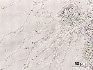
Cryptococcus neoformans is an encapsulated yeast belonging to the class Tremellomycetes and an obligate aerobe that can live in both plants and animals. Its teleomorph is a filamentous fungus, formerly referred to Filobasidiella neoformans. In its yeast state, it is often found in bird excrement. Cryptococcus neoformans can cause disease in apparently immunocompetent, as well as immunocompromised, hosts.

<span class="mw-page-title-main">Cryptococcosis</span> Potentially fatal fungal disease

Cryptococcosis is a potentially fatal fungal infection of mainly the lungs, presenting as a pneumonia, and brain, where it appears as a meningitis. Cough, difficulty breathing, chest pain and fever are seen when the lungs are infected. When the brain is infected, symptoms include headache, fever, neck pain, nausea and vomiting, light sensitivity and confusion or changes in behavior. It can also affect other parts of the body including skin, where it may appear as several fluid-filled nodules with dead tissue.

<i>Candida</i> (fungus) Genus of ascomycete fungi

Candida is a genus of yeasts. It is the most common cause of fungal infections worldwide and the largest genus of medically important yeast.

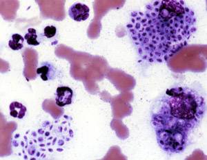
Bartonellosis is an infectious disease produced by bacteria of the genus Bartonella. Bartonella species cause diseases such as Carrión's disease, trench fever, cat-scratch disease, bacillary angiomatosis, peliosis hepatis, chronic bacteremia, endocarditis, chronic lymphadenopathy, and neurological disorders.

<span class="mw-page-title-main">Sporotrichosis</span> Medical condition

Sporotrichosis, also known as rose handler's disease, is a fungal infection that may be localised to skin, lungs, bone and joint, or become systemic. It presents with firm painless nodules that later ulcerate. Following initial exposure to Sporothrix schenckii, the disease typically progresses over a period of a week to several months. Serious complications may develop in people who have a weakened immune system.

<span class="mw-page-title-main">Protothecosis</span> Medical condition

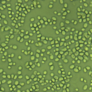
Protothecosis, otherwise known as Algaemia, is a disease found in dogs, cats, cattle, and humans caused by a type of green alga known as Prototheca that lacks chlorophyll and enters the human or animal bloodstream. It and its close relative Helicosporidium are unusual in that they are actually green algae that have become parasites. The two most common species are Prototheca wickerhamii and Prototheca zopfii. Both are known to cause disease in dogs, while most human cases are caused by P. wickerhami. Prototheca is found worldwide in sewage and soil. Infection is rare despite high exposure, and can be related to a defective immune system. In dogs, females and Collies are most commonly affected.

Pichia kudriavzevii is a budding yeast involved in chocolate production. P. kudriavzevii is an emerging fungal nosocomial pathogen primarily found in the immunocompromised and those with hematological malignancies. It has natural resistance to fluconazole, a standard antifungal agent. It is most often found in patients who have had prior fluconazole exposure, sparking debate and conflicting evidence as to whether fluconazole should be used prophylactically. Mortality due to P. kudriavzevii fungemia is much higher than the more common C. albicans. Other Candida species that also fit this profile are C. parapsilosis, C. glabrata, C. tropicalis, C. guillermondii and C. rugosa.

<i>Nakaseomyces glabratus</i> Species of fungus

Nakaseomyces glabratus is a species of haploid yeast of the genus Nakaseomyces, previously known as Candida glabrata. Despite the fact that no sexual life cycle has been documented for this species, N. glabratus strains of both mating types are commonly found. N. glabrata is generally a commensal of human mucosal tissues, but in today's era of wider human immunodeficiency from various causes, N. glabratus is often the second or third most common cause of candidiasis as an opportunistic pathogen. Infections caused by N. glabratus can affect the urogenital tract or even cause systemic infections by entrance of the fungal cells in the bloodstream (Candidemia), especially prevalent in immunocompromised patients.

<i>Geotrichum</i> Genus of fungi

Geotrichum is a genus of fungi found worldwide in soil, water, air, and sewage, as well as in plants, cereals, and dairy products; it is also commonly found in normal human flora and is isolated from sputum and feces. It was first described in 1809 by Johann Heinrich Friedrich Link.

<i>Setosphaeria rostrata</i> Pathogenic fungus

Setosphaeria rostrata is a heat tolerant fungus with an asexual reproductive form (anamorph) known as Exserohilum rostratum. This fungus is a common plant pathogen, causing leaf spots as well as crown rot and root rot in grasses. It is also found in soils and on textiles in subtropical and tropical regions. Exserohilum rostratum is one of the 35 Exserohilum species implicated uncommonly as opportunistic pathogens of humans where it is an etiologic agent of sinusitis, keratitis, skin lesions and an often fatal meningoencephalitis. Infections caused by this species are most often seen in regions with hot climates like Israel, India and the southern USA.

<i>Paracoccidioides brasiliensis</i> Species of fungus

Paracoccidioides brasiliensis is a dimorphic fungus and one of the two species that cause paracoccidioidomycosis. The fungus has been affiliated with the family Ajellomycetaceae although a sexual state or teleomorph has not yet been found.

Lacazia is a genus of fungi containing the single species Lacazia loboi, which is responsible for Lobo's disease. It is a member of the order Onygenales.

<span class="mw-page-title-main">Dimorphic fungus</span> Fungi that can exist as mold or yeast

Dimorphic fungi are fungi that can exist in the form of both mold and yeast. This is usually brought about by change in temperature and the fungi are also described as thermally dimorphic fungi. An example is Talaromyces marneffei, a human pathogen that grows as a mold at room temperature, and as a yeast at human body temperature.

<span class="mw-page-title-main">Blastocystosis</span> Medical condition

Blastocystosis refers to a medical condition caused by infection with Blastocystis. Blastocystis is a protozoal, single-celled parasite that inhabits the gastrointestinal tracts of humans and other animals. Many different types of Blastocystis exist, and they can infect humans, farm animals, birds, rodents, amphibians, reptiles, fish, and even cockroaches. Blastocystosis has been found to be a possible risk factor for development of irritable bowel syndrome.

<i>Fonsecaea pedrosoi</i> Species of fungus

Fonsecaea pedrosoi is a fungal species in the family Herpotrichiellaceae, and the major causative agent of chromoblastomycosis. This species is commonly found in tropical and sub-tropical regions, especially in South America, where it grows as a soil saprotroph. Farming activities in the endemic zone are a risk factor for the development of chromoblastomycosis.

<i>Lichtheimia corymbifera</i> Species of fungus

Lichtheimia corymbifera is a thermophilic fungus in the phylum Zygomycota. It normally lives as a saprotrophic mold, but can also be an opportunistic pathogen known to cause pulmonary, CNS, rhinocerebral, or cutaneous infections in animals and humans with impaired immunity.

<i>Rhinocladiella mackenziei</i> Species of fungus

Rhinocladiella mackenziei is a deeply pigmented mold that is a common cause of human cerebral phaeohyphomycosis. Rhinocladiella mackenziei was believed to be endemic solely to the Middle East, due to the first cases of infection being limited to the region. However, cases of R. mackenziei infection are increasingly reported from regions outside the Middle East. This pathogen is unique in that the majority of cases have been reported from immunologically normal people.

<span class="mw-page-title-main">Trichosporon asteroides</span> Fungus of the genus Trichosporon

Trichosporon asteroides is an asexual basidiomycetous fungus first described from human skin but now mainly isolated from blood and urine. T. asteroides is a hyphal fungus with a characteristically yeast-like appearance due to the presence of slimy arthroconidia. Infections by this species usually respond to treatment with azoles and amphotericin B.

Arthrographis kalrae is an ascomycetous fungus responsible for human nail infections described in 1938 by Cochet as A. langeronii. A. kalrae is considered a weak pathogen of animals including human restricted to the outermost keratinized layers of tissue. Infections caused by this species are normally responsive to commonly used antifungal drugs with only very rare exceptions.

Sporothrix brasiliensis is a fungus that is commonly found in soil. It is an emerging fungal pathogen that is causing disease in humans and cats mainly in Brazil and other countries in South America.

References

  1. 1 2 3 4 5 6 7 8 9 10 11 12 13 14 15 Barros MB, de Almeida Paes R, Schubach AO (October 2011). "Sporothrix schenckii and Sporotrichosis". Clinical Microbiology Reviews. 24 (4): 633–54. doi:10.1128/cmr.00007-11. PMC   3194828 . PMID   21976602.
  2. 1 2 3 4 Vásquez-del-Mercado E, Arenas R, Padilla-Desgarenes C (July 2012). "Sporotrichosis". Clin. Dermatol. 30 (4): 437–43. doi:10.1016/j.clindermatol.2011.09.017. PMID   22682194.
  3. Rodriguez-Caban J, Gonzalez-Velazquez W, Perez-Sanchez L, Gonzalez-Mendez R, Rodriguez-del Valle N (2011). "Calcium/calmodulin kinase1 and its relation to thermotolerance and HSP90 in Sporothrix schenckii: an RNAi and yeast two-hybrid study". BMC Microbiol. 11: 162. doi: 10.1186/1471-2180-11-162 . PMC   3146815 . PMID   21745372.
  4. Toledo MS, Tagliari L, Suzuki E, Silva CM, Straus AH, Takahashi HK (2010). "Effect of anti-glycosphingolipid monoclonal antibodies in pathogenic fungal growth and differentiation. Characterization of monoclonal antibody MEST-3 directed to Manpalpha1→3Manpalpha1→2IPC". BMC Microbiol. 10: 47. doi: 10.1186/1471-2180-10-47 . PMC   2831884 . PMID   20156351.
  5. 1 2 3 Morris-Jones R, Youngchim S, Gomez BL, et al. (July 2003). "Synthesis of melanin-like pigments by Sporothrix schenckii in vitro and during mammalian infection". Infect. Immun. 71 (7): 4026–33. doi:10.1128/iai.71.7.4026-4033.2003. PMC   161969 . PMID   12819091.
  6. Mycology Online - University of Adelaide
  7. Pappas PG, Tellez I, Deep AE, Nolasco D, Holgado W, Bustamante B (January 2000). "Sporotrichosis in Peru: description of an area of hyperendemicity". Clin. Infect. Dis. 30 (1): 65–70. doi: 10.1086/313607 . PMID   10619735.
  8. Marimon R, Gené J, Cano J, Trilles L, Dos Santos Lazéra M, Guarro J (September 2006). "Molecular phylogeny of Sporothrix schenckii". J. Clin. Microbiol. 44 (9): 3251–6. doi:10.1128/jcm.00081-06. PMC   1594699 . PMID   16954256.
  9. 1 2 Dixon DM, Salkin IF, Duncan RA, et al. (June 1991). "Isolation and characterization of Sporothrix schenckii from clinical and environmental sources associated with the largest U.S. epidemic of sporotrichosis". J. Clin. Microbiol. 29 (6): 1106–13. doi:10.1128/jcm.29.6.1106-1113.1991. PMC   269953 . PMID   1864926.
  10. Centers for Disease Control (CDC) (October 1982). "Sporotrichosis associated with Wisconsin sphagnum moss". MMWR Morb. Mortal. Wkly. Rep. 31 (40): 542–4. PMID   6817054.
  11. Centers for Disease Control (CDC) (October 1988). "Multistate outbreak of sporotrichosis in seedling handlers, 1988". MMWR Morb. Mortal. Wkly. Rep. 37 (42): 652–3. PMID   2971870.
  12. Barros MB, Schubach Ade O, do Valle AC, et al. (February 2004). "Cat-transmitted sporotrichosis epidemic in Rio de Janeiro, Brazil: description of a series of cases". Clin. Infect. Dis. 38 (4): 529–35. doi: 10.1086/381200 . PMID   14765346.
  13. 1 2 3 4 5 6 Kauffman CA, Bustamante B, Chapman SW, Pappas PG (November 2007). "Clinical practice guidelines for the management of sporotrichosis: 2007 update by the Infectious Diseases Society of America". Clin. Infect. Dis. 45 (10): 1255–65. doi: 10.1086/522765 . PMID   17968818.
  14. 1 2 al-Tawfiq JA, Wools KK (June 1998). "Disseminated sporotrichosis and Sporothrix schenckii fungemia as the initial presentation of human immunodeficiency virus infection". Clin. Infect. Dis. 26 (6): 1403–6. doi: 10.1086/516356 . PMID   9636870.
  15. Galhardo MC, Silva MT, Lima MA, et al. (June 2010). "Sporothrix schenckii meningitis in AIDS during immune reconstitution syndrome". J. Neurol. Neurosurg. Psychiatry. 81 (6): 696–9. doi:10.1136/jnnp.2009.173187. PMID   20392979. S2CID   8752653.
  16. 1 2 3 4 5 "Sporotrichosis: Rose Handler's Disease" (PDF). Iowa State University. February 2017.
  17. "Sporotrichosis: aka The Rose Thorn Disease" (PDF). American Rose Society. Archived from the original (PDF) on 26 June 2013.
  18. 1 2 3 4 Miller SD (May 2016). Vinson RP, Libow LF (eds.). "Dermatologic Manifestations of Sporotrichosis Workup". Medscape.
  19. Welsh RD (October 2003). "Sporotrichosis" (PDF). Vet Med Today: Zoonosis Update. 223 (8). American Veterinary Medical Association: 1123–6. doi:10.2460/javma.2003.223.1123. PMID   14584741.
  20. 1 2 3 Sporothrix spp. Archived 2013-04-14 at archive.today Doctor Fungus
  21. Ben-Ami R, Lewis RE, Raad II, Kontoyiannis DP (April 2009). "Phaeohyphomycosis in a tertiary care cancer center". Clin. Infect. Dis. 48 (8): 1033–41. doi: 10.1086/597400 . PMID   19267655.
  22. Singh MF, Fernandes SR, Samara AM (February 2004). "Sporothrix schenckii infection mimicking sarcoidosis". Rheumatology (Oxford). 43 (2): 248–9. doi: 10.1093/rheumatology/keh010 . PMID   14739470.
  23. Ishizaki H, Nakamura Y, Kariya H, Iwatsu T, Wheat R (1976). "Delayed hypersensitivity cross-reactions between Sporothrix schenckii and Ceratocystis species in sporotrichotic patients". J. Clin. Microbiol. 3 (6): 545–7. doi:10.1128/jcm.3.6.545-547.1976. PMC   274353 . PMID   59734.
  24. Atkins SD, Clark IM (2004). "Fungal molecular diagnostics: a mini review" (PDF). Journal of Applied Genetics. 45 (1): 3–15. PMID   14960763. S2CID   5658182. Archived from the original (PDF) on 2019-05-05.
  25. Hu S, Chung WH, Hung SI, et al. (April 2003). "Detection of Sporothrix schenckii in clinical samples by a nested PCR assay". J. Clin. Microbiol. 41 (4): 1414–8. doi:10.1128/jcm.41.4.1414-1418.2003. PMC   153868 . PMID   12682123.
  26. Almeida-Paes R, Pimenta MA, Pizzini CV, et al. (March 2007). "Use of mycelial-phase Sporothrix schenckii exoantigens in an enzyme-linked immunosorbent assay for diagnosis of sporotrichosis by antibody detection". Clin. Vaccine Immunol. 14 (3): 244–9. doi:10.1128/cvi.00430-06. PMC   1828849 . PMID   17215334.
  27. Trilles L, Fernández-Torres B, Dos Santos Lazéra M, et al. (September 2005). "In vitro antifungal susceptibilities of Sporothrix schenckii in two growth phases". Antimicrob. Agents Chemother. 49 (9): 3952–4. doi:10.1128/aac.49.9.3952-3954.2005. PMC   1195444 . PMID   16127080.
  28. 1 2 Sterling JB, Heymann WR (October 2000). "Potassium iodide in dermatology: a 19th century drug for the 21st century-uses, pharmacology, adverse effects, and contraindications". J. Am. Acad. Dermatol. 43 (4): 691–7. doi:10.1067/mjd.2000.107247. PMID   11004629.
  29. Revankar SG, Sutton DA (October 2010). "Melanized fungi in human disease". Clin. Microbiol. Rev. 23 (4): 884–928. doi:10.1128/cmr.00019-10. PMC   2952981 . PMID   20930077.
  30. Romero-Martinez R, Wheeler M, Guerrero-Plata A, Rico G, Torres-Guerrero H (June 2000). "Biosynthesis and functions of melanin in Sporothrix schenckii". Infect. Immun. 68 (6): 3696–703. doi:10.1128/iai.68.6.3696-3703.2000. PMC   97661 . PMID   10816530.
  31. Lima OC, Bouchara JP, Renier G, Marot-Leblond A, Chabasse D, Lopes-Bezerra LM (September 2004). "Immunofluorescence and flow cytometry analysis of fibronectin and laminin binding to Sporothrix schenckii yeast cells and conidia". Microb. Pathog. 37 (3): 131–40. doi:10.1016/j.micpath.2004.06.005. PMID   15351036.
  32. 1 2 3 4 Hogan LH, Klein BS, Levitz SM (October 1996). "Virulence factors of medically important fungi". Clin. Microbiol. Rev. 9 (4): 469–88. doi:10.1128/CMR.9.4.469. PMC   172905 . PMID   8894347.
  33. Tsuboi R, Sanada T, Ogawa H (July 1988). "Influence of culture medium pH and proteinase inhibitors on extracellular proteinase activity and cell growth of Sporothrix schenckii". J. Clin. Microbiol. 26 (7): 1431–3. doi:10.1128/jcm.26.7.1431-1433.1988. PMC   266631 . PMID   3045155.
  34. 1 2 3 4 5 6 Carlos IZ, Sassá MF, da Graça Sgarbi DB, Placeres MC, Maia DC (July 2009). "Current research on the immune response to experimental sporotrichosis". Mycopathologia. 168 (1): 1–10. doi:10.1007/s11046-009-9190-z. PMID   19241140. S2CID   39610.
  35. Oda LM, Kubelka CF, Alviano CS, Travassos LR (February 1983). "Ingestion of yeast forms of Sporothrix schenckii by mouse peritoneal macrophages". Infect. Immun. 39 (2): 497–504. doi:10.1128/iai.39.2.497-504.1983. PMC   347978 . PMID   6832808.
  36. Scott EN, Muchmore HG (February 1989). "Immunoblot analysis of antibody responses to Sporothrix schenckii". J. Clin. Microbiol. 27 (2): 300–4. doi:10.1128/jcm.27.2.300-304.1989. PMC   267296 . PMID   2915023.

Further reading