Candida is a genus of yeasts and is the most common cause of fungal infections worldwide and the largest genus of medically important yeast.

Malassezia is a genus of fungi. It is the sole genus in family Malasseziaceae, which is the only family in order Malasseziales, itself the single member of class Malasseziomycetes. Malassezia species are naturally found on the skin surfaces of many animals, including humans. In occasional opportunistic infections, some species can cause hypopigmentation or hyperpigmentation on the trunk and other locations in humans. Allergy tests for these fungi are available.

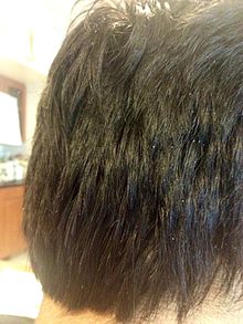
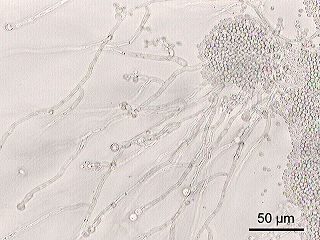
White piedra is a mycosis of the hair caused by several species of fungi in the genus Trichosporon. It is characterized by soft nodules composed of yeast cells and arthroconidia that encompass hair shafts.
Exophiala jeanselmei is a saprotrophic fungus in the family Herpotrichiellaceae. Four varieties have been discovered: Exophiala jeanselmei var. heteromorpha, E. jeanselmei var. lecanii-corni, E. jeanselmei var. jeanselmei, and E. jeanselmei var. castellanii. Other species in the genus Exophiala such as E. dermatitidis and E. spinifera have been reported to have similar annellidic conidiogenesis and may therefore be difficult to differentiate.

Lomentospora prolificans is an emerging opportunistic fungal pathogen that causes a wide variety of infections in immunologically normal and immunosuppressed people and animals. It is resistant to most antifungal drugs and infections are often fatal. Drugs targeting the Class II dihydroorotate dehydrogenase (DHODH) proteins of L. prolificans, Scedosporium, Aspergillus and other deadly moulds are the basis for at least one new therapy, Olorofim, which is currently in phase 2b clinical trials and has received breakthrough status by FDA. For information on all DHODH proteins, please see Dihydroorotate dehydrogenase.

The Sporidiobolales are an order of fungi in the subdivision Pucciniomycotina. The order contains a single family, the Sporidiobolaceae, which currently contains three genera. Most species are known only from their yeast states. Hyphal states produce teliospores from which auricularioid basidia emerge, bearing basidiospores. Species occur worldwide and have been isolated from a wide variety of substrates. Two species, Rhodotorula mucilaginosa and R. glutinis, have been known to cause disease in humans.
Malassezia dermatis is a fungus that can cause opportunistic infections in animals.
Malassezia equina is a fungus first isolated in horses, which can cause opportunistic infections in animals. Its type strain is MA146=CBS 9969. This species will not grow without any lipid supplementation. It grows slowly and forms small colonies. In the lab, colonies will not grow at temperatures of 40 °C, differing from M. sympodialis-related species, such M. dermatis and M. nana, which can grow at this temperature. Malassezia caprae cells are ovoidal.
Malassezia caprae is a fungus first isolated in goats, which can cause opportunistic infections in animals. Its type strain is MA383=CBS 10434. This species will not grow without any lipid supplementation. It grows slowly and forms small colonies. In the lab, colonies will not grow at temperatures of 40 °C, differing from M. sympodialis-related species, such M. dermatis and M. nana, which can grow at this temperature. Malassezia caprae cells are ellipsoidal to more or less spherical.
Malassezia nana is a fungus that can cause opportunistic infections in animals. It was first isolated from animals in Japan and Brazil. M. nana resembles M. dermatis and M. sympodialis, but is distinguished from these species by its inability to use Kolliphor EL (Sigma) as the sole lipid source and to hydrolyse aesculin. The type strain of M. nana is NUSV 1003T(=CBS 9557T=JCM 12085T).
Malassezia pachydermatis is a zoophilic yeast in the division Basidiomycota. It was first isolated in 1925 by Fred Weidman, and it was named pachydermatis after the original sample taken from an Indian rhinoceros with severe exfoliative dermatitis. Within the genus Malassezia, M. pachydermatis is most closely related to the species M. furfur. A commensal fungus, it can be found within the microflora of healthy mammals such as humans, cats and dogs, However, it is capable of acting as an opportunistic pathogen under special circumstances and has been seen to cause skin and ear infections, most often occurring in canines.

Lichtheimia corymbifera is a thermophilic fungus in the phylum Zygomycota. It normally lives as a saprotrophic mold, but can also be an opportunistic pathogen known to cause pulmonary, CNS, rhinocerebral, or cutaneous infections in animals and humans with impaired immunity.
Nakaseomyces bracarensis is an anamorphic yeast species with type strain 153MT.

Neoscytalidium dimidiatum was first described in 1933 as Hendersonula toruloidea from diseased orchard trees in Egypt. Decades later, it was determined to be a causative agent of human dermatomycosis-like infections and foot infections predominantly in tropical areas; however the fungus is considered to be widespread. A newer name, Scytalidium dimidiatum, was applied to a synanamorph of Nattrassia mangiferae, otherwise known as Neofusicoccum mangiferae. Substantial confusion has arisen in the literature on this fungus resulting from the use of multiple different names including Torula dimidiata, Fusicoccum dimidiatum, Scytalidium dimidiatum, and Hendersonula toruloidea. Additionally, Scytalidium lignicola and Scytalidium lignicolum are often considered earlier names of N. dimidiatum.

Rhinocladiella mackenziei is a deeply pigmented mold that is a common cause of human cerebral phaeohyphomycosis. Rhinocladiella mackenziei was believed to be endemic solely to the Middle East, due to the first cases of infection being limited to the region. However, cases of R. mackenziei infection are increasingly reported from regions outside the Middle East. This pathogen is unique in that the majority of cases have been reported from immunologically normal people.

Trichosporon asteroides is an asexual basidiomycetous fungus first described from human skin but now mainly isolated from blood and urine. T. asteroides is a hyphal fungus with a characteristically yeast-like appearance due to the presence of slimy arthroconidia. Infections by this species usually respond to treatment with azoles and amphotericin B.
Malassezia arunalokei is a species of yeast-like fungus that was identified as a new to science in 2016. It has been isolated from the skin of both seborrheic dermatitis patients and healthy individuals from India. A combination of several phenotypic characteristics distinguish this species from others in genus Malassezia. These include: colony morphology ; the absence of activity from the enzyme catalase; growth at 37 °C (99 °F); and the precipitation that results when grown with the chemicals Tween 20 or Cremophor EL.
The Trichosporonaceae are a family of fungi in the order Trichosporonales. The family currently contains six genera. Species are not known to produce basidiocarps, but exist as yeasts or produce septate hyphae with arthroconidia. Several species are human pathogens.
Apiotrichum mycotoxinivorans is a yeast species purportedly useful in the detoxification of various mycotoxins. It was first isolated from the hindgut of the termite Mastotermes darwiniensis. It has been shown to detoxify mycotoxins such as ochratoxin A and zearalenone. It can occasionally become a human pathogen.
Apiotrichum is a genus of fungi in the family Trichosporonaceae. Species are only known from their yeast states, most of which were formerly referred to the genus Trichosporon. Twenty species have been described worldwide. Apiotrichum mycotoxinivorans is an occasional human pathogen.