Hyperimmunoglobulin E syndrome

Last updated
Hyper-IgE syndrome
Other namesHIES
Specialty Immunology   OOjs UI icon edit-ltr-progressive.svg

Hyperimmunoglobulinemia E syndrome [1] (HIES), of which the autosomal dominant form is called Job's syndrome [1] or Buckley syndrome, [1] is a heterogeneous group of immune disorders. Job's is also very rare at about 300 cases currently in the literature.

Contents

Presentation

It is characterized by recurrent "cold" staphylococcal infections (due to impaired recruitment of neutrophils), [2] unusual eczema-like skin rashes, severe lung infections that result in pneumatoceles (balloon-like lesions that may be filled with air or pus or scar tissue) and very high (> 2000 IU/mL or 4800 mcg/L) [3] concentrations of the serum antibody IgE. Inheritance can be autosomal dominant or autosomal recessive. [4] Many patients with autosomal dominant STAT3 hyper-IgE syndrome have characteristic facial and dental abnormalities, fail to lose their primary teeth, and have two sets of teeth simultaneously. [5]

Pathophysiology

Abnormal neutrophil chemotaxis due to decreased production of interferon gamma by T lymphocytes is thought to cause the disease. [6]

Both autosomal dominant and recessive inheritance have been described: [7] [8]

Autosomal dominant:

Autosomal recessive:

Diagnosis

Elevated IgE is the hallmark of HIES. An IgE level greater than 2,000 IU/mL is often considered diagnostic. [17] However, patients younger than 6 months of age may have very low to non-detectable IgE levels. Eosinophilia is also a common finding with greater than 90% of patients having eosinophil elevations greater than two standard deviations above the normal mean. [18] Genetic testing is available for STAT3 (Job's Syndrome), DOCK8 (DOCK8 Immunodeficiency or DIDS), PGM3 (PGM3 deficiency), SPINK5 (Netherton Syndrome - NTS), and TYK2 genetic defects. [19]

Types

HIES often appears early in life with recurrent staphylococcal and candidal infections, pneumonias, and eczematoid skin. [20]

Treatment

Most patients with hyper IgE syndrome are treated with long-term antibiotic therapy to prevent staphylococcal infections. Good skin care is also important in patients with hyper IgE syndrome. High-dose intravenous gamma-globulin has also been suggested for the treatment of severe eczema in patients with HIES and atopic dermatitis. [21]

History

HIES was first described by Davis et al. in 1966 in two girls with red hair, chronic dermatitis, and recurrent staphylococcal abscesses and pneumonias. [22] They named the disease after the biblical figure Job, whose body was covered with boils by Satan. In 1972, Buckley et al. described two boys with similar symptoms as well as coarse facies, eosinophilia, and elevated serum IgE levels. These two syndromes are thought to be the same and are under the broad category of HIES. [23]

See also

Related Research Articles

Immunodeficiency, also known as immunocompromisation, is a state in which the immune system's ability to fight infectious diseases and cancer is compromised or entirely absent. Most cases are acquired ("secondary") due to extrinsic factors that affect the patient's immune system. Examples of these extrinsic factors include HIV infection and environmental factors, such as nutrition. Immunocompromisation may also be due to genetic diseases/flaws such as SCID.

<span class="mw-page-title-main">Immunodeficiency–centromeric instability–facial anomalies syndrome</span> Medical condition

ICF syndrome is a very rare autosomal recessive immune disorder.

<span class="mw-page-title-main">Wiskott–Aldrich syndrome</span> Medical condition

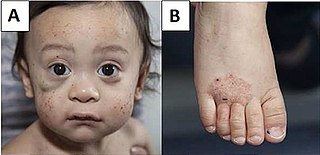
Wiskott–Aldrich syndrome (WAS) is a rare X-linked recessive disease characterized by eczema, thrombocytopenia, immune deficiency, and bloody diarrhea. It is also sometimes called the eczema-thrombocytopenia-immunodeficiency syndrome in keeping with Aldrich's original description in 1954. The WAS-related disorders of X-linked thrombocytopenia (XLT) and X-linked congenital neutropenia (XLN) may present with similar but less severe symptoms and are caused by mutations of the same gene.

<span class="mw-page-title-main">X-linked agammaglobulinemia</span> Medical condition

X-linked agammaglobulinemia (XLA) is a rare genetic disorder discovered in 1952 that affects the body's ability to fight infection. As the form of agammaglobulinemia that is X-linked, it is much more common in males. In people with XLA, the white blood cell formation process does not generate mature B cells, which manifests as a complete or near-complete lack of proteins called gamma globulins, including antibodies, in their bloodstream. B cells are part of the immune system and normally manufacture antibodies, which defend the body from infections by sustaining a humoral immunity response. Patients with untreated XLA are prone to develop serious and even fatal infections. A mutation occurs at the Bruton's tyrosine kinase (Btk) gene that leads to a severe block in B cell development and a reduced immunoglobulin production in the serum. Btk is particularly responsible for mediating B cell development and maturation through a signaling effect on the B cell receptor BCR. Patients typically present in early childhood with recurrent infections, in particular with extracellular, encapsulated bacteria. XLA is deemed to have a relatively low incidence of disease, with an occurrence rate of approximately 1 in 200,000 live births and a frequency of about 1 in 100,000 male newborns. It has no ethnic predisposition. XLA is treated by infusion of human antibody. Treatment with pooled gamma globulin cannot restore a functional population of B cells, but it is sufficient to reduce the severity and number of infections due to the passive immunity granted by the exogenous antibodies.

Hypogammaglobulinemia is an immune system disorder in which not enough gamma globulins are produced in the blood. This results in a lower antibody count, which impairs the immune system, increasing risk of infection. Hypogammaglobulinemia may result from a variety of primary genetic immune system defects, such as common variable immunodeficiency, or it may be caused by secondary effects such as medication, blood cancer, or poor nutrition, or loss of gamma globulins in urine, as in nonselective glomerular proteinuria. Patients with hypogammaglobulinemia have reduced immune function; important considerations include avoiding use of live vaccines, and take precautionary measures when traveling to regions with endemic disease or poor sanitation such as receiving immunizations, taking antibiotics abroad, drinking only safe or boiled water, arranging appropriate medical cover in advance of travel, and ensuring continuation of any immunoglobulin infusions needed.

<span class="mw-page-title-main">Hyper IgM syndrome</span> Primary immune deficiency disorders

Hyper IgM syndrome is a rare primary immune deficiency disorders characterized by low or absent levels of serum IgG, IgA, IgE and normal or increased levels of serum IgM.

<span class="mw-page-title-main">Hyper-IgM syndrome type 2</span> Primary immune deficiency disorder

Hyper IgM Syndrome Type 2 is a rare disease. Unlike other hyper-IgM syndromes, Type 2 patients identified thus far did not present with a history of opportunistic infections. One would expect opportunistic infections in any immunodeficiency syndrome. The responsible genetic lesion is in the AICDA gene found at 12p13.

<span class="mw-page-title-main">Hyper-IgM syndrome type 5</span> Primary immune deficiency disorder

The fifth type of hyper-IgM syndrome has been characterized in three patients from France and Japan. The symptoms are similar to hyper IgM syndrome type 2, but the AICDA gene is intact.

<span class="mw-page-title-main">Netherton syndrome</span> Medical condition

Netherton syndrome is a severe, autosomal recessive form of ichthyosis associated with mutations in the SPINK5 gene. It is named after Earl W. Netherton (1910–1985), an American dermatologist who discovered it in 1958.

An immune disorder is a dysfunction of the immune system. These disorders can be characterized in several different ways:

<span class="mw-page-title-main">Dedicator of cytokinesis protein 8</span> Protein found in humans

Dedicator of cytokinesis protein 8 (Dock8) is a large protein encoded in the human by the DOCK8 gene, involved in intracellular signalling networks. It is a member of the DOCK-C subfamily of the DOCK family of guanine nucleotide exchange factors (GEFs) which function as activators of small G-proteins.

<span class="mw-page-title-main">Hyper-IgM syndrome type 3</span> Primary immune deficiency disorder

Hyper-IgM syndrome type 3 is a form of hyper IgM syndrome characterized by mutations of the CD40 gene. In this type, Immature B cells cannot receive signal 2 from helper T cells which is necessary to mature into mature B cells.

<span class="mw-page-title-main">Hyper-IgM syndrome type 4</span> Medical condition

Hyper-IgM syndrome type 4 is a form of Hyper IgM syndrome which is a defect in class switch recombination downstream of the AICDA gene that does not impair somatic hypermutation.

Hans Dieter Ochs, is an immunologist and pediatrician. He is Professor of Pediatrics, Division of Immunology, Department of Pediatrics, University of Washington School of Medicine, Seattle.

<span class="mw-page-title-main">DOCK8 deficiency</span> Medical condition

DOCK8 deficiency, also called DOCK8 immunodeficiency syndrome, is the autosomal recessive form of hyperimmunoglobulin E syndrome, a genetic disorder characterized by elevated immunoglobulin E levels, eosinophilia, and recurrent infections with staphylococcus and viruses. It is caused by a mutation in the DOCK8 gene.

PGM3 deficiency is a rare genetic disorder of the immune system associated with diminished phosphoglucomutase 3 function. PGM3 is an enzyme which in humans is encoded by gene PGM3. This disorder manifests as severe atopy, immune deficiency, autoimmunity, intellectual disability, and hypomyelination. In 2014, Investigators Atfa Sassi at the Pasteur Institute of Tunis, Sandra Lazaroski at the University Medical Center Freiburg, and Gang Wu at the Imperial College London, identified PGM3 mutations in nine patients from four consanguineous families. In the same year, a researchers from the laboratories of Joshua Milner and Helen Su at the National Institute of Allergy and Infectious Disease at the U.S. National Institutes of Health described PGM3 deficiency in eight additional patients from two families.

STAT3 GOF is a rare genetic disorder of the immune system. Signal transducer and activator of transcription 3 (STAT3) is a transcription factor which is encoded by the STAT3 gene in humans. Germline gain-of-function (GOF) mutations in the gene STAT3 causes this early-onset autoimmune disease characterized by lymphadenopathy, autoimmune cytopenias, multiorgan autoimmunity, infections, eczema, and short stature. Investigations conducted by Sarah E Flanagan and Mark Russell from the Institute of Biomedical and Clinical Science, University of Exeter Medical School, Emma Haapaniemi from the Institute of Biomedical and Clinical Science, University of Exeter Medical Schoolby, and Joshua Milner from the National Institute of Allergy and Infectious Disease, National Institutes of Health have described this condition in 19 patients.

P14 deficiency is a rare autosomal recessive disease characterized as a primary immunodeficiency syndrome. This disease was first identified within a white Mennonite family by Professor Bodo Grimbacher and Professor Christoph Klein’s teams in 2006. Four out of 15 offspring in this family showed symptoms including short stature, recurrent infection of Streptococcus pneumonia, and dysfunction of cells that contain specific lysosome-related organelles, including cytotoxic T cells, melanocytes, and neutrophil granulocytes.

<span class="mw-page-title-main">Hypohidrotic ectodermal dysplasia with immune deficiency</span> Medical condition

Hypohidrotic/anhidrotic ectodermal dysplasia with immune deficiency is a rare genetic condition characterized by a combination of the features of ectodermal dysplasia alongside immunodeficiency.

References

  1. 1 2 3 Rapini, Ronald P.; Bolognia, Jean L.; Jorizzo, Joseph L. (2007). Dermatology: 2-Volume Set. St. Louis: Mosby. ISBN   978-1-4160-2999-1.
  2. " hyperimmunoglobulinemia E syndrome " at Dorland's Medical Dictionary
  3. "Hyper-IgE Syndrome". Merck Manual. Merck Sharp & Dohme Corp. Retrieved 3 August 2021.
  4. Dermatologic Manifestations of Job Syndrome at eMedicine
  5. 1 2 Freeman, Alexandra F.; Holland, Steven M. (2008). "The Hyper-IgE Syndromes". Immunology and Allergy Clinics of North America. Elsevier BV. 28 (2): 277–291. doi:10.1016/j.iac.2008.01.005. ISSN   0889-8561. PMC   2683262 . PMID   18424333.
  6. Borges WG, Augustine NH, Hill HR (February 2000). "Defective interleukin-12/interferon-gamma pathway in patients with hyperimmunoglobulinemia E syndrome". The Journal of Pediatrics. 136 (2): 176–80. doi:10.1016/S0022-3476(00)70098-9. PMID   10657822.
  7. 1 2 3 Rael EL, Marshall RT, McClain JJ (July 2012). "The Hyper-IgE Syndromes: Lessons in Nature, From Bench to Bedside". The World Allergy Organization Journal. 5 (7): 79–87. doi:10.1097/WOX.0b013e31825a73b2. PMC   3651150 . PMID   23283142.
  8. Freeman AF, Holland SM (May 2009). "Clinical manifestations, etiology, and pathogenesis of the hyper-IgE syndromes". Pediatric Research. 65 (5 Pt 2): 32R–37R. doi:10.1203/PDR.0b013e31819dc8c5. PMC   2919366 . PMID   19190525.
  9. Rael EL, Marshall RT, McClain JJ (July 2012). "The Hyper-IgE Syndromes: Lessons in Nature, From Bench to Bedside". The World Allergy Organization Journal. 5 (7): 79–87. doi:10.1097/WOX.0b013e31825a73b2. PMC   3651150 . PMID   23283142.
  10. Holland SM, DeLeo FR, Elloumi HZ, Hsu AP, Uzel G, Brodsky N, et al. (October 2007). "STAT3 mutations in the hyper-IgE syndrome". The New England Journal of Medicine. 357 (16): 1608–19. doi: 10.1056/NEJMoa073687 . PMID   17881745.
  11. Hafsi, Wissem; Yarrarapu, Siva Naga S. (2021), "Job Syndrome", StatPearls, Treasure Island (FL): StatPearls Publishing, PMID   30247822 , retrieved 2021-11-28
  12. Zhang Q, Davis JC, Lamborn IT, Freeman AF, Jing H, Favreau AJ, et al. (November 2009). "Combined immunodeficiency associated with DOCK8 mutations". The New England Journal of Medicine. 361 (21): 2046–55. doi:10.1056/NEJMoa0905506. PMC   2965730 . PMID   19776401.
  13. Boos AC, Hagl B, Schlesinger A, Halm BE, Ballenberger N, Pinarci M, et al. (2014-05-20). "Atopic dermatitis, STAT3- and DOCK8-hyper-IgE syndromes differ in IgE-based sensitization pattern". Allergy. 69 (7): 943–953. doi:10.1111/all.12416. PMID   24840882.
  14. Yang L, Fliegauf M, Grimbacher B (December 2014). "Hyper-IgE syndromes: reviewing PGM3 deficiency". Current Opinion in Pediatrics. 26 (6): 697–703. doi:10.1097/MOP.0000000000000158. PMID   25365149. S2CID   24566050.
  15. Minegishi Y, Saito M, Morio T, Watanabe K, Agematsu K, Tsuchiya S, et al. (November 2006). "Human tyrosine kinase 2 deficiency reveals its requisite roles in multiple cytokine signals involved in innate and acquired immunity". Immunity. 25 (5): 745–55. doi: 10.1016/j.immuni.2006.09.009 . PMID   17088085.
  16. Kreins AY, Ciancanelli MJ, Okada S, Kong XF, Ramírez-Alejo N, Kilic SS, et al. (September 2015). "Human TYK2 deficiency: Mycobacterial and viral infections without hyper-IgE syndrome". The Journal of Experimental Medicine. 212 (10): 1641–62. doi:10.1084/jem.20140280. PMC   4577846 . PMID   26304966.
  17. Ochs, HD; Notarangelo, LD (2010). Williams Hematology: Chapter 82. Immunodeficiency Diseases (8th ed.). New York: McGraw-Hill Medical. ISBN   9780071621519.
  18. 1 2 Grimbacher B, Holland SM, Gallin JI, Greenberg F, Hill SC, Malech HL, et al. (March 1999). "Hyper-IgE syndrome with recurrent infections--an autosomal dominant multisystem disorder". The New England Journal of Medicine. 340 (9): 692–702. doi: 10.1056/NEJM199903043400904 . PMID   10053178.
  19. Yaakoubi, Roukaya; Mekki, Najla; Ben-Mustapha, Imen; Ben-Khemis, Leila; Bouaziz, Asma; Ben Fraj, Ilhem; Ammar, Jamel; Hamzaoui, Agnès; Turki, Hamida; Boussofara, Lobna; Denguezli, Mohamed; Haddad, Samir; Ouederni, Monia; Bejaoui, Mohamed; Chan, Koon Wing; Lau, Yu Lung; Mellouli, Fethi; Barbouche, Mohamed-Ridha; Ben-Ali, Meriem (January 10, 2023). "Diagnostic challenge in a series of eleven patients with hyper IgE syndromes". Frontiers in Immunology. Frontiers Media SA. 13. doi: 10.3389/fimmu.2022.1057679 . ISSN   1664-3224. PMC   9871884 . PMID   36703986.
  20. "Hyper IgE syndrome". Immune Deficiency Foundation. August 9, 2023. Retrieved October 14, 2023.
  21. Kimata H (March 1995). "High-dose intravenous gamma-globulin treatment for hyperimmunoglobulinemia E syndrome". The Journal of Allergy and Clinical Immunology. 95 (3): 771–4. doi:10.1016/S0091-6749(95)70185-0. PMID   7897163.
  22. Davis SD, Schaller J, Wedgwood RJ (May 1966). "Job's Syndrome. Recurrent, "cold", staphylococcal abscesses". Lancet. 1 (7445): 1013–5. doi:10.1016/S0140-6736(66)90119-X. PMID   4161105.
  23. Buckley RH, Wray BB, Belmaker EZ (January 1972). "Extreme hyperimmunoglobulinemia E and undue susceptibility to infection". Pediatrics. 49 (1): 59–70. doi:10.1542/peds.49.1.59. PMID   5059313. S2CID   245107427.

Further reading