Related Research Articles

Radiography is an imaging technique using X-rays, gamma rays, or similar ionizing radiation and non-ionizing radiation to view the internal form of an object. Applications of radiography include medical radiography and industrial radiography. Similar techniques are used in airport security. To create an image in conventional radiography, a beam of X-rays is produced by an X-ray generator and is projected toward the object. A certain amount of the X-rays or other radiation is absorbed by the object, dependent on the object's density and structural composition. The X-rays that pass through the object are captured behind the object by a detector. The generation of flat two dimensional images by this technique is called projectional radiography. In computed tomography an X-ray source and its associated detectors rotate around the subject which itself moves through the conical X-ray beam produced. Any given point within the subject is crossed from many directions by many different beams at different times. Information regarding attenuation of these beams is collated and subjected to computation to generate two dimensional images in three planes which can be further processed to produce a three dimensional image.

Radiology is the medical discipline that uses medical imaging to diagnose and treat diseases within the bodies of animals and humans.
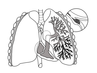
Pulmonary embolism (PE) is a blockage of an artery in the lungs by a substance that has moved from elsewhere in the body through the bloodstream (embolism). Symptoms of a PE may include shortness of breath, chest pain particularly upon breathing in, and coughing up blood. Symptoms of a blood clot in the leg may also be present, such as a red, warm, swollen, and painful leg. Signs of a PE include low blood oxygen levels, rapid breathing, rapid heart rate, and sometimes a mild fever. Severe cases can lead to passing out, abnormally low blood pressure, obstructive shock, and sudden death.

Clearing the cervical spine is the process by which medical professionals determine whether cervical spine injuries exist, mainly regarding cervical fracture. It is generally performed in cases of major trauma. This process can take place in the emergency department or in the field by appropriately trained EMS personnel.

A cervical fracture, commonly called a broken neck, is a fracture of any of the seven cervical vertebrae in the neck. Examples of common causes in humans are traffic collisions and diving into shallow water. Abnormal movement of neck bones or pieces of bone can cause a spinal cord injury resulting in loss of sensation, paralysis, or usually instant death.

In medicine, the Ottawa ankle rules are a set of guidelines for clinicians to help decide if a patient with foot or ankle pain should be offered X-rays to diagnose a possible bone fracture. Before the introduction of the rules most patients with ankle injuries would have been imaged. However the vast majority of patients with unclear ankle injuries do not have bone fractures. As a result, many unnecessary X-rays were taken, which was costly, time consuming and a slight health risk due to radiation exposure.

An ankle fracture is a break of one or more of the bones that make up the ankle joint. Symptoms may include pain, swelling, bruising, and an inability to walk on the injured leg. Complications may include an associated high ankle sprain, compartment syndrome, stiffness, malunion, and post-traumatic arthritis.

Blunt trauma, also known as blunt force trauma or non-penetrating trauma, is physical trauma or impactful force to a body part, often occurring with road traffic collisions, direct blows, assaults, injuries during sports, and particularly in the elderly who fall. It is contrasted with penetrating trauma which occurs when an object pierces the skin and enters a tissue of the body, creating an open wound and bruise.
Heterotopic ossification (HO) is the process by which bone tissue forms outside of the skeleton.
An acute abdomen refers to a sudden, severe abdominal pain. It is in many cases a medical emergency, requiring urgent and specific diagnosis. Several causes need immediate surgical treatment.

Transient synovitis of hip is a self-limiting condition in which there is an inflammation of the inner lining of the capsule of the hip joint. The term irritable hip refers to the syndrome of acute hip pain, joint stiffness, limp or non-weightbearing, indicative of an underlying condition such as transient synovitis or orthopedic infections. In everyday clinical practice however, irritable hip is commonly used as a synonym for transient synovitis. It should not be confused with sciatica, a condition describing hip and lower back pain much more common to adults than transient synovitis but with similar signs and symptoms.
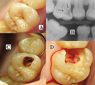
Dental radiographs are commonly called X-rays. Dentists use radiographs for many reasons: to find hidden dental structures, malignant or benign masses, bone loss, and cavities.

Projectional radiography, also known as conventional radiography, is a form of radiography and medical imaging that produces two-dimensional images by x-ray radiation. The image acquisition is generally performed by radiographers, and the images are often examined by radiologists. Both the procedure and any resultant images are often simply called "X-ray". Plain radiography or roentgenography generally refers to projectional radiography. Plain radiography can also refer to radiography without a radiocontrast agent or radiography that generates single static images, as contrasted to fluoroscopy, which are technically also projectional.

Recursive partitioning is a statistical method for multivariable analysis. Recursive partitioning creates a decision tree that strives to correctly classify members of the population by splitting it into sub-populations based on several dichotomous independent variables. The process is termed recursive because each sub-population may in turn be split an indefinite number of times until the splitting process terminates after a particular stopping criterion is reached.
A clinical prediction rule or clinical probability assessment specifies how to use medical signs, symptoms, and other findings to estimate the probability of a specific disease or clinical outcome.
March fracture, is the fracture of the distal third of one of the metatarsals occurring because of recurrent stress. It is more common in soldiers, but also occurs in hikers, organists, people whose duties entail much standing. March fractures most commonly occur in the second and third metatarsal bones of the foot. It is a common cause of foot pain, especially when people suddenly increase their activities.

A panoramic radiograph is a panoramic scanning dental X-ray of the upper and lower jaw. It shows a two-dimensional view of a half-circle from ear to ear. Panoramic radiography is a form of focal plane tomography; thus, images of multiple planes are taken to make up the composite panoramic image, where the maxilla and mandible are in the focal trough and the structures that are superficial and deep to the trough are blurred.
The Pittsburgh knee rules are medical rules created to ascertain whether a knee injury requires the use of X-ray to assess a fracture.

Computed tomography of the head uses a series of X-rays in a CT scan of the head taken from many different directions; the resulting data is transformed into a series of cross sections of the brain using a computer program. CT images of the head are used to investigate and diagnose brain injuries and other neurological conditions, as well as other conditions involving the skull or sinuses.; it used to guide some brain surgery procedures as well. CT scans expose the person getting them to ionizing radiation which has a risk of eventually causing cancer; some people have allergic reactions to contrast agents that are used in some CT procedures.

EOS imaging is a medical device company based in Paris, France that designs, develops, and markets EOSedge™ and the EOS system, innovative, orthopedic medical imaging systems, associated with several orthopedic solutions along the patient care pathway – from diagnosis to post-operative treatments. The EOS platform targets musculoskeletal disorders and orthopedic surgical care through 2D X-ray scans and 3D skeletal models from stereo-radiographic images of patients in a seated or standing position.
References
- ↑ "Ottawa Knee Rule - Emergency Medicine Research - Ottawa Hospital Research Institute".
- ↑ Stiell IG, Wells GA, Hoag RH, Sivilotti ML, Cacciotti TF, Verbeek PR, Greenway KT, McDowell I, Cwinn AA, Greenberg GH, Nichol G, Michael JA (1997). "Implementation of the Ottawa Knee Rule for the use of radiography in acute knee injuries". JAMA. 278 (27): 2075–9. doi:10.1001/jama.1997.03550230051036. PMID 9403421.
- ↑ Cheung TC, Tank Y, Breederveld RS, Tuinebreijer WE, de Lange-de Klerk ES, Derksen RJ (2013). "Diagnostic accuracy and reproducibility of the Ottawa Knee Rule vs the Pittsburgh Decision Rule". Am J Emerg Med. 31 (4): 641–5. doi:10.1016/j.ajem.2012.11.003. PMID 23399332.
| | This medical diagnostic article is a stub. You can help Wikipedia by expanding it. |