Pulse oximetry is a noninvasive method for monitoring a person's blood oxygen saturation. Peripheral oxygen saturation (SpO2) readings are typically within 2% accuracy (within 4% accuracy in 95% of cases) of the more accurate (and invasive) reading of arterial oxygen saturation (SaO2) from arterial blood gas analysis.[1] But the two are correlated well enough that the safe, convenient, noninvasive, inexpensive pulse oximetry method is valuable for measuring oxygen saturation in clinical use.[citation needed]
A standard pulse oximeter passes two wavelengths of light through tissue to a photodetector. Taking advantage of the pulsate flow of arterial blood, it measures the change in absorbance over the course of a cardiac cycle, allowing it to determine the absorbance due to arterial blood alone, excluding unchanging absorbance due to venous blood, skin, bone, muscle, fat, and, in many cases, nail polish.[2] The two wavelengths measure the quantities of bound (oxygenated) and unbound (non-oxygenated) hemoglobin, and from their ratio, the percentage of bound hemoglobin is computed. The most common approach is transmissive pulse oximetry. In this approach, one side of a thin part of the patient's body, usually a fingertip or earlobe, is illuminated, and the photodetector is on the other side. Fingertips and earlobes have disproportionately high blood flow relative to their size, in order to keep warm, but this will be lacking in hypothermic patients.[1] Other convenient sites include an infant's foot or an unconscious patient's cheek or tongue.
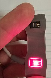
Reflectance pulse oximetry is a less common alternative, placing the photodetector on the same surface as the illumination. This method does not require a thin section of the person's body and therefore may be used almost anywhere on the body, such as the forehead, chest, or feet, but it still has some limitations. Vasodilation and pooling of venous blood in the head due to compromised venous return to the heart can cause a combination of arterial and venous pulsations in the forehead region and lead to spurious SpO2 results. Such conditions occur while undergoing anaesthesia with endotracheal intubation and mechanical ventilation or in patients in the Trendelenburg position.[3]
Medical uses
A pulse oximeter probe applied to a person's finger
A pulse oximeter is a medical device that indirectly monitors the oxygen saturation of a patient's blood (as opposed to measuring oxygen saturation directly through a blood sample) and changes in blood volume in the skin, producing a photoplethysmogram that may be further processed into other measurements.[4] The pulse oximeter may be incorporated into a multiparameter patient monitor. Most monitors also display the pulse rate. Portable, battery-operated pulse oximeters are also available for transport or home blood-oxygen monitoring.[5]
Advantages
Pulse oximetry is particularly convenient for noninvasive continuous measurement of blood oxygen saturation. In contrast, blood gas levels must otherwise be determined in a laboratory on a drawn blood sample. Pulse oximetry is useful in any setting where a patient's oxygenation is unstable, including intensive care, operating, recovery, emergency and hospital ward settings, pilots in unpressurized aircraft, for assessment of any patient's oxygenation, and determining the effectiveness of or need for supplemental oxygen. Although a pulse oximeter is used to monitor oxygenation, it cannot determine the metabolism of oxygen, or the amount of oxygen being used by a patient. For this purpose, it is necessary to also measure carbon dioxide (CO2) levels. It is possible that it can also be used to detect abnormalities in ventilation. However, the use of a pulse oximeter to detect hypoventilation is impaired with the use of supplemental oxygen, as it is only when patients breathe room air that abnormalities in respiratory function can be detected reliably with its use. Therefore, the routine administration of supplemental oxygen may be unwarranted if the patient is able to maintain adequate oxygenation in room air, since it can result in hypoventilation going undetected.[6]
Because of their simplicity of use and the ability to provide continuous and immediate oxygen saturation values, pulse oximeters are of critical importance in emergency medicine and are also very useful for patients with respiratory or cardiac problems,[7] especially COPD, or for diagnosis of some sleep disorders such as apnea and hypopnea.[8] For patients with obstructive sleep apnea, pulse oximetry readings will be in the 70–90% range for much of the time spent attempting to sleep.[9]
Portable battery-operated pulse oximeters are useful for pilots operating in non-pressurized aircraft above 10,000 feet (3,000m) or 12,500 feet (3,800m) in the U.S.[10] where supplemental oxygen is required. Portable pulse oximeters are also useful for mountain climbers and athletes whose oxygen levels may decrease at high altitudes or with exercise. Some portable pulse oximeters employ software that charts a patient's blood oxygen and pulse, serving as a reminder to check blood oxygen levels.[citation needed]
Connectivity advancements have made it possible for patients to have their blood oxygen saturation continuously monitored without a cabled connection to a hospital monitor, without sacrificing the flow of patient data back to bedside monitors and centralized patient surveillance systems.[11]
For patients with COVID-19, pulse oximetry helps with early detection of silent hypoxia, in which the patients still look and feel comfortable, but their SpO2 is dangerously low.[5] This happens to patients either in the hospital or at home. Low SpO2 may indicate severe COVID-19-related pneumonia, requiring a ventilator.[12]
Safety
Continuous monitoring with pulse oximetry is generally considered safe for most patients for up to 8 hours. However, prolonged use in certain types of patients can cause burns due to the heat emitted by the infrared LED, which reaches up to 43°C. Additionally, pulse oximeters occasionally develop electrical faults which causes them to heat up above this temperature. Patients at greater risk include those with delicate or fragile skin, such as infants, particularly premature infants, and the elderly. Additional risks for injury include lack of pain response where the probe is placed, such as having an insensate limb, or being unconscious or under anesthesia, or having communication difficulties. Patients who are at high risk for injury should be have the site of their probe moved frequently, i.e. every hour, whereas patients who are at lower risk should have theirs moved every 2-4 hours.[13]
Limitations
Fundamental limitations
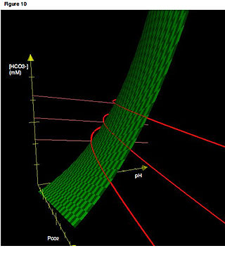
Pulse oximetry solely measures hemoglobin saturation, not ventilation and is not a complete measure of respiratory sufficiency. It is not a substitute for blood gases checked in a laboratory, because it gives no indication of base deficit, carbon dioxide levels, blood pH, or bicarbonate (HCO3−) concentration. The metabolism of oxygen can be readily measured by monitoring expired CO2, but saturation figures give no information about blood oxygen content. Most of the oxygen in the blood is carried by hemoglobin; in severe anemia, the blood contains less hemoglobin, which despite being saturated cannot carry as much oxygen.[citation needed]
Pulse oximetry also is not a complete measure of circulatory oxygen sufficiency. If there is insufficient bloodflow or insufficient hemoglobin in the blood (anemia), tissues can suffer hypoxia despite high arterial oxygen saturation.
Since pulse oximetry measures only the percentage of bound hemoglobin, a falsely high or falsely low reading will occur when hemoglobin binds to something other than oxygen:
Hemoglobin has a higher affinity to carbon monoxide than it does to oxygen. Therefore, in cases of carbon monoxide poisoning, most hemoglobin might be bound not to oxygen but to carbon monoxide. A pulse oximeter would correctly report most hemoglobin to be bound, but nevertheless the patient would be in a state of hypoxemia and subsequently hypoxia (low cellular oxygen level).
Cyanide poisoning gives a high reading because it reduces oxygen extraction from arterial blood. In this case, the reading is not false, as arterial blood oxygen is indeed high early in cyanide poisoning: the patient is not hypoxemic, but is hypoxic.
Methemoglobinemia characteristically causes pulse oximetry readings in the mid-80s.
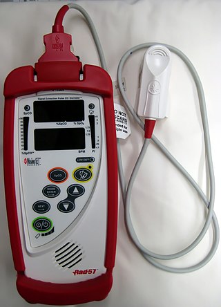
A noninvasive method that allows continuous measurement of the dyshemoglobins is the pulse CO-oximeter, which was built in 2005 by Masimo.[15] By using additional wavelengths,[16] it provides clinicians a way to measure the dyshemoglobins, carboxyhemoglobin, and methemoglobin along with total hemoglobin.[17]
Conditions affecting accuracy
Because pulse oximeter devices are calibrated for healthy subjects, their accuracy is poor for critically ill patients and preterm newborns.[1] Erroneously low readings may be caused by hypoperfusion of the extremity being used for monitoring (often due to a limb being cold or from vasoconstriction secondary to the use of vasopressor agents); incorrect sensor application; highly calloused skin; or movement (such as shivering), especially during hypoperfusion. To ensure accuracy, the sensor should return a steady pulse and/or pulse waveform. Pulse oximetry technologies differ in their abilities to provide accurate data during conditions of motion and low perfusion.[18][19]Obesity, hypotension (low blood pressure), and some hemoglobin variants can reduce the accuracy of the results.[8] Some home pulse oximeters have low sampling rates, which can significantly underestimate dips in blood oxygen levels.[8] The accuracy of pulse oximetry deteriorates considerably for readings below 80%.[9] Research has suggested that error rates in common pulse oximeter devices may be higher for adults with dark skin color, leading to claims of encoding systemic racism in countries with multi-racial populations such as the United States.[20][21] The issue was first identified decades ago; one of the earliest studies on this topic occurred in 1976, which reported reading errors in dark-skinned patients that reflected lower blood oxygen saturation values.[22] Further studies indicate that while accuracy with dark skin is good at higher, healthy saturation levels, some devices overestimate the saturation at lower levels, which may lead to hypoxia not being detected.[23] A study that reviewed thousands of cases of occult hypoxemia, where patients were found to have oxygen saturation below 88% per arterial blood gas measurement despite pulse oximeter readings indicating 92% to 96% oxygen saturation, found that black patients were three times as likely as white patients to have their low oxygen saturation missed by pulse oximeters.[24] Another research study investigated patients in the hospital with COVID-19 and found that occult hypoxemia occurred in 28.5% of black patients compared to only 17.2% of white patients.[25] There has been research to indicate that black COVID-19 patients were 29% less likely to receive supplemental oxygen in a timely manner and three times more likely to have hypoxemia.[26] A further study, which used a MIMIC-IV critical care dataset of both pulse oximeter readings and oxygen saturation levels detected in blood samples, demonstrated that black, Hispanic, and Asian patients had higher SpO2 readings than white patients for a given blood oxygen saturation level measured in blood samples.[27] As a result, black, Hispanic, and Asian patients also received lower rates of supplemental oxygen than white patients.[27] It is suggested that melanin can interfere with the absorption of light used to measure the level of oxygenated blood, often measured from a person's finger.[27] Further studies and computer simulations show that the increased amounts of melanin found in people with darker skin scatter the photons of light used by the pulse oximeters, decreasing the accuracy of the measurements. As the studies used to calibrate the devices typically oversample people with lighter skin, the parameters for pulse oximeters are set based on information that is not equitably balanced to account for diverse skin colors.[28] This inaccuracy can lead to potentially missing people who need treatment, as pulse oximetry is used for the screening of sleep apnea and other types of sleep-disordered breathing,[8] which in the United States are conditions more prevalent among minorities.[29][30][31] This bias is a significant concern, as a 2% decrease is important for respiratory rehabilitation, studies of sleep apnea, and athletes performing physical efforts; it can lead to severe complications for the patient, requiring an external oxygen supply or even hospitalization.[32] Another concern regarding pulse oximetry bias is that insurance companies and hospital systems increasingly use these numbers to inform their decisions. Pulse oximetry measurements are used to identify candidates for reimbursement.[33] Similarly, pulse oximetry data is being incorporated into algorithms for clinicians. Early Warning Scores, which provide a record for analyzing a patient's clinical status and alerting clinicians if needed, incorporate algorithms with pulse oximetry information and can result in misinformed patient records.[33]
Equipment
Consumer pulse oximeters
In addition to pulse oximeters for professional use, many inexpensive "consumer" models are available. Opinions vary about the reliability of consumer oximeters; a typical comment is "The research data on home monitors has been mixed, but they tend to be accurate within a few percentage points".[34] Some smart watches with activity tracking incorporate an oximeter function. An article on such devices, in the context of diagnosing COVID-19 infection, quoted João Paulo Cunha of the University of Porto, Portugal: "these sensors are not precise, that's the main limitation ... the ones that you wear are only for the consumer level, not for the clinical level".[35] Pulse oximeters used for diagnosis of conditions such as COVID-19 should be Class IIB medical grade oximeters. Class IIB oximeters can be used on patients of all skin colors, low pigmentation and in the presence of motion.[citation needed] When a pulse oximeter is shared between two patients, it should be either cleaned with alcohol wipes after each use or a disposable probe or finger cover to be used to prevent cross-infection.[36]
According to a report by iData Research, the US pulse oximetry monitoring market for equipment and sensors was over $700 million in 2011.[37]
Mobile apps
Mobile app pulse oximeters use the flashlight and the camera of the phone, instead of infrared light used in conventional pulse oximeters. However, apps do not generate as accurate readings because the camera cannot measure the light reflection at two wavelengths, so the oxygen saturation readings that are obtained through an app on a smartphone are inconsistent for clinical use. At least one study has suggested these are not reliable relative to clinical pulse oximeters.[38]
Mechanism
Absorption spectra of oxygenated hemoglobin (HbO2) and deoxygenated hemoglobin (Hb) for red and infrared wavelengthsSimplified principle of operation of a transmissive LED pulse oximeterThe inner side of a pulse oximeter
A blood-oxygen monitor displays the percentage of blood that is loaded with oxygen. More specifically, it uses light spectrometry to measure what percentage of hemoglobin, the protein in blood that carries oxygen, is loaded. Acceptable normal SaO2 ranges for patients without pulmonary pathology are from 95 to 99 percent.[citation needed] For a person breathing room air at or near sea level, an estimate of arterial pO2 can be made from the blood-oxygen monitor "saturation of peripheral oxygen" (SpO2) reading.[citation needed]
Mode of operation
A typical pulse oximeter uses an electronic processor and a pair of small light-emitting diodes (LEDs) facing a photodiode through a translucent part of the patient's body, usually a fingertip or an earlobe. One LED is red, with wavelength of 660nm, and the other is infrared with a wavelength of 940nm. Absorption of light at these wavelengths differs significantly between blood loaded with oxygen and blood lacking oxygen. Oxygenated hemoglobin absorbs more infrared light and allows more red light to pass through. Deoxygenated hemoglobin allows more infrared light to pass through and absorbs more red light. The LEDs sequence through their cycle of one on, then the other, then both off about thirty times per second which allows the photodiode to respond to the red and infrared light separately and also adjust for the ambient light baseline.[39]
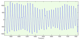
The amount of light that is transmitted (in other words, that is not absorbed) is measured, and separate normalized signals are produced for each wavelength. These signals fluctuate in time because the amount of arterial blood that is present increases (literally pulses) with each heartbeat. By subtracting the minimum transmitted light from the transmitted light in each wavelength, the effects of other tissues are corrected for, generating a continuous signal for pulsatile arterial blood.[40] The ratio of the red light measurement to the infrared light measurement is then calculated by the processor (which represents the ratio of oxygenated hemoglobin to deoxygenated hemoglobin), and this ratio is then converted to SpO2 by the processor via a lookup table[40] based on the Beer–Lambert law.[39] The signal separation also serves other purposes: a plethysmograph waveform ("pleth wave") representing the pulsatile signal is usually displayed for a visual indication of the pulses as well as signal quality,[4] and a numeric ratio between the pulsatile and baseline absorbance ("perfusion index") can be used to evaluate perfusion.[41]
where HbO2 is oxygenated hemoglobin (oxyhemoglobin) and Hb is deoxygenated hemoglobin.
Due to changes in blood volumes in the skin, a plethysmographic variation can be seen in the light signal received (transmittance) by the sensor on an oximeter. The variation can be described as a periodic function, which in turn can be split into a DC component (the peak value)[lower-alpha 1] and an AC component (peak minus trough).[42] The ratio of the AC component to the DC component, expressed as a percentage, is known as the (peripheral) perfusion index (Pi) for a pulse, and typically has a range of 0.02% to 20%.[43] An earlier measurement called the pulse oximetry plethysmographic (POP) only measures the "AC" component, and is derived manually from monitor pixels.[41][44]
Pleth variability index (PVI) is a measure of the variability of the perfusion index, which occurs during breathing cycles. Mathematically it is calculated as (Pimax − Pimin)/Pimax × 100%, where the maximum and minimum Pi values are from one or many breathing cycles.[42] It has been shown to be a useful, noninvasive indicator of continuous fluid responsiveness for patients undergoing fluid management.[41]Pulse oximetry plethysmographic waveform amplitude (ΔPOP) is an analogous earlier technique for use on the manually-derived POP, calculated as (POPmax − POPmin)/(POPmax + POPmin)×2.[44]
History
In 1935, German physician Karl Matthes (1905–1962) developed the first two-wavelength ear O2 saturation meter with red and green filters (later red and infrared filters). It was the first device to measure O2 saturation.[45]
The original oximeter was made by Glenn Allan Millikan in the 1940s.[46] In 1943[47] and as published in 1949,[48]Earl Wood added a pressure capsule to squeeze blood out of the ear so as to obtain an absolute O2 saturation value when blood was readmitted. The concept is similar to today's conventional pulse oximetry, but was difficult to implement because of unstable photocells and light sources; today this method is not used clinically. In 1964 Shaw assembled the first absolute reading ear oximeter, which used eight wavelengths of light.[citation needed]
The first pulse oximetry was developed in 1972 by Japanese bioengineers Takuo Aoyagi and Michio Kishi at Japanese medical electronic equipment manufacturer Nihon Kohden, using the ratio of red to infrared light absorption of pulsating components at the measuring site. Nihon Kohden manufactured the first pulse oximeter, Ear Oximeter OLV-5100. Surgeon Susumu Nakajima and his associates first tested the device in patients, reporting it in 1975.[49] However, Nihon Kohden suspended the development of pulse oximetry and did not apply for a basic patent of pulse oximetry except in Japan, which facilitated further development and utilization of pulse oximetry later in U.S. In 1977, Minolta commercialized the first finger pulse oximeter OXIMET MET-1471. In the U.S., the first pulse oximetry was commercialized by Biox in 1980.[49][50][51]
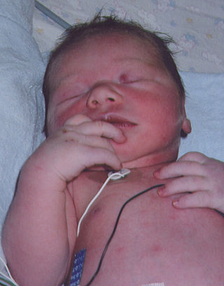
By 1987, the standard of care for the administration of a general anesthetic in the U.S. included pulse oximetry. From the operating room, the use of pulse oximetry rapidly spread throughout the hospital, first to recovery rooms, and then to intensive care units. Pulse oximetry was of particular value in the neonatal unit where the patients do not thrive with inadequate oxygenation, but too much oxygen and fluctuations in oxygen concentration can lead to vision impairment or blindness from retinopathy of prematurity (ROP). Furthermore, obtaining an arterial blood gas from a neonatal patient is painful to the patient and a major cause of neonatal anemia.[52] Motion artifact can be a significant limitation to pulse oximetry monitoring, resulting in frequent false alarms and loss of data. This is because during motion and low peripheral perfusion, many pulse oximeters cannot distinguish between pulsating arterial blood and moving venous blood, leading to underestimation of oxygen saturation. Early studies of pulse oximetry performance during subject motion made clear the vulnerabilities of conventional pulse oximetry technologies to motion artifact.[18][53]
In 1995, Masimo introduced Signal Extraction Technology (SET) that could measure accurately during patient motion and low perfusion by separating the arterial signal from the venous and other signals. Since then, pulse oximetry manufacturers have developed new algorithms to reduce some false alarms during motion,[54] such as extending averaging times or freezing values on the screen, but they do not claim to measure changing conditions during motion and low perfusion. So there are still important differences in performance of pulse oximeters during challenging conditions.[19] Also in 1995, Masimo introduced perfusion index, quantifying the amplitude of the peripheral plethysmograph waveform. Perfusion index has been shown to help clinicians predict illness severity and early adverse respiratory outcomes in neonates,[55][56][57] predict low superior vena cava flow in very low birth weight infants,[58] provide an early indicator of sympathectomy after epidural anesthesia,[59] and improve detection of critical congenital heart disease in newborns.[60]
Published papers have compared signal extraction technology to other pulse oximetry technologies and have demonstrated consistently favorable results for signal extraction technology.[18][19][61] Signal extraction technology pulse oximetry performance has also been shown to translate into helping clinicians improve patient outcomes. In one study, retinopathy of prematurity (eye damage) was reduced by 58% in very low birth weight neonates at a center using signal extraction technology, while there was no decrease in retinopathy of prematurity at another center with the same clinicians using the same protocol but with non-signal extraction technology.[62] Other studies have shown that signal extraction technology pulse oximetry results in fewer arterial blood gas measurements, faster oxygen weaning time, lower sensor utilization, and lower length of stay.[63] The measure-through motion and low perfusion capabilities it has also allow it to be used in previously unmonitored areas such as the general floor, where false alarms have plagued conventional pulse oximetry. As evidence of this, a landmark study was published in 2010 showing that clinicians at Dartmouth-Hitchcock Medical Center using signal extraction technology pulse oximetry on the general floor were able to decrease rapid response team activations, ICU transfers, and ICU days.[64] In 2020, a follow-up retrospective study at the same institution showed that over ten years of using pulse oximetry with signal extraction technology, coupled with a patient surveillance system, there were zero patient deaths and no patients were harmed by opioid-induced respiratory depression while continuous monitoring was in use.[65]
In 2007, Masimo introduced the first measurement of the pleth variability index (PVI), which multiple clinical studies have shown provides a new method for automatic, noninvasive assessment of a patient's ability to respond to fluid administration.[41][66][67] Appropriate fluid levels are vital to reducing postoperative risks and improving patient outcomes: fluid volumes that are too low (under-hydration) or too high (over-hydration) have been shown to decrease wound healing and increase the risk of infection or cardiac complications.[68] Recently, the National Health Service in the United Kingdom and the French Anesthesia and Critical Care Society listed PVI monitoring as part of their suggested strategies for intra-operative fluid management.[69][70]
In 2011, an expert workgroup recommended newborn screening with pulse oximetry to increase the detection of critical congenital heart disease (CCHD).[71] The CCHD workgroup cited the results of two large, prospective studies of 59,876 subjects that exclusively used signal extraction technology to increase the identification of CCHD with minimal false positives.[72][73] The CCHD workgroup recommended newborn screening be performed with motion tolerant pulse oximetry that has also been validated in low perfusion conditions. In 2011, the US Secretary of Health and Human Services added pulse oximetry to the recommended uniform screening panel.[74] Before the evidence for screening using signal extraction technology, less than 1% of newborns in the United States were screened. Today, The Newborn Foundation has documented near universal screening in the United States and international screening is rapidly expanding.[75] In 2014, a third large study of 122,738 newborns that also exclusively used signal extraction technology showed similar, positive results as the first two large studies.[76]
High-resolution pulse oximetry (HRPO) has been developed for in-home sleep apnea screening and testing in patients for whom it is impractical to perform polysomnography.[77][78] It stores and records both pulse rate and SpO2 in 1 second intervals and has been shown in one study to help to detect sleep disordered breathing in surgical patients.[79]
See also
Arterial blood gas– A test of blood taken from an artery that measures the amounts of certain dissolved gasesPages displaying short descriptions of redirect targets
Capnography– Monitoring of the concentration of carbon dioxide in respiratory gases
Integrated pulmonary index– single value that describes the patient’s respiratory statusPages displaying wikidata descriptions as a fallback
Respiratory monitoring– Method to mechanically assist or replace spontaneous breathingPages displaying short descriptions of redirect targets
Medical equipment– Device to be used for medical purposesPages displaying short descriptions of redirect targets
↑ This definition used by Masimo varies from the mean value used in signal processing; it is meant to measure the pulsatile arterial blood absorbance over the baseline absorbance.
Related Research Articles
Hypoxia is a condition in which the body or a region of the body is deprived of adequate oxygen supply at the tissue level. Hypoxia may be classified as either generalized, affecting the whole body, or local, affecting a region of the body. Although hypoxia is often a pathological condition, variations in arterial oxygen concentrations can be part of the normal physiology, for example, during strenuous physical exercise.
Respiratory failure results from inadequate gas exchange by the respiratory system, meaning that the arterial oxygen, carbon dioxide, or both cannot be kept at normal levels. A drop in the oxygen carried in the blood is known as hypoxemia; a rise in arterial carbon dioxide levels is called hypercapnia. Respiratory failure is classified as either Type 1 or Type 2, based on whether there is a high carbon dioxide level, and can be acute or chronic. In clinical trials, the definition of respiratory failure usually includes increased respiratory rate, abnormal blood gases, and evidence of increased work of breathing. Respiratory failure causes an altered state of consciousness due to ischemia in the brain.
An arterial blood gas (ABG) test, or arterial blood gas analysis (ABGA) measures the amounts of arterial gases, such as oxygen and carbon dioxide. An ABG test requires that a small volume of blood be drawn from the radial artery with a syringe and a thin needle, but sometimes the femoral artery in the groin or another site is used. The blood can also be drawn from an arterial catheter.
Cyanosis is the change of body tissue color to a bluish-purple hue, as a result of decrease in the amount of oxygen bound to the hemoglobin in the red blood cells of the capillary bed. Cyanosis is apparent usually in the body tissues covered with thin skin, including the mucous membranes, lips, nail beds, and ear lobes. Some medications may cause discoloration such as medications containing amiodarone or silver. Furthermore, mongolian spots, large birthmarks, and the consumption of food products with blue or purple dyes can also result in the bluish skin tissue discoloration and may be mistaken for cyanosis. Appropriate physical examination and history taking is a crucial part to diagnose cyanosis. Management of cyanosis involves treating the main cause, as cyanosis isn’t a disease, it is a symptom.
Extracorporeal membrane oxygenation (ECMO), is a form of extracorporeal life support, providing prolonged cardiac and respiratory support to persons whose heart and lungs are unable to provide an adequate amount of oxygen, gas exchange or blood supply (perfusion) to sustain life. The technology for ECMO is largely derived from cardiopulmonary bypass, which provides shorter-term support with arrested native circulation. The device used is a membrane oxygenator, also known as an artificial lung.
A photoplethysmogram (PPG) is an optically obtained plethysmogram that can be used to detect blood volume changes in the microvascular bed of tissue. A PPG is often obtained by using a pulse oximeter which illuminates the skin and measures changes in light absorption. A conventional pulse oximeter monitors the perfusion of blood to the dermis and subcutaneous tissue of the skin.
Blue baby syndrome can refer to conditions that cause cyanosis, or blueness of the skin, in babies as a result of low oxygen levels in the blood. This term has traditionally been applied to cyanosis as a result of:.
Cyanotic heart disease, which is a category of congenital heart defect that results in low levels of oxygen in the blood. This can be caused by either reduced blood flow to the lungs or mixing of oxygenated and deoxygenated blood.
Methemoglobinemia, which is a disease defined by high levels of methemoglobin in the blood. Increased levels of methemoglobin prevent oxygen from being released into the tissues and result in hypoxemia.
Oxygen saturation is a relative measure of the concentration of oxygen that is dissolved or carried in a given medium as a proportion of the maximal concentration that can be dissolved in that medium at the given temperature. It can be measured with a dissolved oxygen probe such as an oxygen sensor or an optode in liquid media, usually water. The standard unit of oxygen saturation is percent (%).
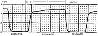
Capnography is the monitoring of the concentration or partial pressure of carbon dioxide (CO 2) in the respiratory gases. Its main development has been as a monitoring tool for use during anesthesia and intensive care. It is usually presented as a graph of CO 2 (measured in kilopascals, "kPa" or millimeters of mercury, "mmHg") plotted against time, or, less commonly, but more usefully, expired volume (known as volumetric capnography). The plot may also show the inspired CO 2, which is of interest when rebreathing systems are being used. When the measurement is taken at the end of a breath (exhaling), it is called "end tidal" CO 2 (PETCO2).
Somnology is the scientific study of sleep. It includes clinical study and treatment of sleep disorders and irregularities. Sleep medicine is a subset of somnology.
Hypoxemia is an abnormally low level of oxygen in the blood. More specifically, it is oxygen deficiency in arterial blood. Hypoxemia has many causes, and often causes hypoxia as the blood is not supplying enough oxygen to the tissues of the body.
A CO-oximeter is a device that measures the oxygen carrying state of hemoglobin in a blood specimen, including oxygen-carrying hemoglobin (O2Hb), non-oxygen-carrying but normal hemoglobin (HHb), as well as the dyshemoglobins such as carboxyhemoglobin (COHb) and methemoglobin (MetHb). The use of 'CO' rather than 'Co' or 'co' is more appropriate since this designation represents a device that measures carbon monoxide (CO) bound to hemoglobin, as distinguished from simple oximetry which measures hemoglobin bound to molecular oxygen—O2Hb—or hemoglobin capable of binding to molecular oxygen—HHb. Simpler oximeters may report oxygen saturation alone, i.e. the ratio of oxyhemoglobin to total 'bindable' hemoglobin. CO-oximetry is useful in defining the causes for hypoxemia, or hypoxia,.
The Haldane effect is a property of hemoglobin first described by John Scott Haldane, within which oxygenation of blood in the lungs displaces carbon dioxide from hemoglobin, increasing the removal of carbon dioxide. Consequently, oxygenated blood has a reduced affinity for carbon dioxide. Thus, the Haldane effect describes the ability of hemoglobin to carry increased amounts of carbon dioxide (CO2) in the deoxygenated state as opposed to the oxygenated state. Vice versa, it is true that a high concentration of CO2 facilitates dissociation of oxyhemoglobin, though this is the result of two distinct processes (Bohr effect and Margaria-Green effect) and should be distinguished from Haldane effect.
A pulmonary shunt is the passage of deoxygenated blood from the right side of the heart to the left without participation in gas exchange in the pulmonary capillaries. It is a pathological condition that results when the alveoli of parts of the lungs are perfused with blood as normal, but ventilation fails to supply the perfused region. In other words, the ventilation/perfusion ratio of those areas is zero.
The Alveolar–arterial gradient, is a measure of the difference between the alveolar concentration (A) of oxygen and the arterial (a) concentration of oxygen. It is a useful parameter for narrowing the differential diagnosis of hypoxemia.
Masimo Corporation is a health technology and consumer electronics company based in Irvine, California. The company primarily manufactures patient monitoring devices and technologies, including non-invasive sensors using optical technology, patient management, and telehealth platforms. In 2022, the company expanded into home audio by acquiring Sound United, and began to manufacture health-oriented wearable devices.
A hypoxicator is a medical device intended to provide a stimulus for the adaptation of an individual's cardiovascular system by means of breathing reduced oxygen hypoxic air and triggering mechanisms of compensation. The aim of intermittent hypoxic training or hypoxic therapy conducted with such a device is to obtain benefits in physical performance and wellbeing through improved oxygen metabolism.
Oxygen saturation is the fraction of oxygen-saturated haemoglobin relative to total haemoglobin in the blood. The human body requires and regulates a very precise and specific balance of oxygen in the blood. Normal arterial blood oxygen saturation levels in humans are 96–100 percent. If the level is below 90 percent, it is considered low and called hypoxemia. Arterial blood oxygen levels below 80 percent may compromise organ function, such as the brain and heart, and should be promptly addressed. Continued low oxygen levels may lead to respiratory or cardiac arrest. Oxygen therapy may be used to assist in raising blood oxygen levels. Oxygenation occurs when oxygen molecules enter the tissues of the body. For example, blood is oxygenated in the lungs, where oxygen molecules travel from the air and into the blood. Oxygenation is commonly used to refer to medical oxygen saturation.
Blood gas tension refers to the partial pressure of gases in blood. There are several significant purposes for measuring gas tension. The most common gas tensions measured are oxygen tension (PxO2), carbon dioxide tension (PxCO2) and carbon monoxide tension (PxCO). The subscript x in each symbol represents the source of the gas being measured: "a" meaning arterial, "A" being alveolar, "v" being venous, and "c" being capillary. Blood gas tests (such as arterial blood gas tests) measure these partial pressures.
Respiratory compromise describes a deterioration in respiratory function with a high likelihood of rapid progression to respiratory failure and death. Respiratory failure occurs when inadequate gas exchange by the respiratory system occurs, with a low oxygen level or a high carbon dioxide level.
↑ Brand TM, Brand ME, Jay GD (February 2002). "Enamel nail polish does not interfere with pulse oximetry among normoxic volunteers". Journal of Clinical Monitoring and Computing. 17 (2): 93–96. doi:10.1023/A:1016385222568. PMID12212998. S2CID24354030.
↑ Maisel WH, Lewis RJ (October 2010). "Noninvasive measurement of carboxyhemoglobin: how accurate is accurate enough?". Annals of Emergency Medicine. 56 (4): 389–391. doi:10.1016/j.annemergmed.2010.05.025. PMID20646785.
1 2 3 Shah N, Ragaswamy HB, Govindugari K, Estanol L (August 2012). "Performance of three new-generation pulse oximeters during motion and low perfusion in volunteers". Journal of Clinical Anesthesia. 24 (5): 385–391. doi:10.1016/j.jclinane.2011.10.012. PMID22626683.
↑ N A Saunders; A C P Powles; A S Rebuck (Jun 1976). "Ear oximetry: accuracy and practicability in the assessment of arterial oxygenation". American Review of Respiratory Disease. 113 (6): 745–749. doi:10.1164/arrd.1976.113.6.745 (inactive 31 January 2024). PMID937815.{{cite journal}}: CS1 maint: DOI inactive as of January 2024 (link)
↑ Redline S, Tishler PV, Hans MG, Tosteson TD, Strohl KP, Spry K (January 1997). "Racial differences in sleep-disordered breathing in African-Americans and Caucasians". American Journal of Respiratory and Critical Care Medicine. 155 (1): 186–192. doi:10.1164/ajrccm.155.1.9001310. OCLC209489389. PMID9001310.
↑ Matthes K (1935). "Untersuchungen über die Sauerstoffsättigung des menschlichen Arterienblutes" [Studies on the Oxygen Saturation of Arterial Human Blood]. Naunyn-Schmiedeberg's Archives of Pharmacology (in German). 179 (6): 698–711. doi:10.1007/BF01862691. S2CID24678464.
↑ Gilbert, Barry; Haider, Clifton; Schwab, Daniel; Delp, Gary. "Development of a Capability to Measure and Record Physical and Electrical Parameters in Free-Living Subjects, Motivating the Requirement for a Machine to Measure Natural Analytes of Clinical Importance in Blood Samples". SPIE J. Of Biomed. Opt. TBD (Under Consideration).
↑ Jopling MW, Mannheimer PD, Bebout DE (January 2002). "Issues in the laboratory evaluation of pulse oximeter performance". Anesthesia and Analgesia. 94 (1 Suppl): S62–68. PMID11900041.
↑ De Felice C, Leoni L, Tommasini E, Tonni G, Toti P, Del Vecchio A, Ladisa G, Latini G (March 2008). "Maternal pulse oximetry perfusion index as a predictor of early adverse respiratory neonatal outcome after elective cesarean delivery". Pediatric Critical Care Medicine. 9 (2): 203–208. doi:10.1097/pcc.0b013e3181670021. PMID18477934. S2CID24626430.
↑ De Felice C, Latini G, Vacca P, Kopotic RJ (October 2002). "The pulse oximeter perfusion index as a predictor for high illness severity in neonates". European Journal of Pediatrics. 161 (10): 561–562. doi:10.1007/s00431-002-1042-5. PMID12297906. S2CID20910692.
↑ De Felice C, Goldstein MR, Parrini S, Verrotti A, Criscuolo M, Latini G (March 2006). "Early dynamic changes in pulse oximetry signals in preterm newborns with histologic chorioamnionitis". Pediatric Critical Care Medicine. 7 (2): 138–142. doi:10.1097/01.PCC.0000201002.50708.62. PMID16474255. S2CID12780058.
↑ Ginosar Y, Weiniger CF, Meroz Y, Kurz V, Bdolah-Abram T, Babchenko A, Nitzan M, Davidson EM (September 2009). "Pulse oximeter perfusion index as an early indicator of sympathectomy after epidural anesthesia". Acta Anaesthesiologica Scandinavica. 53 (8): 1018–1026. doi:10.1111/j.1399-6576.2009.01968.x. PMID19397502. S2CID24986518.
↑ Granelli A, Ostman-Smith I (October 2007). "Noninvasive peripheral perfusion index as a possible tool for screening for critical left heart obstruction". Acta Paediatrica. 96 (10): 1455–1459. doi:10.1111/j.1651-2227.2007.00439.x. PMID17727691. S2CID6181750.
↑ Durbin CG, Rostow SK (August 2002). "More reliable oximetry reduces the frequency of arterial blood gas analyses and hastens oxygen weaning after cardiac surgery: a prospective, randomized trial of the clinical impact of a new technology". Critical Care Medicine. 30 (8): 1735–1740. doi:10.1097/00003246-200208000-00010. PMID12163785. S2CID10226994.
↑ Zimmermann M, Feibicke T, Keyl C, Prasser C, Moritz S, Graf BM, Wiesenack C (June 2010). "Accuracy of stroke volume variation compared with pleth variability index to predict fluid responsiveness in mechanically ventilated patients undergoing major surgery". European Journal of Anaesthesiology. 27 (6): 555–561. doi:10.1097/EJA.0b013e328335fbd1. PMID20035228. S2CID45041607.
↑ Zhao QM, Ma XJ, Ge XL, Liu F, Yan WL, Wu L, Ye M, Liang XC, Zhang J, Gao Y, Jia B, Huang GY (August 2014). "Pulse oximetry with clinical assessment to screen for congenital heart disease in neonates in China: a prospective study". Lancet. 384 (9945): 747–754. doi:10.1016/S0140-6736(14)60198-7. PMID24768155. S2CID23218716.
This page is based on this Wikipedia article Text is available under the CC BY-SA 4.0 license; additional terms may apply. Images, videos and audio are available under their respective licenses.