A melanocytic nevus is a type of melanocytic tumor that contains nevus cells.

Lichen planus (LP) is a chronic inflammatory and immune mediated disease that affects the skin, nails, hair, and mucous membranes. It is characterized by polygonal, flat-topped, violaceous papules and plaques with overlying, reticulated, fine white scale, commonly affecting dorsal hands, flexural wrists and forearms, trunk, anterior lower legs and oral mucosa. Although there is a broad clinical range of LP manifestations, the skin and oral cavity remain as the major sites of involvement. The cause is unknown, but it is thought to be the result of an autoimmune process with an unknown initial trigger. There is no cure, but many different medications and procedures have been used in efforts to control the symptoms.

Pityriasis rosea is a type of skin rash. Classically, it begins with a single red and slightly scaly area known as a "herald patch". This is then followed, days to weeks later, by a pink whole body rash. It typically lasts less than three months and goes away without treatment. Sometime a fever may occur before the start of the rash or itchiness may be present, but often there are few other symptoms.

Tuberculosis verrucosa cutis is a rash of small, red papular nodules in the skin that may appear 2–4 weeks after inoculation by Mycobacterium tuberculosis in a previously infected and immunocompetent individual.
Erythema toxicum neonatorum is a common rash in neonates. It appears in up to half of newborns carried to term, usually between day 2–5 after birth; it does not occur outside the neonatal period.
Leukemia cutis is the infiltration of neoplastic leukocytes or their precursors into the skin resulting in clinically identifiable cutaneous lesions. This condition may be contrasted with leukemids, which are skin lesions that occur with leukemia, but which are not related to leukemic cell infiltration. Leukemia cutis can occur in most forms of leukemia, including chronic myeloid leukemia, acute lymphoblastic leukemia, chronic lymphocytic leukemia, acute myeloid leukemia, and prolymphocytic leukemia.

Dermatosis papulosa nigra (DPN) is a condition of many small, benign skin lesions on the face, a condition generally presenting on dark-skinned individuals. DPN is extremely common, affecting up to 30% of Black people in the US. From a histological perspective, DPN resembles seborrheic keratoses. The condition may be cosmetically undesirable to some.

A venous lake is a generally solitary, soft, compressible, dark blue to violaceous, 0.2- to 1-cm papule commonly found on sun-exposed surfaces of the vermilion border of the lip, face and ears. Lesions generally occur among the elderly.
Cutis verticis gyrata is a medical condition usually associated with thickening of the scalp. People show visible folds, ridges or creases on the surface of the top of the scalp. The number of folds can vary from two to roughly ten and are typically soft and spongy. These folds cannot be corrected with pressure. The condition typically affects the central and rear regions of the scalp, but sometimes can involve the entire scalp.
Gianotti–Crosti syndrome, also known as infantile papular acrodermatitis, papular acrodermatitis of childhood, and papulovesicular acrolocated syndrome, is a reaction of the skin to a viral infection. Hepatitis B virus and Epstein–Barr virus are the most frequently reported pathogens. Other incriminated viruses are hepatitis A virus, hepatitis C virus, cytomegalovirus, coxsackievirus, adenovirus, enterovirus, rotavirus, rubella virus, HIV, and parainfluenza virus.

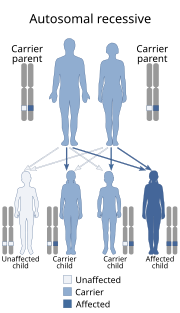
Urbach–Wiethe disease is a rare recessive genetic disorder, with approximately 400 reported cases since its discovery. It was first officially reported in 1929 by Erich Urbach and Camillo Wiethe, although cases may be recognized dating back as early as 1908.
Id reactions are types of acute dermatitis developing after days or weeks at skin locations distant from the initial inflammatory or infectious site. They can be localised or generalised. This is also known as an 'autoeczematous response' and there must be an identifiable initial inflammatory or infectious skin problem which leads to the generalised eczema. Often intensely itchy, the red papules and pustules can also be associated with blisters and scales and are always remote from the primary lesion. It is most commonly a blistering rash with itchy vesicles on the sides of fingers and feet as a reaction to fungal infection on the feet, athlete's foot. Stasis dermatitis, allergic contact dermatitis, acute irritant contact eczema and infective dermatitis have been documented as possible triggers, but the exact cause and mechanism is not fully understood. Several other types of id reactions exist including erythema nodosum, erythema multiforme, Sweet's syndrome and urticaria.
Primary inoculation tuberculosis is a skin condition that develops at the site of inoculation of tubercle bacilli into a tuberculosis-free individual.
Tuberculosis cutis orificialis is a form of cutaneous tuberculosis that occurs at the mucocutaneous borders of the nose, mouth, anus, urinary meatus, and vagina, and on the mucous membrane of the mouth or tongue.
Leukemids are nonspecific skin lesions that occur with leukemia which are not related to leukemic cell infiltration. This condition may be contrasted with leukemia cutis, which is the infiltration of neoplastic leukocytes or their precursors into the skin resulting in clinically identifiable cutaneous lesions.
Dystrophic calcinosis cutis is a cutaneous condition characterized by calcification of the skin resulting from the deposition of calcium and phosphorus, and occurs in a preexisting skin lesion of inflammatory process.
Metastatic calcinosis cutis is a cutaneous condition characterized by calcification of the skin resulting from the deposition of calcium and phosphorus, and associated with an internal malignancy.
Idiopathic scrotal calcinosis is a cutaneous condition characterized by calcification of the skin resulting from the deposition of calcium and phosphorus occurring on the scrotum. However, the levels of calcium and phosphate in the blood are normal. Idiopathic scrotal calcinosis typically affects young males, with an onset between adolescence and early adulthood. The scrotal calcinosis appears, without any symptoms, as yellowish nodules that range in size from 1 mm to several centimeters.

Subepidermal calcified nodule is a cutaneous condition characterized by calcification of the skin resulting from the deposition of calcium and phosphorus, occurring most frequently as one or a few skin lesions on the scalp or face of children. Lesions may also appear on the ear and eyelid.

Osteoma cutis is a cutaneous condition characterized by the presence of bone within the skin in the absence of a preexisting or associated lesion.


