Anna Akhmanova

Last updated
Anna Akhmanova
Annaakhmanova (1).jpg
Anna Akhmanova.
Born (1967-05-11) 11 May 1967 (age 56)
Moscow, Russia
NationalityRussian
CitizenshipThe Netherlands, Russia
Alma mater Moscow State University (masters), Radboud University Nijmegen (doctorate)
Children1
Awards2018 Spinoza Prize
Scientific career
Fields Cell biology
InstitutionsRadboud University Nijmegen, Erasmus University of Rotterdam, Utrecht University
Doctoral advisor Wolfgang Hennig
Other academic advisorsAlexander Mankin
Website cellbiology.science.uu.nl/research-groups/anna-akhmanova-cellular-dynamics/

Anna Sergeevna Akhmanova (born 11 May 1967) is a Russian-born professor of Cell Biology at Utrecht University in the Netherlands. [1] [2] She is best known for her research regarding microtubules and the proteins, called TIPs, that stabilize one specific end of the tubules. Among the awards she has won, she was one of the recipients of the 2018 Spinoza Prize, the highest honor for Dutch scientists.

Contents

Biography

Anna Akhmanova was born on 11 May 1967 in Moscow, Russia, to a family of scientists. [3] [4] Her grandmother was an English and linguistics professor, her father a physics professor, and her mother and now her brother hold PhDs in physics as well. She cites an interest in nature from an early age and that "a career in science was a very natural choice" for herself. [5] She attended Moscow State University, where she studied biology in the country's then-standard five-year program to receive her masters. During this program she studied basic biology, along with cell biology and biochemistry. Her fifth-year research thesis was completed in Alexander Mankin's laboratory where she researched halophilic archaebacteria; she credits Mankin as the person from whom she learned most of her molecular biology knowledge. She received her master's degree in 1989. [5]

After graduating from Moscow State, Akhmanova left Russia to continue her studies in the Netherlands. She had originally looked for doctorate programs in Russia, but however, "the salaries were very low, there was absolutely no funding to do research, and the country as a whole was experiencing problems." [5] During this time, the Soviet policy of perestroika was negatively affecting the university and research programs there, which led to Akhmanova's decision to go to the Netherlands with her young daughter to obtain her PhD. There, she worked at Radboud University Nijmegen (RU) in a lab under Wolfgang Hennig; her research then focused on obtaining mutants of histone genes. [5] She received her PhD in 1997 from RU. [6]

She completed two postdoctoral projects, the first of which was at RU, where she worked with anaerobic organisms for the Department of Microbiology. Her second postdoc was done at the Erasmus University of Rotterdam. She worked in Niels Galjart's lab in the Department of Cell Biology which Frank Grosveld headed; her research focused on gene regulation and transcription. She worked with one transcription factor using two-hybrid screening and was asked by Casper Hoogenraad for help with screening CLIP-115, a microtubule-binding protein that Hoogenraad was working with. Akhmanova and Hoogenraad then created clones for the proteins CLASP and Bicaudal-D, which Akhmanova describes as the proteins that defined her career. [5]

In 2011, Akhmanova and Hoogenraad continued to collaborate on research and moved their laboratories to Utrecht University, where they began running the Division of Cell Biology. [5] As of 2023, she is still a cell biology professor at Utrecht University, where she continues to do research on intracellular transportation, especially involving microtubule proteins. [7]

Research

Akhmanova and her team study the cell cytoskeleton and its effect on human diseases, cell polarization, and vertebrate development. Their main focus is on the microtubules that form part of the cytoskeleton and are essential for many processes, especially cell division. Their research is important for battling disease processes such as cancer, neurodegeneration, and the spread of pathogens throughout the cell. [8]

In terms of methods, the team uses high resolution images of the cells they are studying. They utilize specific assays to measure protein dynamics, reconstitute cytoskeleton processes in vitro, and identify the interactions of different proteins. [8]

The team studies specific proteins that interact on the plus and minus ends of the microtubules, specifically the plus end tracking proteins (+TIPs), which associate with the plus end of the microtubule to regulate its dynamics, and how the +TIPs interact with other structures in the cell. More recently, they have started researching "the biochemical properties and functional roles of the proteins" which organize minus end tracking proteins (-TIPs).4 There is far less information about –TIPs, and they are still not fully understood; however, recent research on CAMSAP, a type of –TIP, has shown that it plays an important role for organizing and stabilizing microtubules during interphase. Akhmanova's group now focus on finding how CAMSAP contributes to the organization and stabilization of non-centrosomal microtubules during cell division. [8]

Another of their projects concerns the mechanisms involved in microtubule-based vesicle transport. They identified several structures that link the microtubule motors, kinesin and dynein, to vesicles, and they developed procedures to show the function of the linkers when gathering[ clarification needed ] motor proteins to associate with membrane organelles. Inside the cell, kinesin and dynein protein motors are required for long-range transport along microtubules. Akhmanova's team focuses mainly on dynein, the motor that moves toward the minus end of the microtubule, and how it is linked to the various organelles and vesicles it transfers. They also study how dynein coordinates with kinesin, the motor that moves toward the plus end of the microtubule, when they are attached to the same organelle or vesicle, and they study the different signaling pathways that affect these motors. As of 2016, they were examining the protein Bicaudal D and its role in dynein-dependent transport, as it has been found to be important for dynein-dependent transport of mRNA in flies and of exocytotic vesicles in mammals. Bicaudal D was also found to be important for the positioning of the centrosomes and nucleus during mitosis, as the positioning is facilitated by dynein and kinesin. [8]

Akhmanova and her team use constitutive exocytosis as a model system for their study of kinesin and dynein. Exocytotic carriers move from the Golgi to the plasma membrane along microtubules. The team has found that the same cortical complexes are used to attach the microtubule to the plasma membrane as are used to attach them to vesicles. From here, the team plans to study how the cortical complexes are made and regulated, how they affect the attachments and dynamics of microtubules, and what the mechanism is that allows them to fuse vesicles. Also, they would like to find more information on the NF-κB signaling pathway as it was found to have proteins, called ELKS, which are found in the cortical complex. They plan to research how the pathway's components interact and how it affects microtubule stabilization and vesicle fusion. [8]

Honours and awards

Akhmanova has received several awards, including the NWO Spinoza Prize in 2018, the ALW Vernieuwingsimpuls VIDI award in 2001, and the VICI award in 2007. [6] In 2013, she and her colleague Marileen Dogterom received a European Research Council Synergy grant of 7.1 million euro. [9] The grant was given for research on cell division and cell movement. [2]

Akhmanova is a member of the European Molecular Biology Organization (2010) [10] and the Royal Netherlands Academy of Arts and Sciences (2015), [11] and she is the chair of the board for the Netherlands Society for Microscopy. She is also on the editorial board for various publications such as eLife, Journal of Cell Science, BMC Cell Biology, The Journal of Biological Chemistry, Traffic, and BioArchitecture. [8]

Listed are some of her awards: [6] [8]

Related Research Articles

<span class="mw-page-title-main">Microtubule</span> Polymer of tubulin that forms part of the cytoskeleton

Microtubules are polymers of tubulin that form part of the cytoskeleton and provide structure and shape to eukaryotic cells. Microtubules can be as long as 50 micrometres, as wide as 23 to 27 nm and have an inner diameter between 11 and 15 nm. They are formed by the polymerization of a dimer of two globular proteins, alpha and beta tubulin into protofilaments that can then associate laterally to form a hollow tube, the microtubule. The most common form of a microtubule consists of 13 protofilaments in the tubular arrangement.

<span class="mw-page-title-main">Cytoskeleton</span> Network of filamentous proteins that forms the internal framework of cells

The cytoskeleton is a complex, dynamic network of interlinking protein filaments present in the cytoplasm of all cells, including those of bacteria and archaea. In eukaryotes, it extends from the cell nucleus to the cell membrane and is composed of similar proteins in the various organisms. It is composed of three main components:microfilaments, intermediate filaments, and microtubules, and these are all capable of rapid growth or disassembly depending on the cell's requirements.
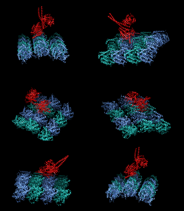
<span class="mw-page-title-main">Kinesin</span> Eukaryotic motor protein

A kinesin is a protein belonging to a class of motor proteins found in eukaryotic cells. Kinesins move along microtubule (MT) filaments and are powered by the hydrolysis of adenosine triphosphate (ATP). The active movement of kinesins supports several cellular functions including mitosis, meiosis and transport of cellular cargo, such as in axonal transport, and intraflagellar transport. Most kinesins walk towards the plus end of a microtubule, which, in most cells, entails transporting cargo such as protein and membrane components from the center of the cell towards the periphery. This form of transport is known as anterograde transport. In contrast, dyneins are motor proteins that move toward the minus end of a microtubule in retrograde transport.

<span class="mw-page-title-main">Dynein</span> Class of enzymes

Dyneins are a family of cytoskeletal motor proteins that move along microtubules in cells. They convert the chemical energy stored in ATP to mechanical work. Dynein transports various cellular cargos, provides forces and displacements important in mitosis, and drives the beat of eukaryotic cilia and flagella. All of these functions rely on dynein's ability to move towards the minus-end of the microtubules, known as retrograde transport; thus, they are called "minus-end directed motors". In contrast, most kinesin motor proteins move toward the microtubules' plus-end, in what is called anterograde transport.

<span class="mw-page-title-main">Axoneme</span>

An axoneme, also called an axial filament is the microtubule-based cytoskeletal structure that forms the core of a cilium or flagellum. Cilia and flagella are found on many cells, organisms, and microorganisms, to provide motility. The axoneme serves as the "skeleton" of these organelles, both giving support to the structure and, in some cases, the ability to bend. Though distinctions of function and length may be made between cilia and flagella, the internal structure of the axoneme is common to both.

In cell biology, microtubule-associated proteins (MAPs) are proteins that interact with the microtubules of the cellular cytoskeleton. MAPs are integral to the stability of the cell and its internal structures and the transport of components within the cell.

<span class="mw-page-title-main">Axonal transport</span> Movement of organelles

Axonal transport, also called axoplasmic transport or axoplasmic flow, is a cellular process responsible for movement of mitochondria, lipids, synaptic vesicles, proteins, and other organelles to and from a neuron's cell body, through the cytoplasm of its axon called the axoplasm. Since some axons are on the order of meters long, neurons cannot rely on diffusion to carry products of the nucleus and organelles to the end of their axons. Axonal transport is also responsible for moving molecules destined for degradation from the axon back to the cell body, where they are broken down by lysosomes.
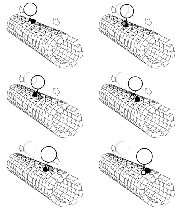
<span class="mw-page-title-main">Motor protein</span> Class of molecular proteins

Motor proteins are a class of molecular motors that can move along the cytoplasm of cells. They convert chemical energy into mechanical work by the hydrolysis of ATP. Flagellar rotation, however, is powered by a proton pump.

<span class="mw-page-title-main">Dynactin</span>

Dynactin is a 23 subunit protein complex that acts as a co-factor for the microtubule motor cytoplasmic dynein-1. It is built around a short filament of actin related protein-1 (Arp1).

<span class="mw-page-title-main">CLIP1</span> Protein-coding gene in the species Homo sapiens

CAP-GLY domain containing linker protein 1, also known as CLIP1, is a protein which in humans is encoded by the CLIP1 gene.

<span class="mw-page-title-main">BICD1</span> Protein-coding gene in humans

Bicaudal D cargo adaptor 1 is a protein that in humans is encoded by the BICD1 gene.
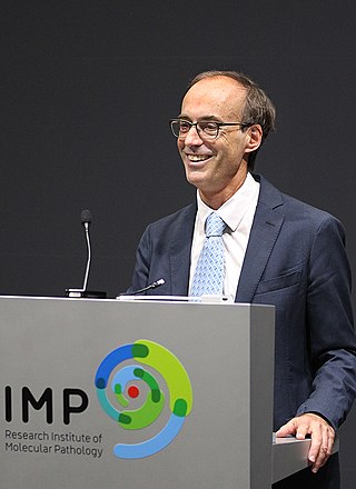
<span class="mw-page-title-main">Ronald Vale</span> American biochemist

Ronald David Vale ForMemRS is an American biochemist and cell biologist. He is a professor at the Department of Cellular and Molecular Pharmacology, University of California, San Francisco. His research is focused on motor proteins, particularly kinesin and dynein. He was awarded the Canada Gairdner International Award for Biomedical Research in 2019, the Shaw Prize in Life Science and Medicine in 2017 together with Ian Gibbons, and the Albert Lasker Award for Basic Medical Research in 2012 alongside Michael Sheetz and James Spudich. He is a fellow of the American Academy of Arts and Sciences and a member of the National Academy of Sciences. He was the president of the American Society for Cell Biology in 2012. He has also been an investigator at the Howard Hughes Medical Institute since 1995. In 2019, Vale was named executive director of the Janelia Research Campus and a vice president of HHMI; his appointment began in early 2020.

<span class="mw-page-title-main">Intracellular transport</span> Directed movement of vesicles and substances within a cell

Intracellular transport is the movement of vesicles and substances within a cell. Intracellular transport is required for maintaining homeostasis within the cell by responding to physiological signals. Proteins synthesized in the cytosol are distributed to their respective organelles, according to their specific amino acid’s sorting sequence. Eukaryotic cells transport packets of components to particular intracellular locations by attaching them to molecular motors that haul them along microtubules and actin filaments. Since intracellular transport heavily relies on microtubules for movement, the components of the cytoskeleton play a vital role in trafficking vesicles between organelles and the plasma membrane by providing mechanical support. Through this pathway, it is possible to facilitate the movement of essential molecules such as membrane‐bounded vesicles and organelles, mRNA, and chromosomes.

<span class="mw-page-title-main">Microtubule plus-end tracking protein</span>

Microtubule plus-end/positive-end tracking proteins or +TIPs are a type of microtubule associated protein (MAP) which accumulate at the plus ends of microtubules. +TIPs are arranged in diverse groups which are classified based on their structural components; however, all classifications are distinguished by their specific accumulation at the plus end of microtubules and their ability to maintain interactions between themselves and other +TIPs regardless of type. +TIPs can be either membrane bound or cytoplasmic, depending on the type of +TIPs. Most +TIPs track the ends of extending microtubules in a non-autonomous manner.

<span class="mw-page-title-main">Marileen Dogterom</span> Dutch biophysicist

Marileen Dogterom is a Dutch biophysicist and professor at the Kavli Institute of Nanoscience at Delft University of Technology. She published in Science, Cell, and Nature and is notable for her research of the cell cytoskeleton. For this research, she was awarded the 2018 Spinoza Prize.

<span class="mw-page-title-main">Neurotubule</span>

Neurotubules are microtubules found in neurons in nervous tissues. Along with neurofilaments and microfilaments, they form the cytoskeleton of neurons. Neurotubules are undivided hollow cylinders that are made up of tubulin protein polymers and arrays parallel to the plasma membrane in neurons. Neurotubules have an outer diameter of about 23 nm and an inner diameter, also known as the central core, of about 12 nm. The wall of the neurotubules is about 5 nm in width. There is a non-opaque clear zone surrounding the neurotubule and it is about 40 nm in diameter. Like microtubules, neurotubules are greatly dynamic and the length of them can be adjusted by polymerization and depolymerization of tubulin.

<span class="mw-page-title-main">Casper Hoogenraad</span> Dutch biologist

Casper Hoogenraad is a Dutch Cell Biologist who specializes in molecular neuroscience. The focus of his research is the basic molecular and cellular mechanisms that regulate the development and function of the brain. As of January 2020, he serves as Vice President of Neuroscience at Genentech Research and Early Development.

Jennifer L. Ross is an American physicist who is Professor and Chair of the Department of Physics at Syracuse University. Her research considers active biological condensed matter physics. She was elected fellow of the American Physical Society in 2018 and American Association for the Advancement of Science in 2022.

<span class="mw-page-title-main">Erika Holzbaur</span> American biologist

Erika L F. Holzbaur is an American biologist who is the William Maul Measey Professor of Physiology at University of Pennsylvania Perelman School of Medicine. Her research considers the dynamics of organelle motility along cytoskeleton of cells. She is particularly interested in the molecular mechanisms that underpin neurodegenerative diseases.

J. Richard McIntosh is a Distinguished Professor Emeritus in Molecular, Cellular, and Developmental Biology at the University of Colorado Boulder. McIntosh first graduated from Harvard with a BA in Physics in 1961, and again with a Ph.D. in Biophysics in 1968. He began his teaching career at Harvard but has spent most of his career at the University of Colorado Boulder. At the University of Colorado Boulder, McIntosh taught biology courses at both the undergraduate and graduate levels. Additionally, he created an undergraduate course in the biology of cancer towards the last several years of his teaching career. McIntosh's research career looks at a variety of things, including different parts of mitosis, microtubules, and motor proteins.

References

  1. "Academy selects sixteen new members". Royal Netherlands Academy of Arts and Sciences. Retrieved 2 August 2015.
  2. 1 2 "Anna Akhmanova" (in Dutch). Kennisbits.nl. Archived from the original on 17 November 2015. Retrieved 2 August 2015.
  3. "Prof.dr. A.S. Akhmanova (1967 - )". Catalogus Professorum Academiæ Rheno-Traiectinæ, Utrecht University. Retrieved 8 May 2020.
  4. Sedwick, Caitlin (17 October 2011). "Anna Akhmanova: Great tips on microtubules". Journal of Cell Biology. 195 (2): 168–9. doi:10.1083/jcb.1952pi. PMC   3198163 . PMID   22006946.
  5. 1 2 3 4 5 6 Sedwick, Caitlin (2011-10-17). "Anna Akhmanova: Great tips on microtubules". J Cell Biol. 195 (2): 168–169. doi:10.1083/jcb.1952pi. ISSN   0021-9525. PMC   3198163 . PMID   22006946.
  6. 1 2 3 "Anna Akhmanova – Cellular Dynamics – Institute of Biodynamics and Biocomplexity". Institute of Biodynamics and Biocomplexity. Retrieved 2016-11-27.
  7. "Anna Akhmanova". Utrecht University. 2014-12-01. Retrieved 2016-11-27.
  8. 1 2 3 4 5 6 7 "Anna Akhmanova: Cellular Dynamics – Cell Biology Utrecht University". Cell Biology. Retrieved 2016-11-27.
  9. "ERC Synergy grant for Marileen Dogterom and Anna Akhmanova". AMOLF. 16 December 2013. Retrieved 2 August 2015.
  10. "Anna Akhmanova". European Molecular Biology Organization. Retrieved 8 May 2020.
  11. "Anna Akhmanova". Royal Netherlands Academy of Arts and Sciences. Archived from the original on 26 January 2019.
  12. "NWO Spinoza Prize 2018". Netherlands Organisation for Scientific Research. Archived from the original on 11 April 2020.